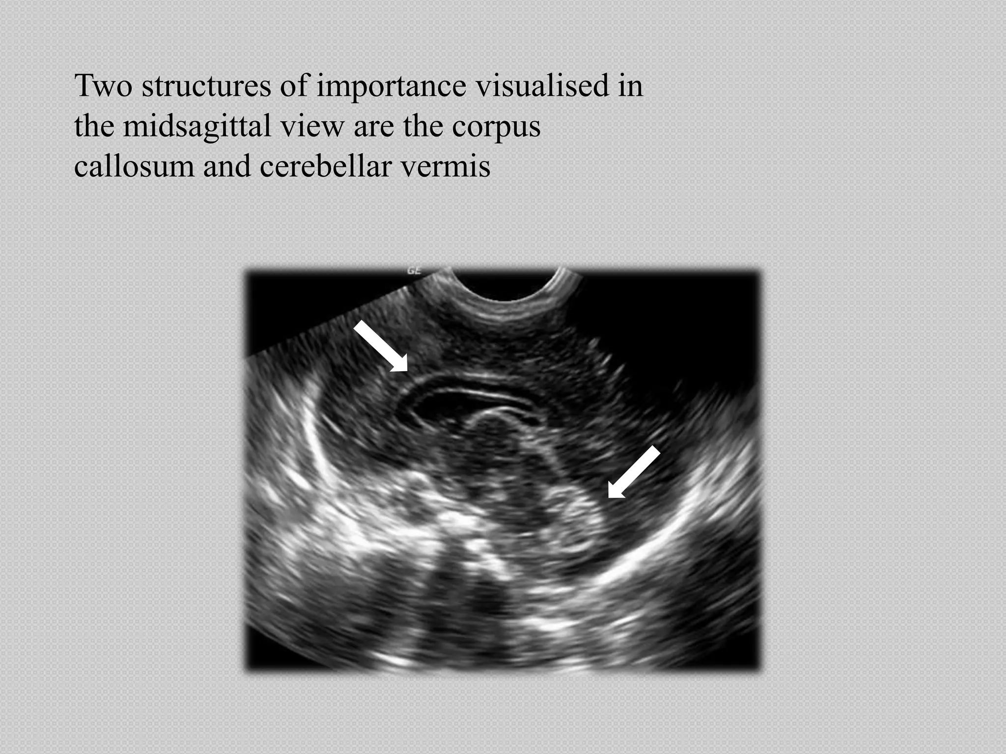
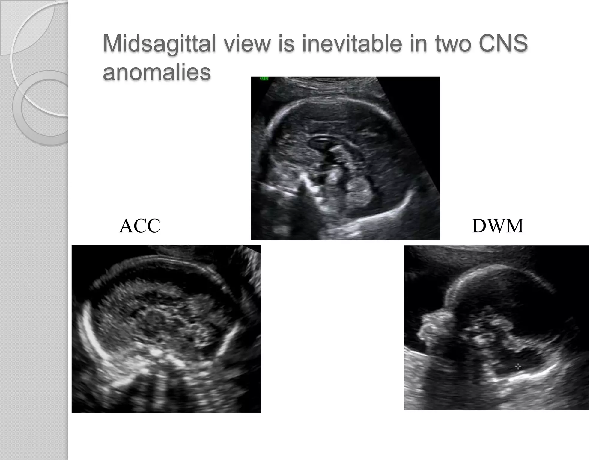
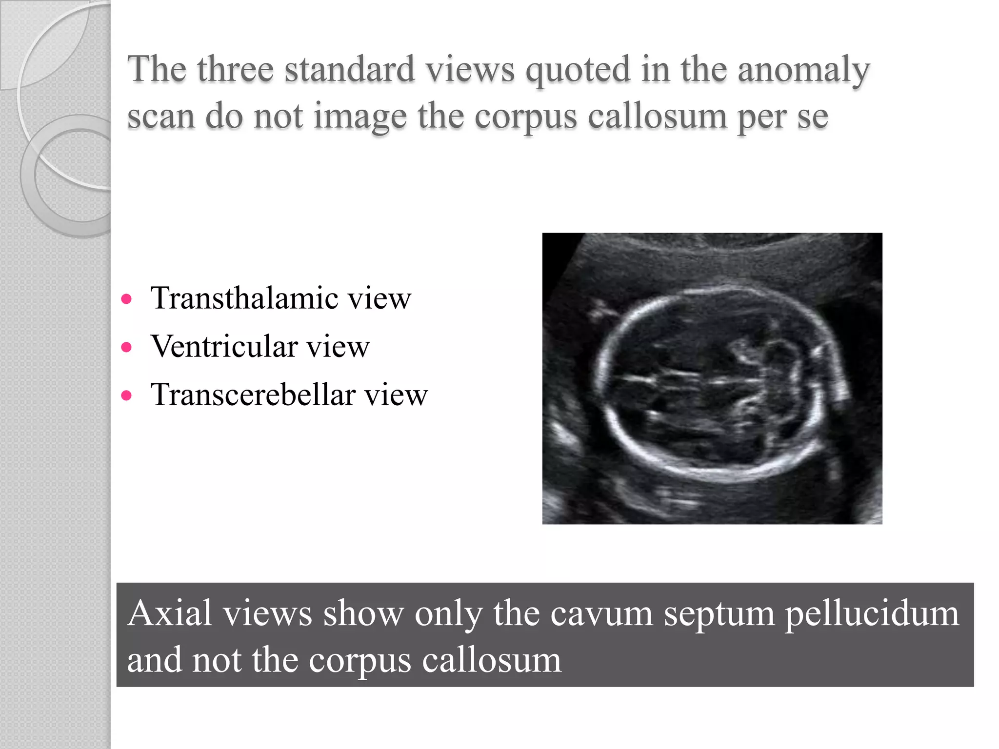
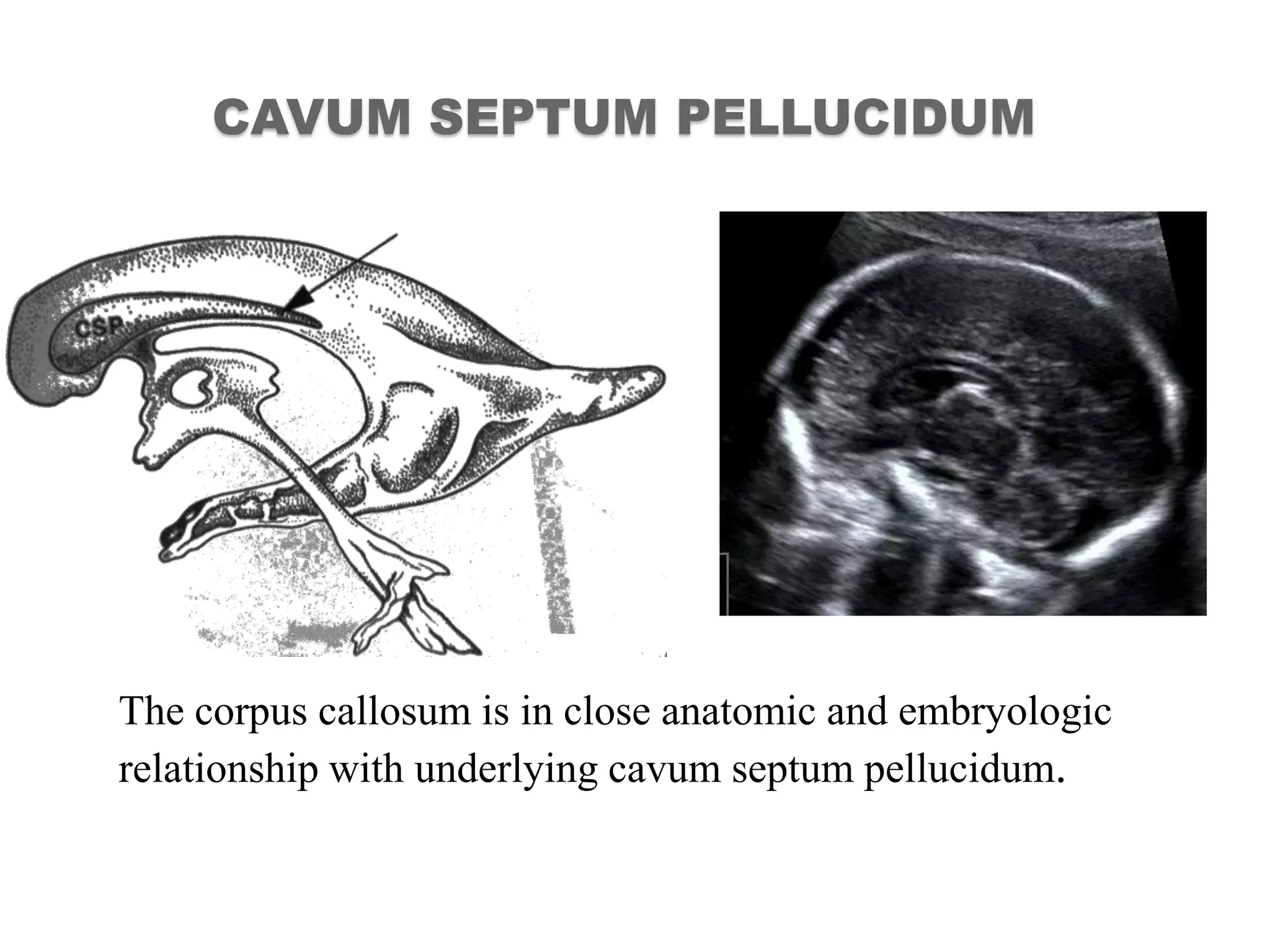
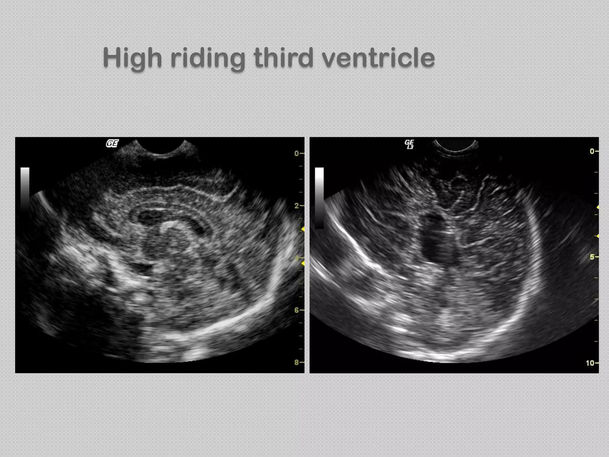
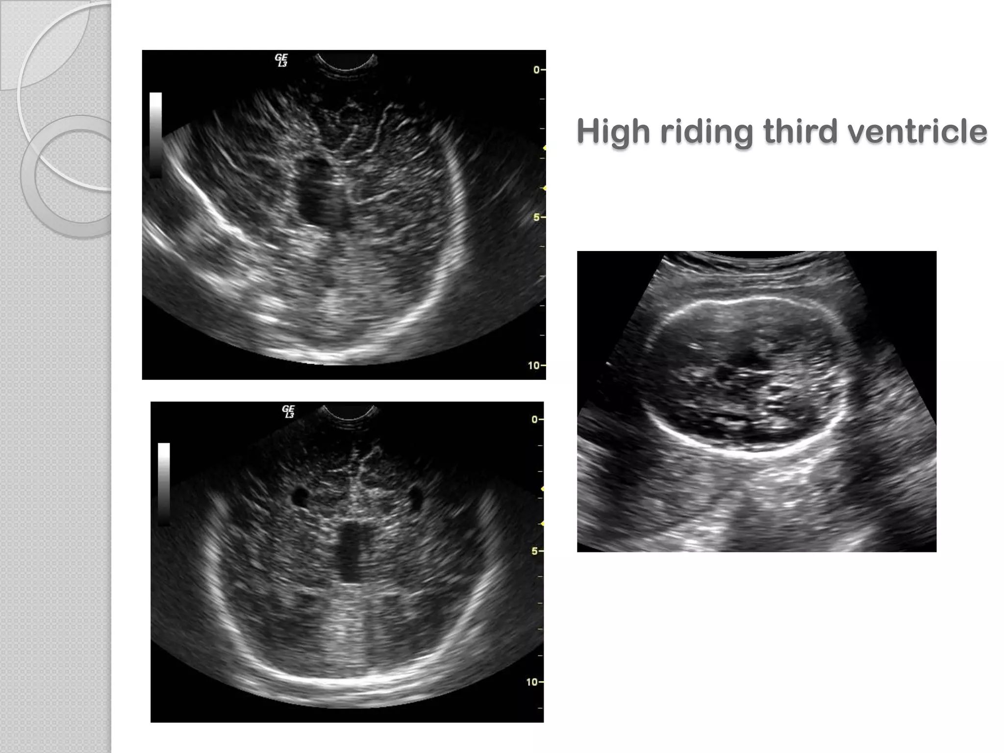
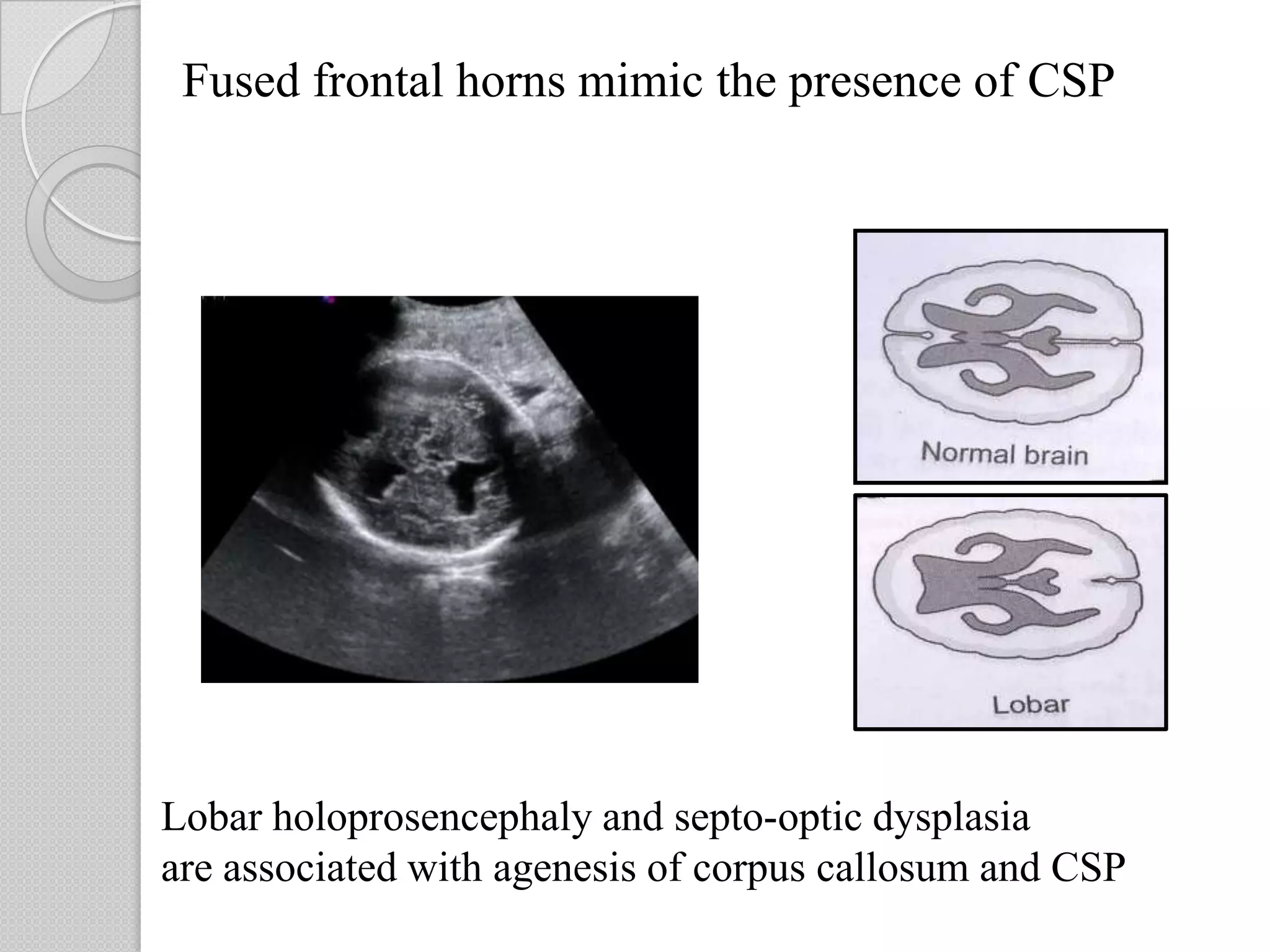
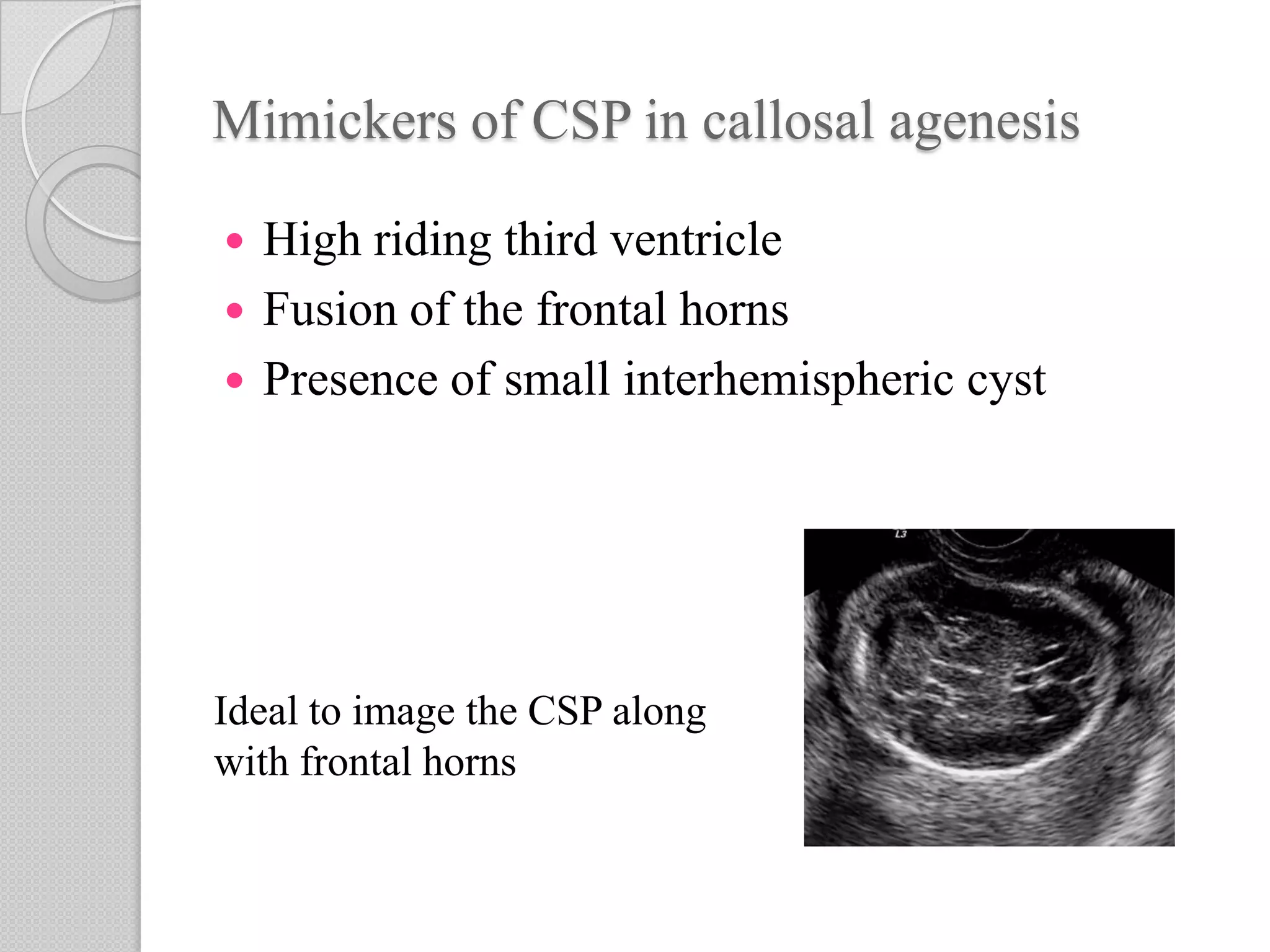
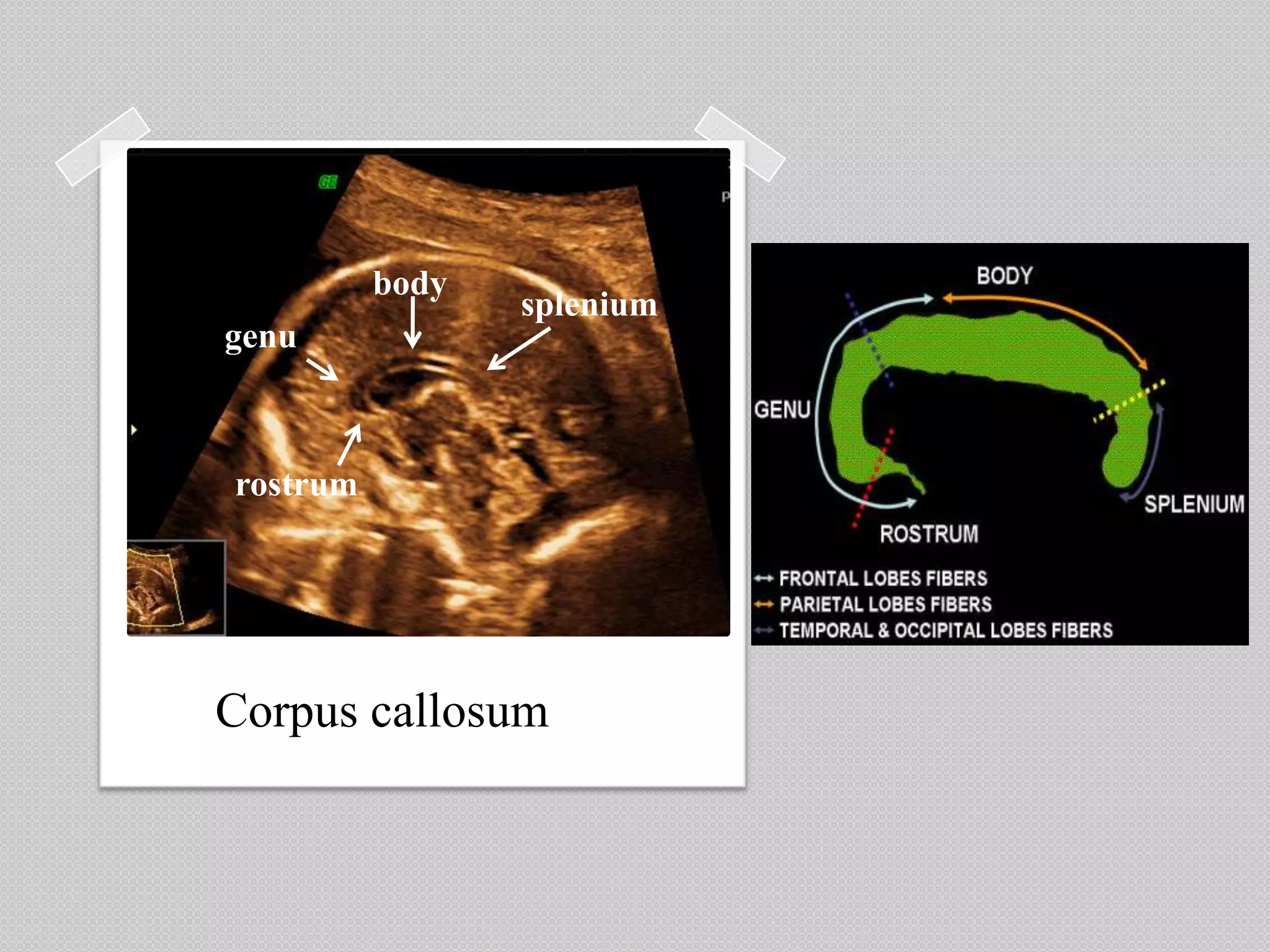
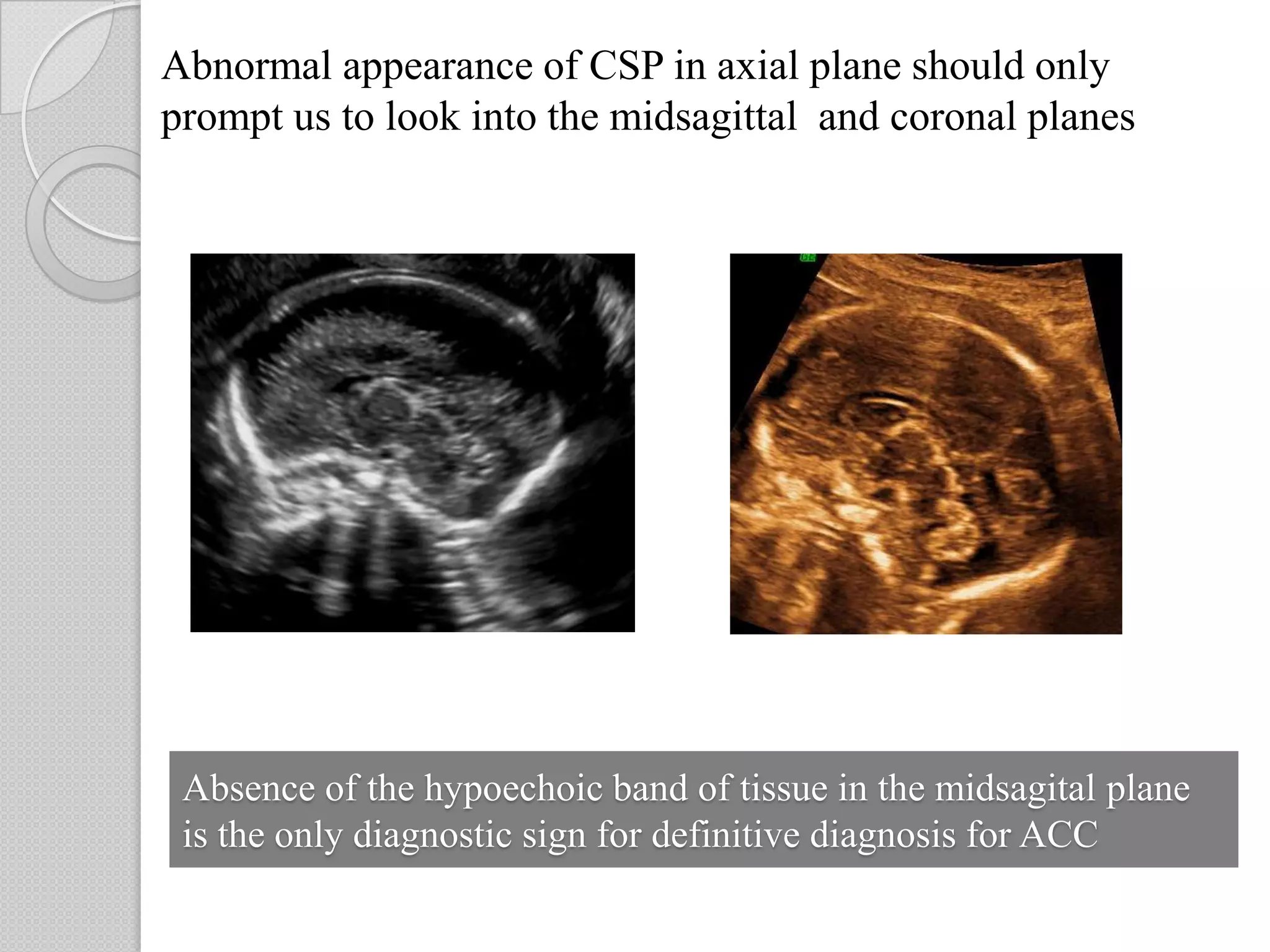
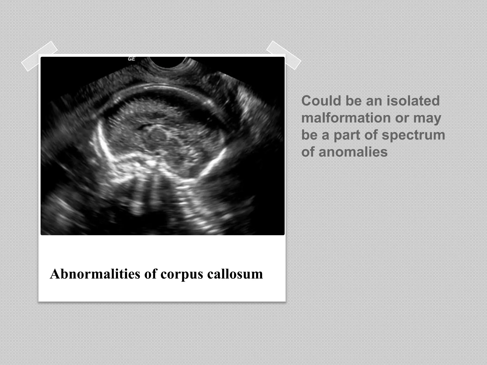
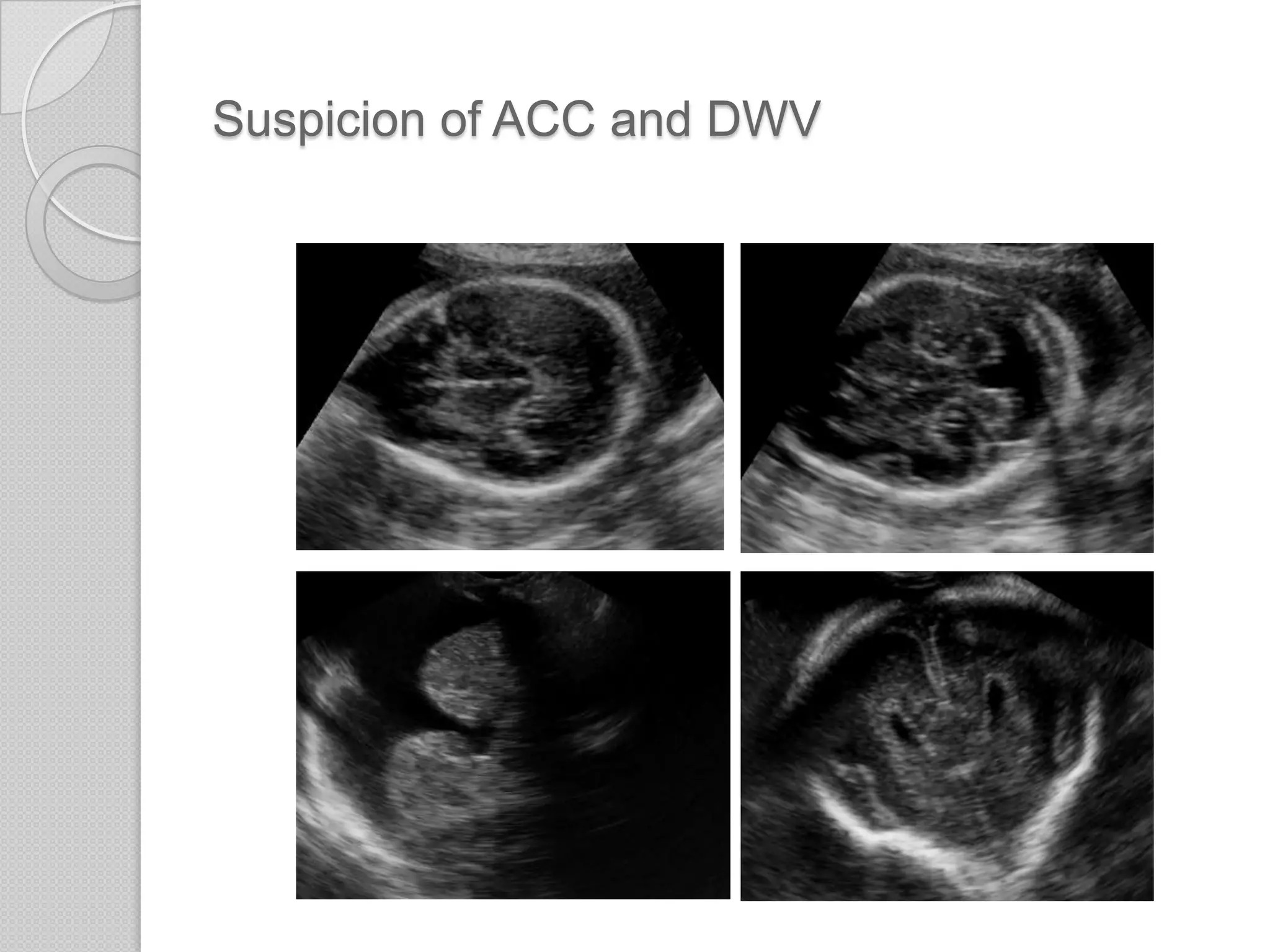
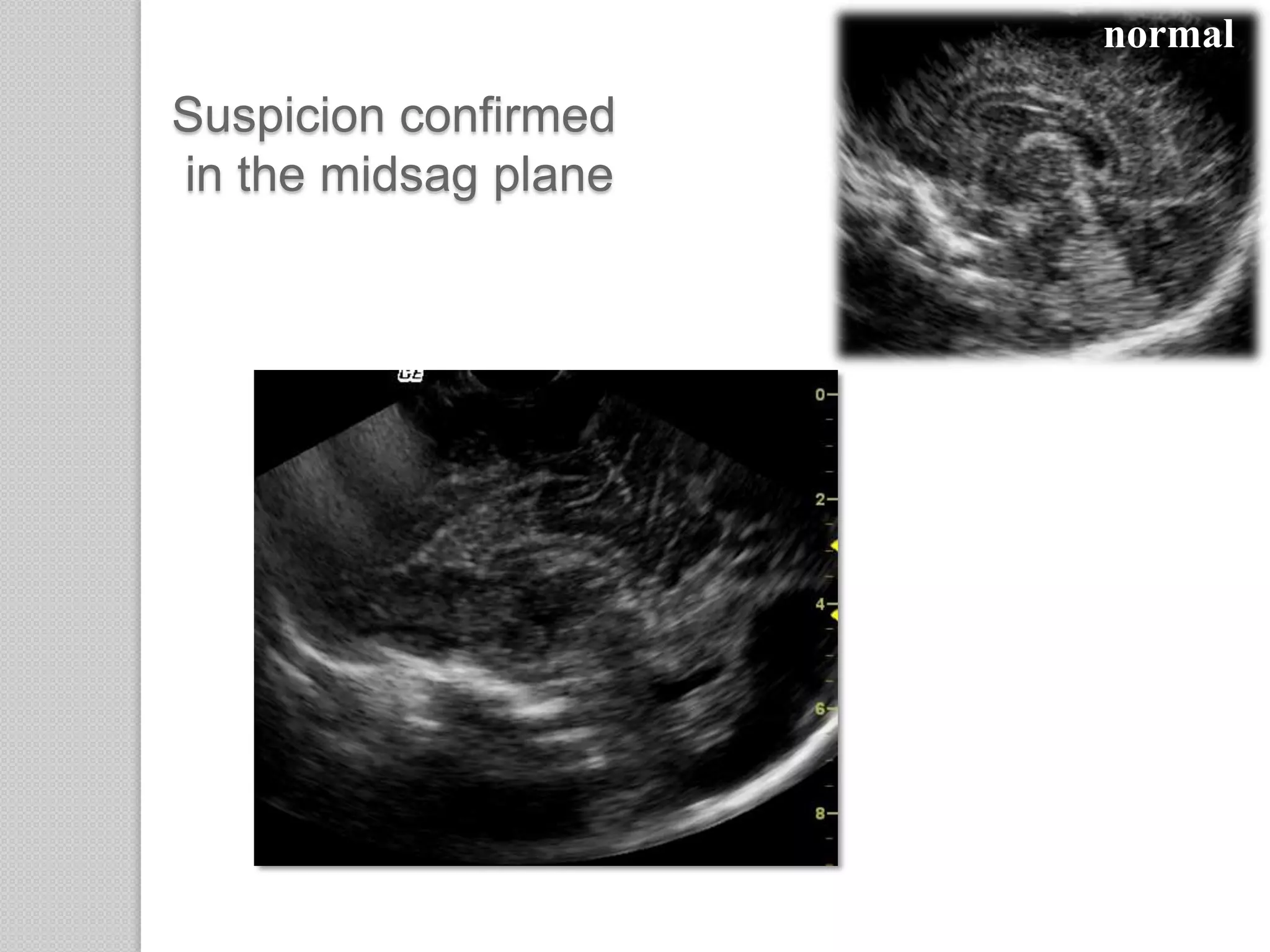
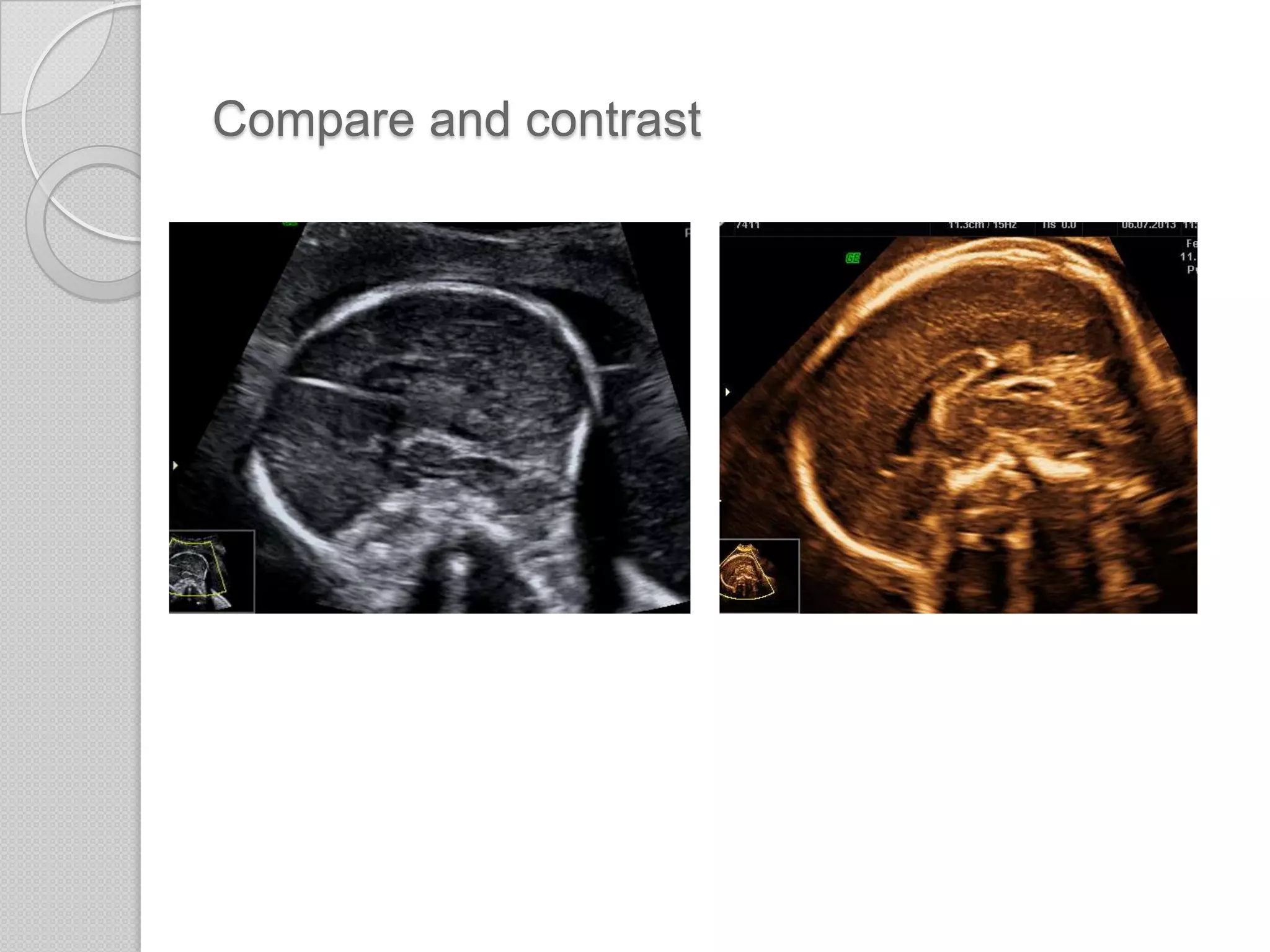
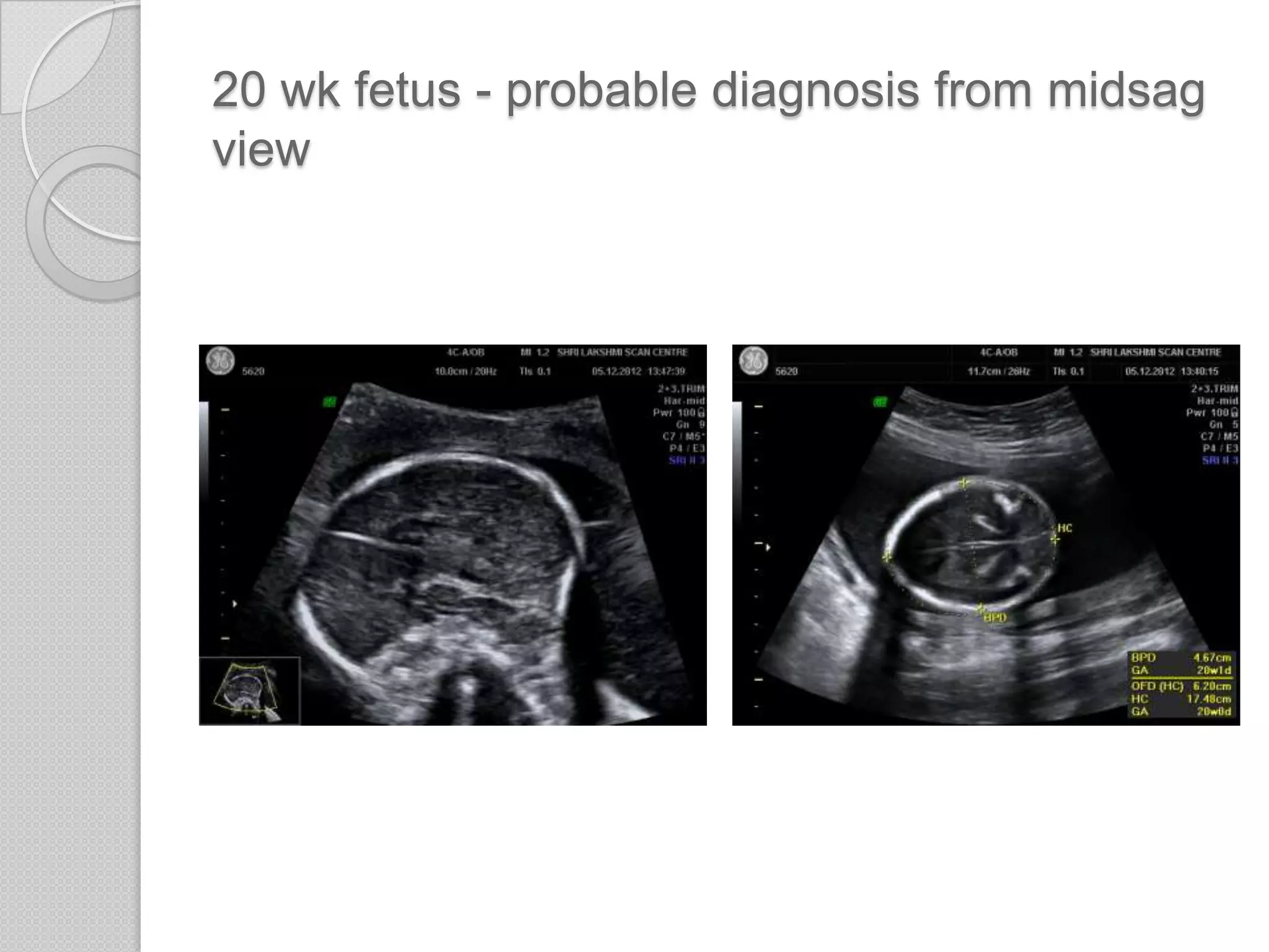
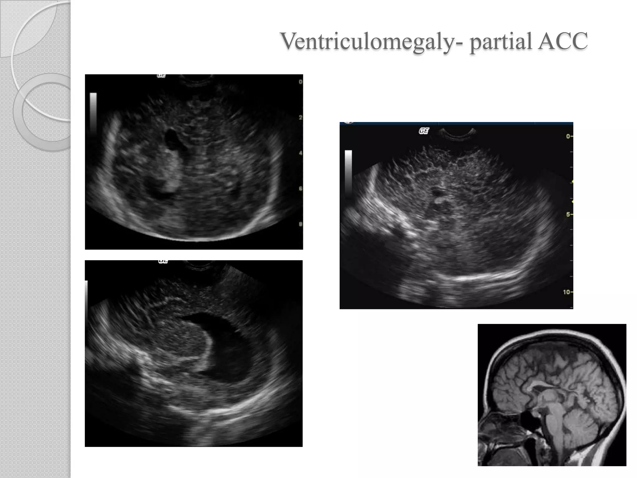
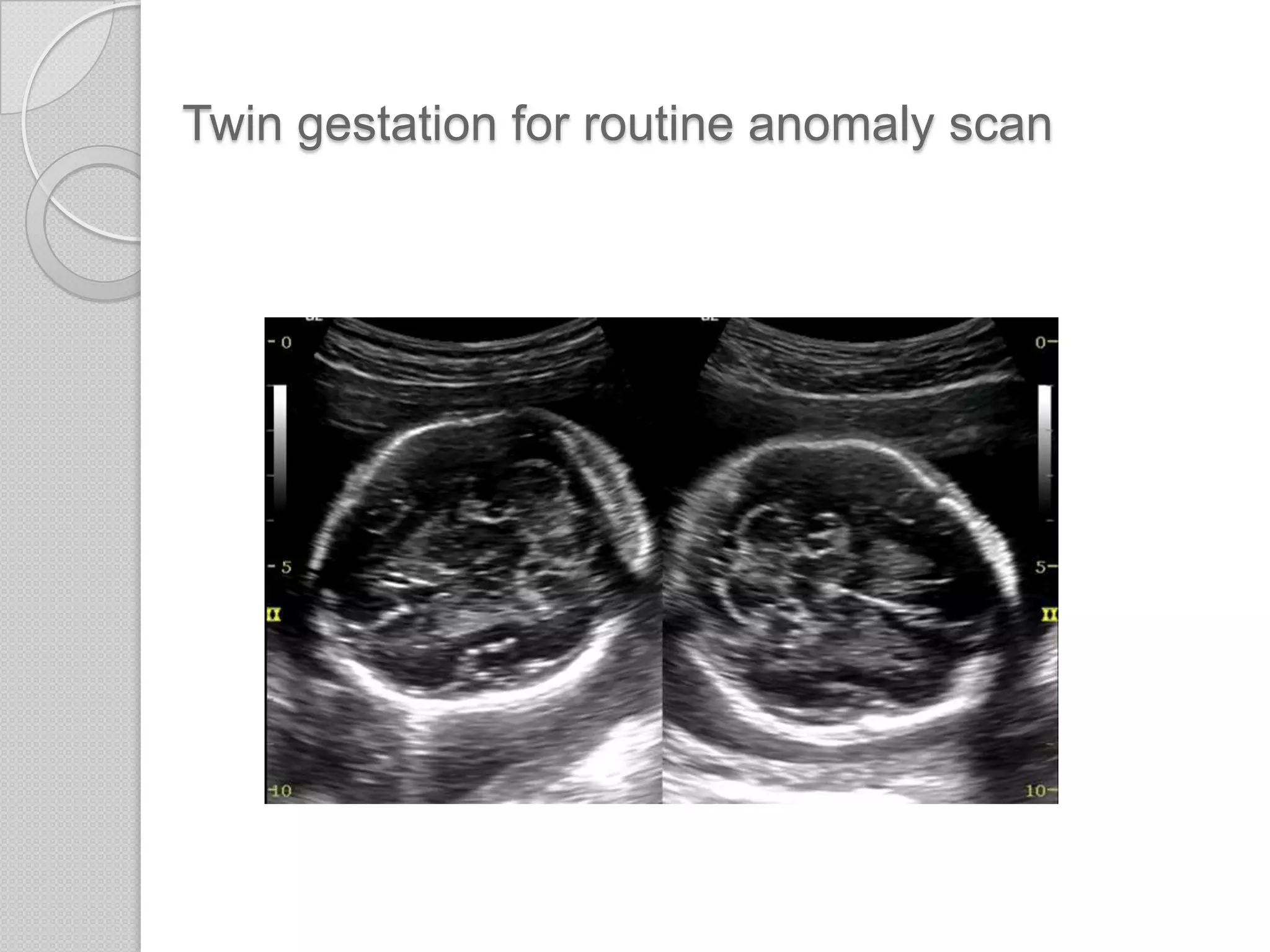
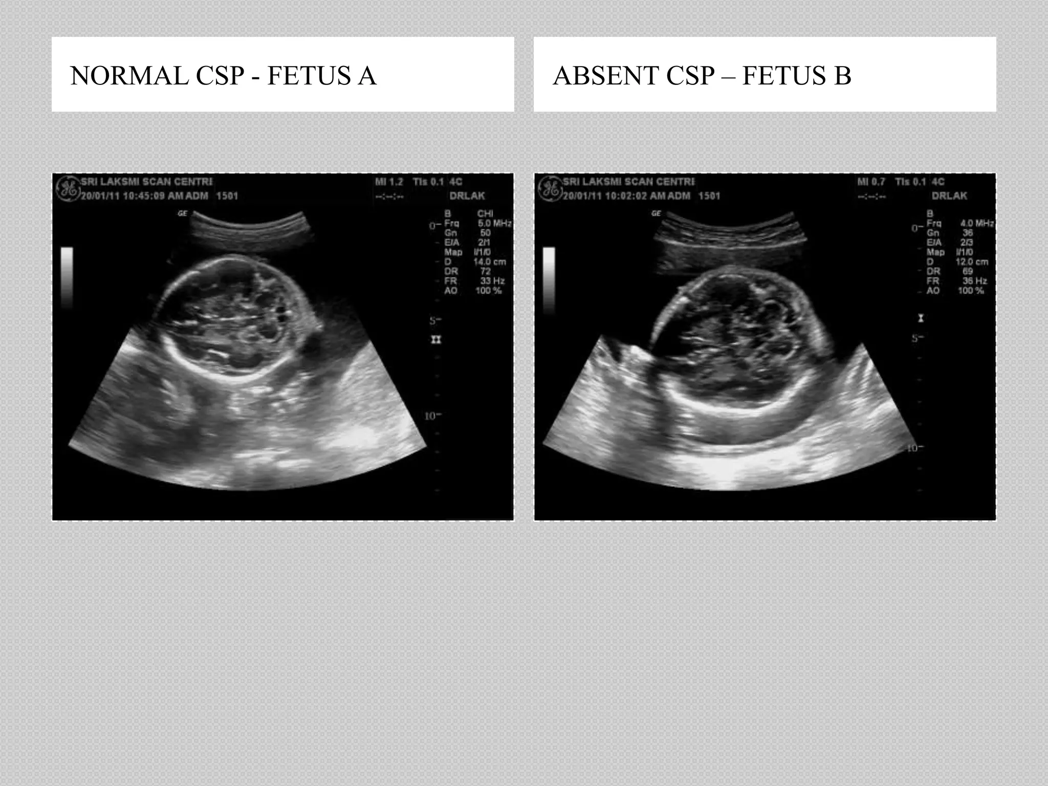
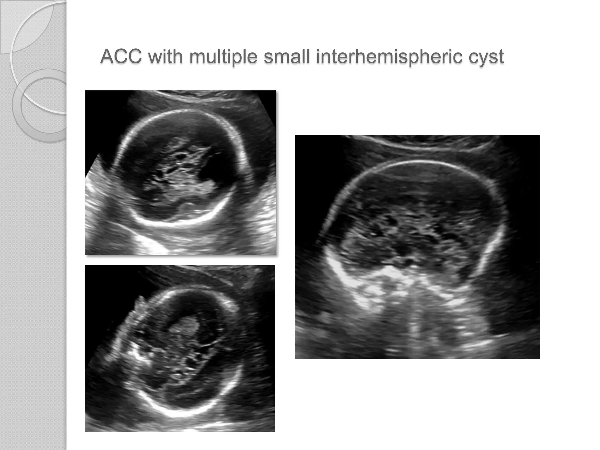
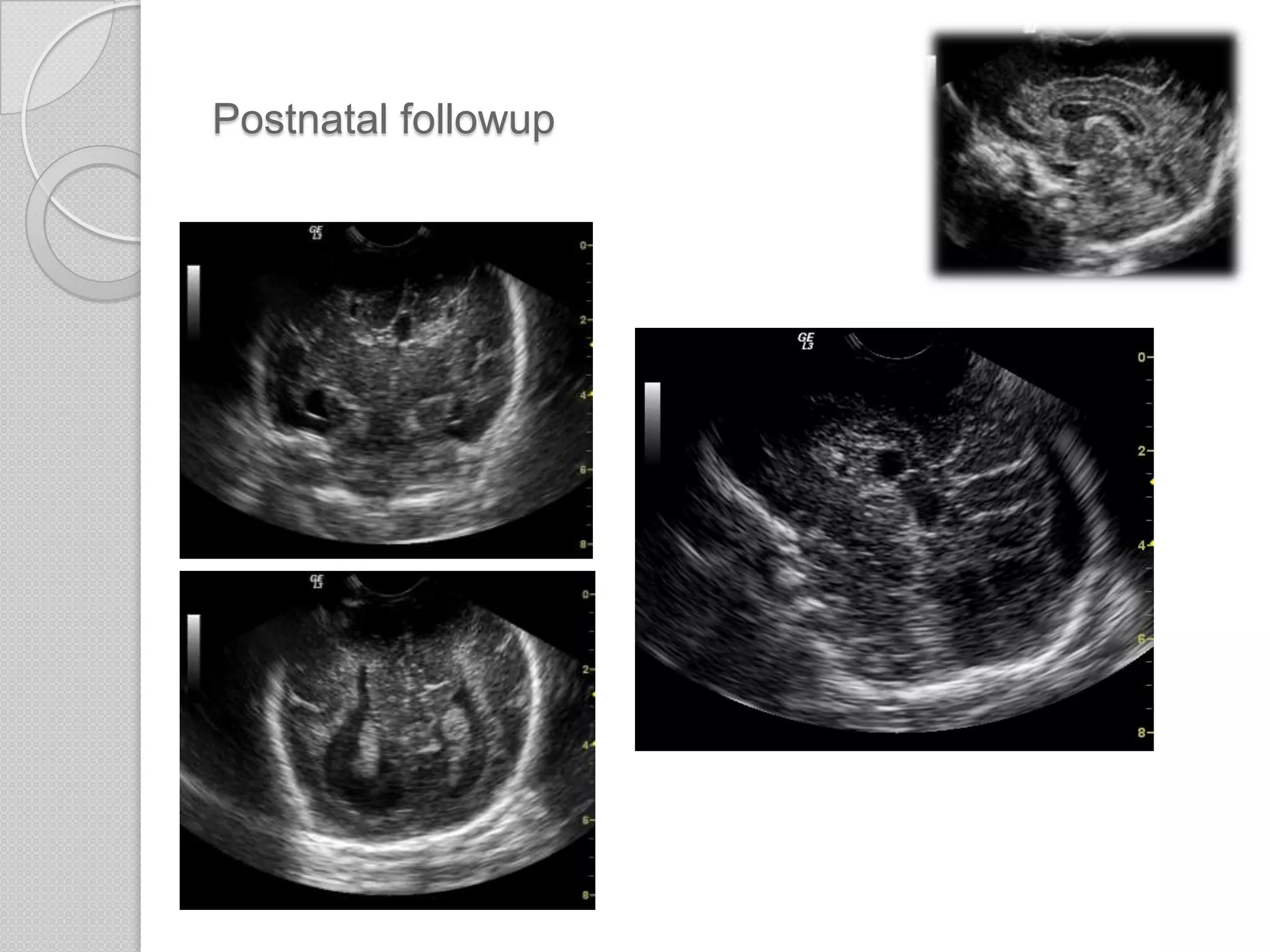
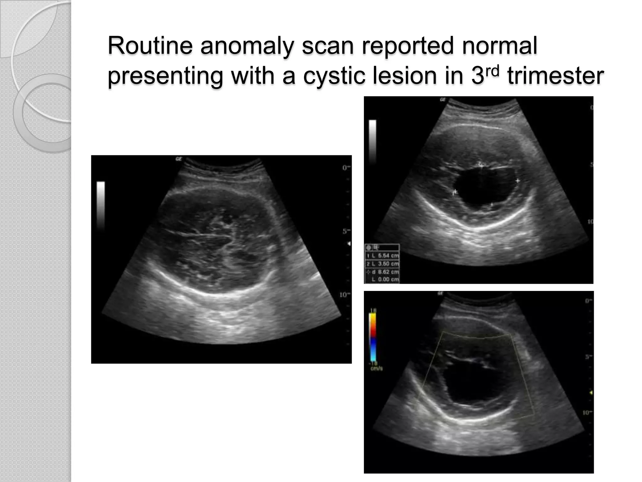
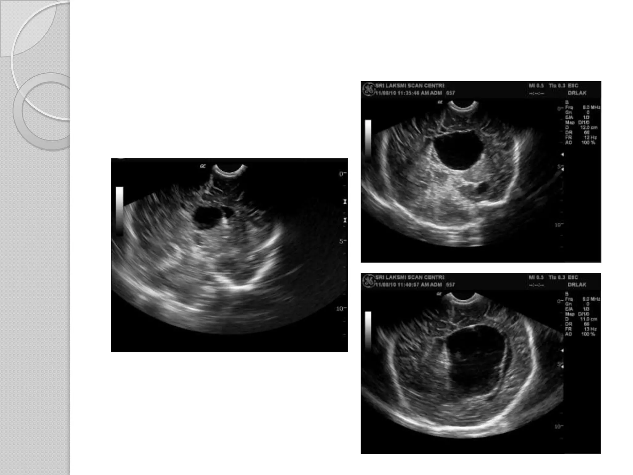
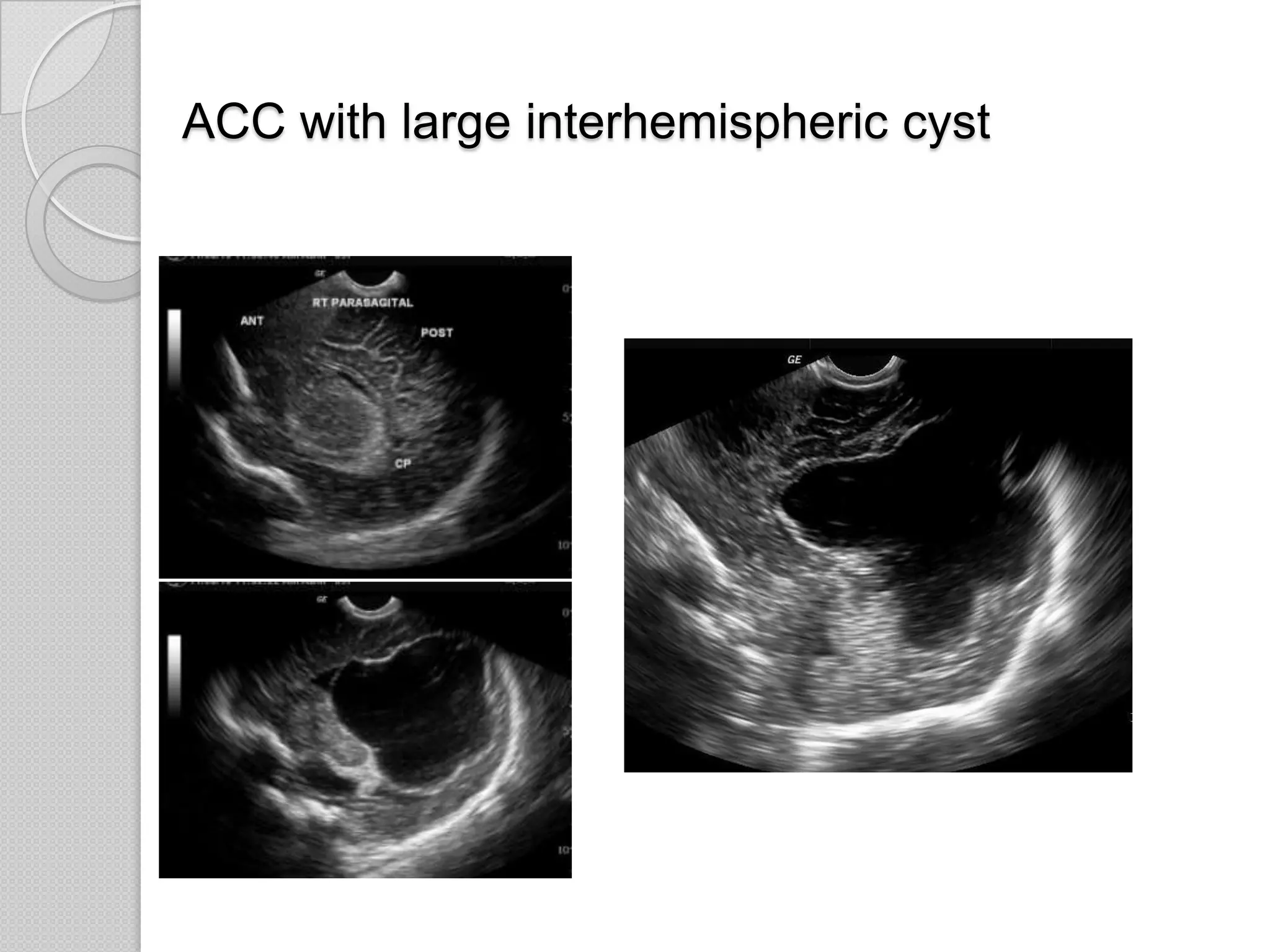
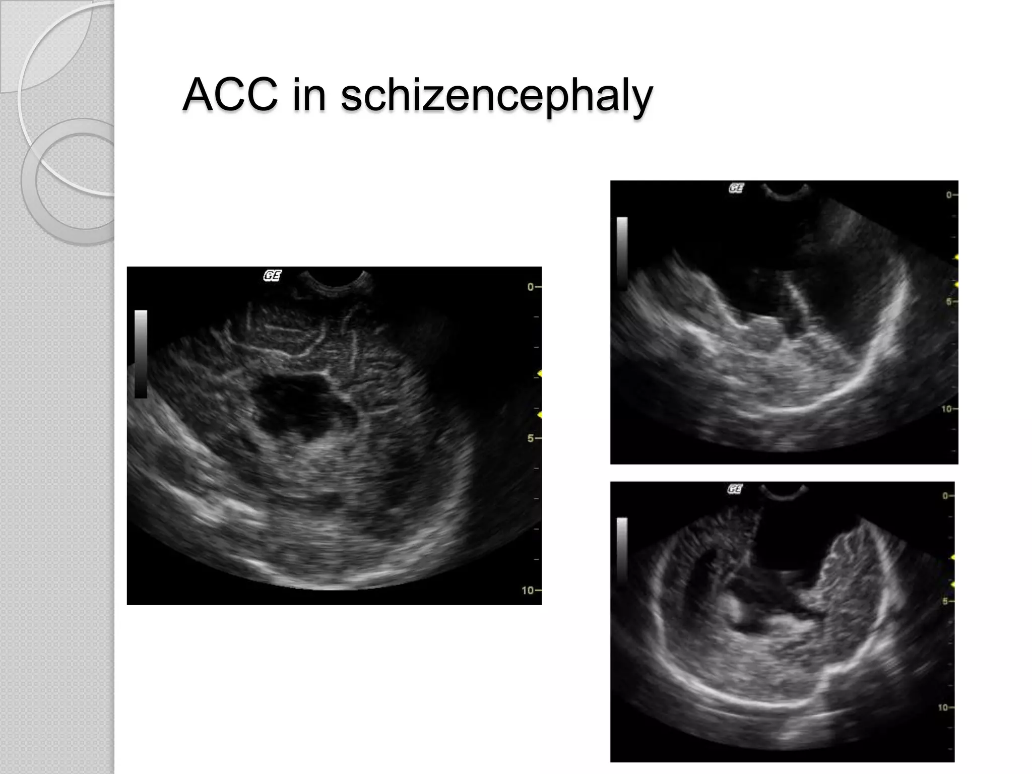
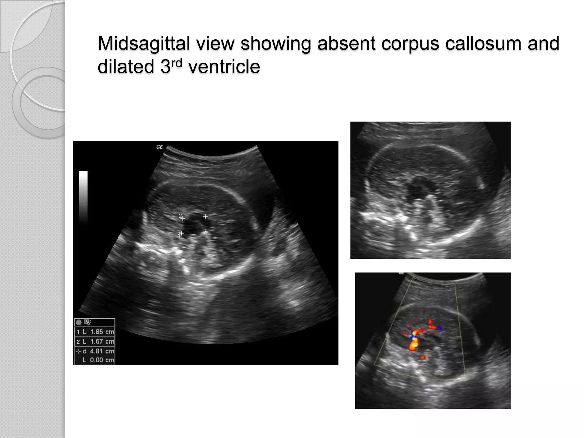
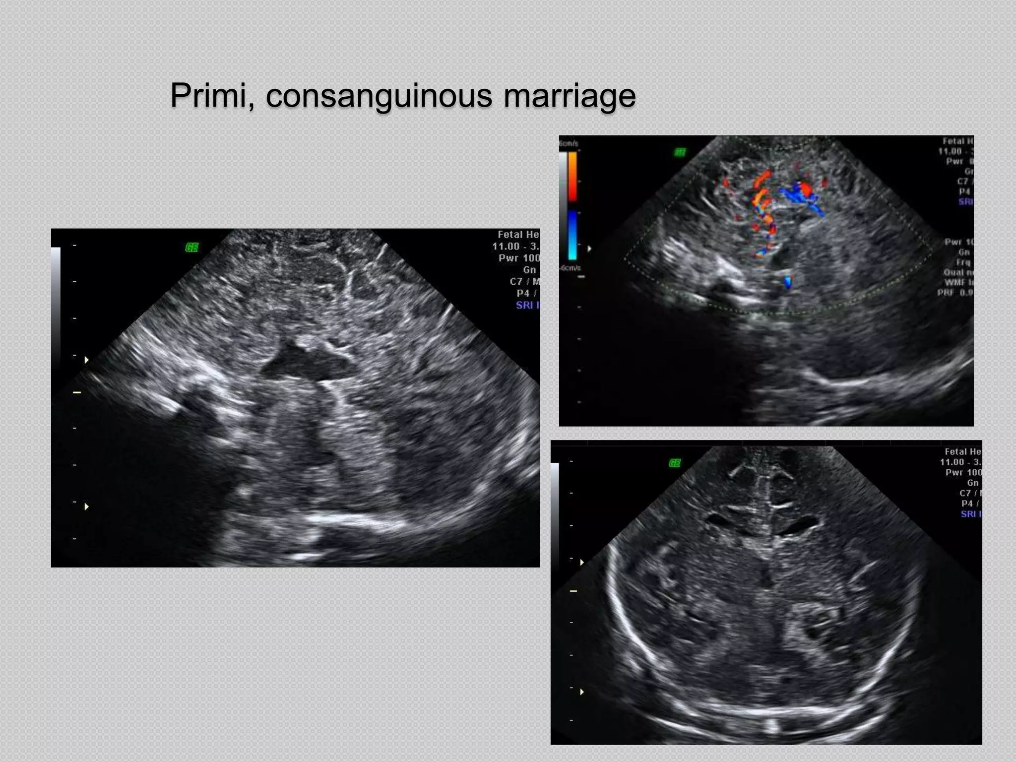
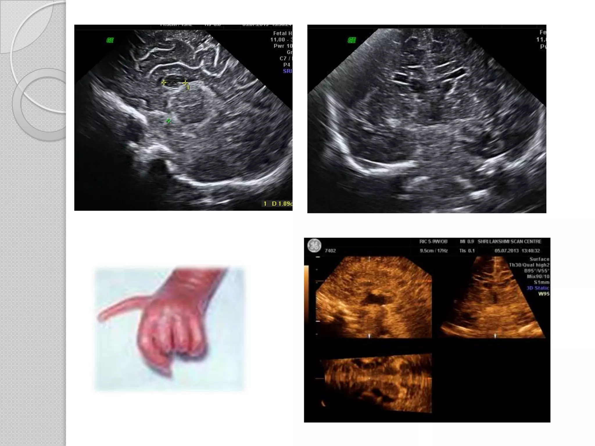
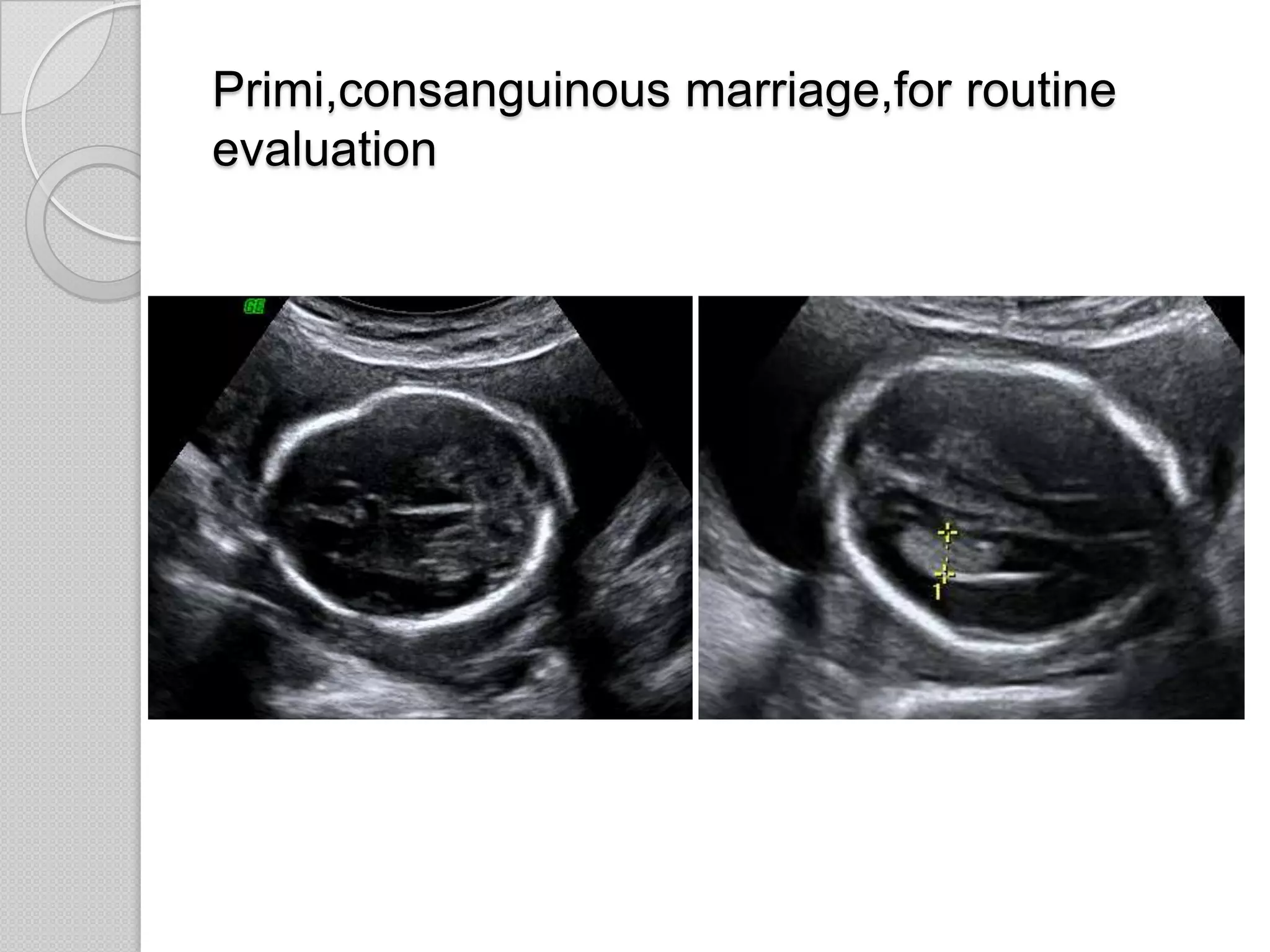
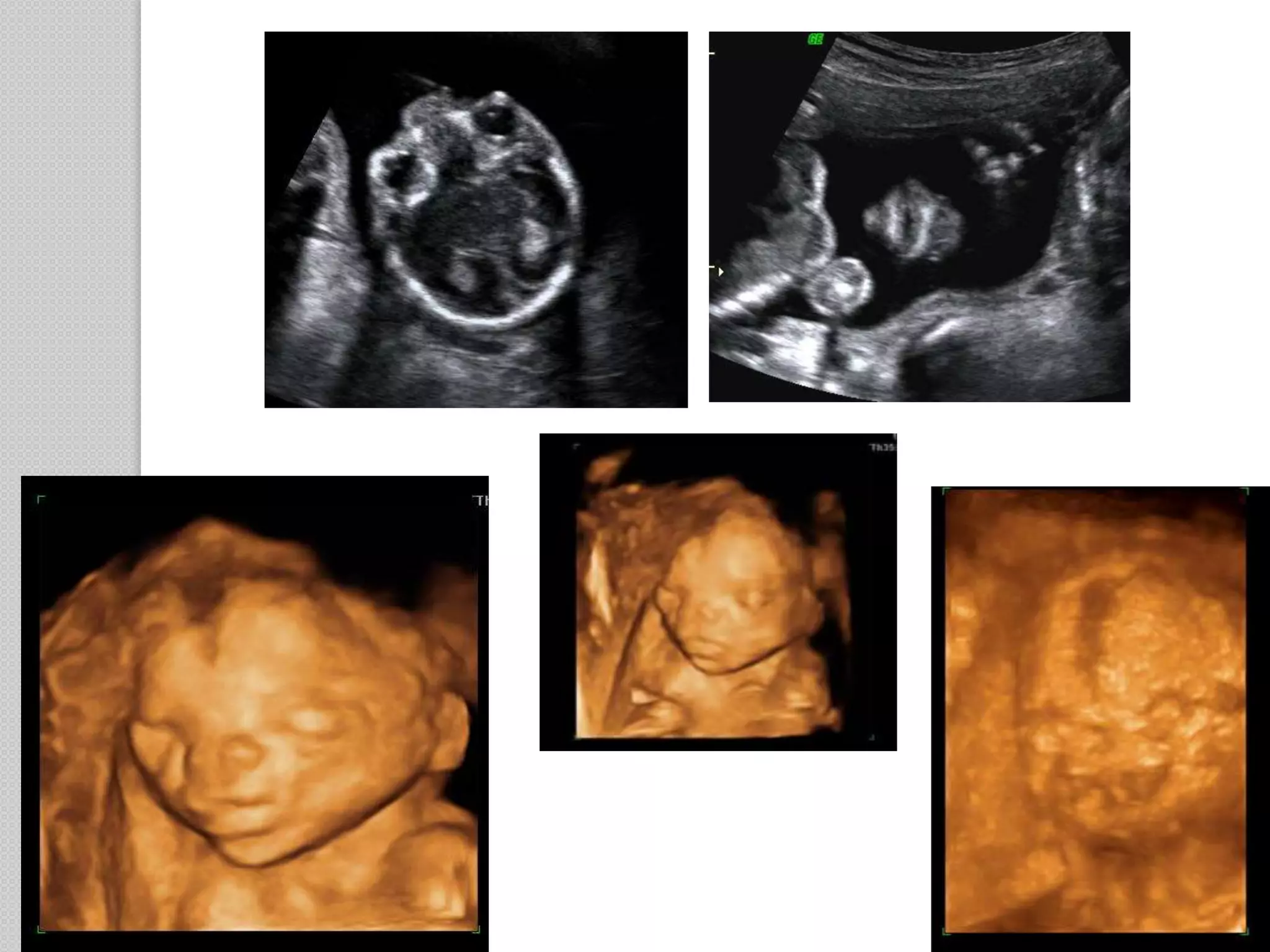
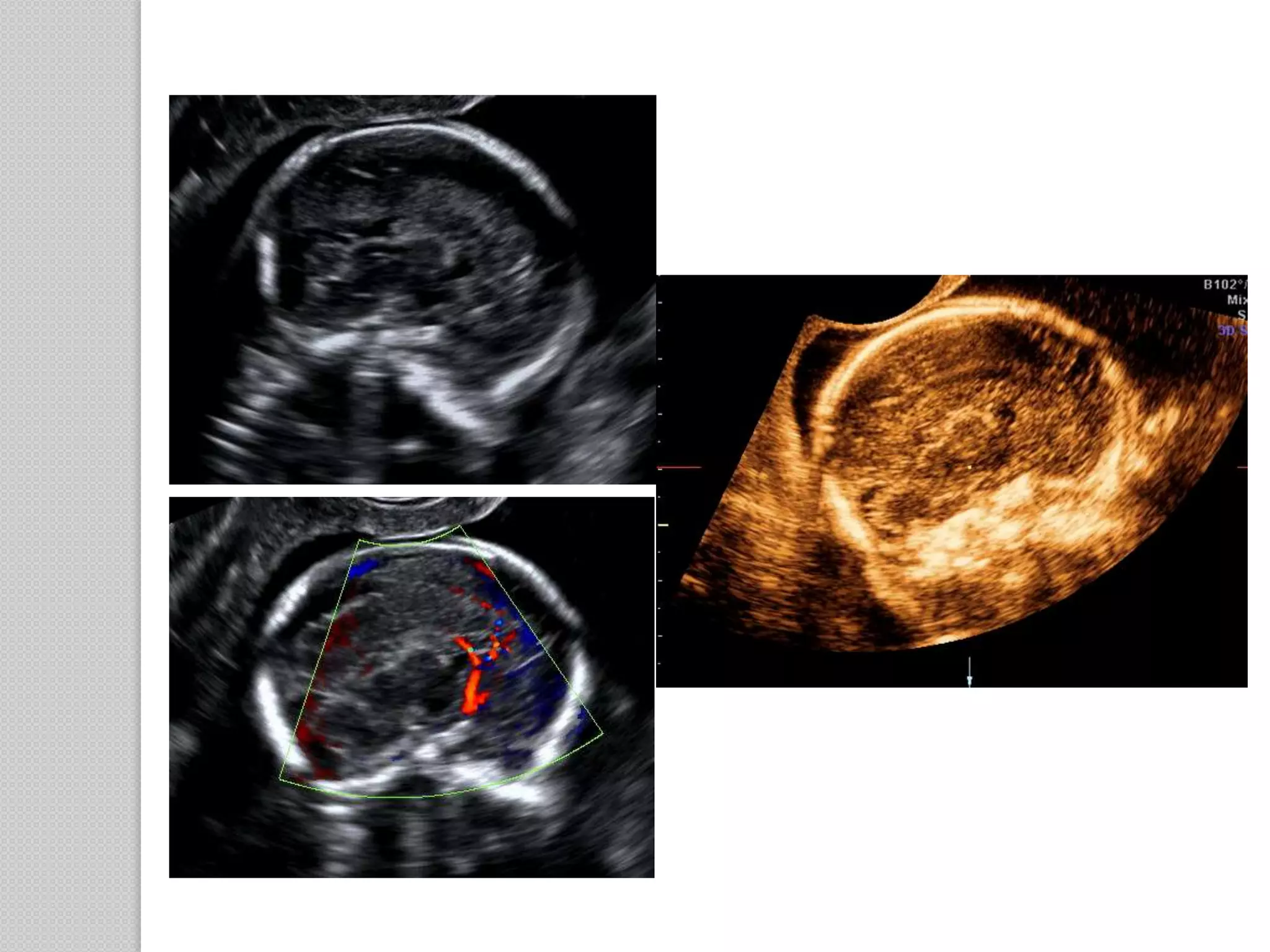
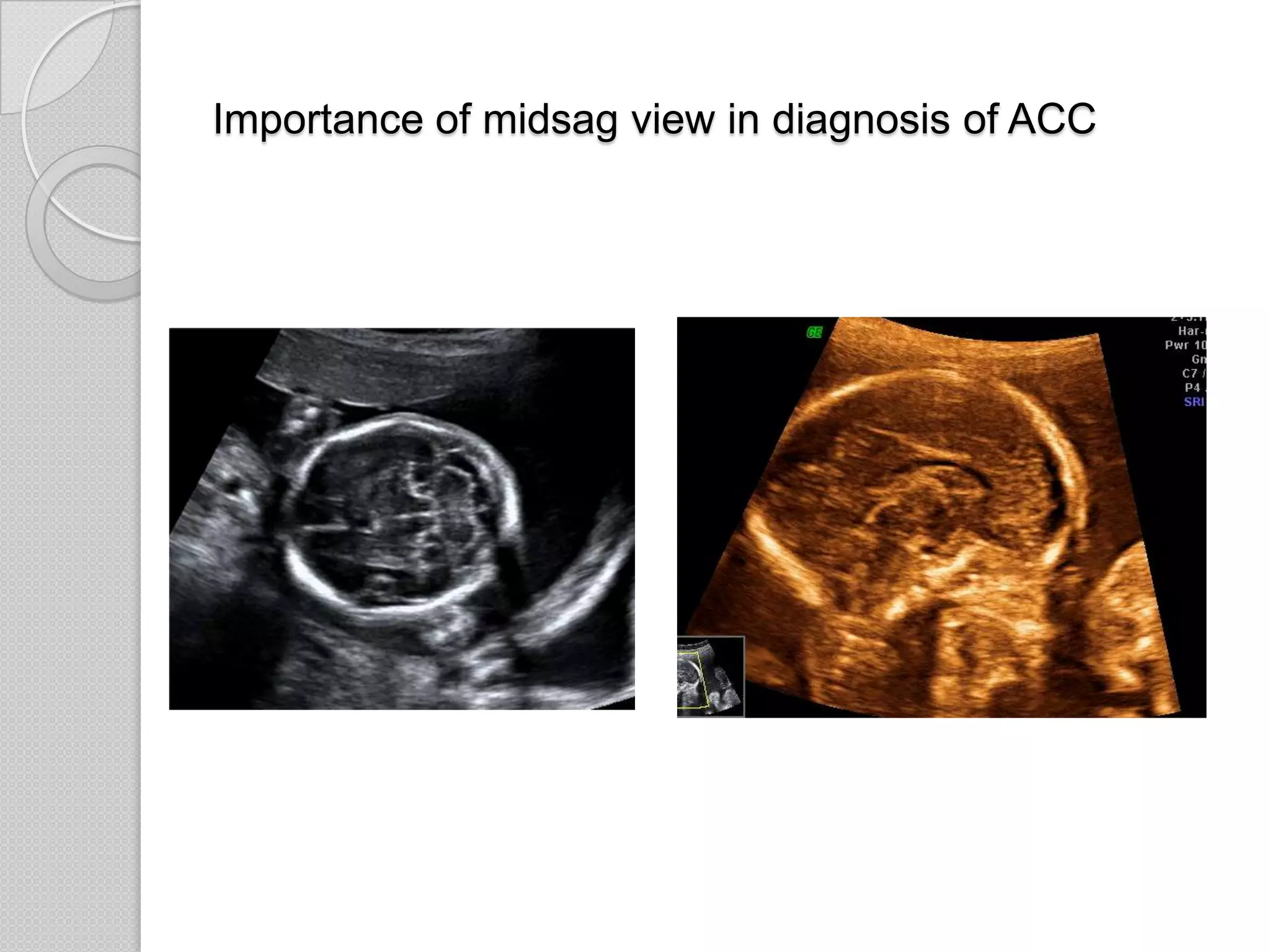
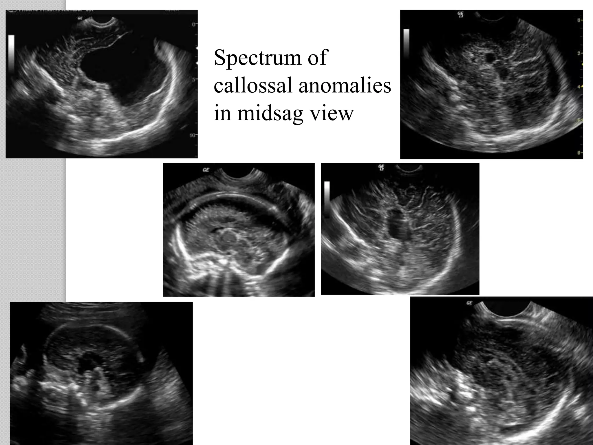
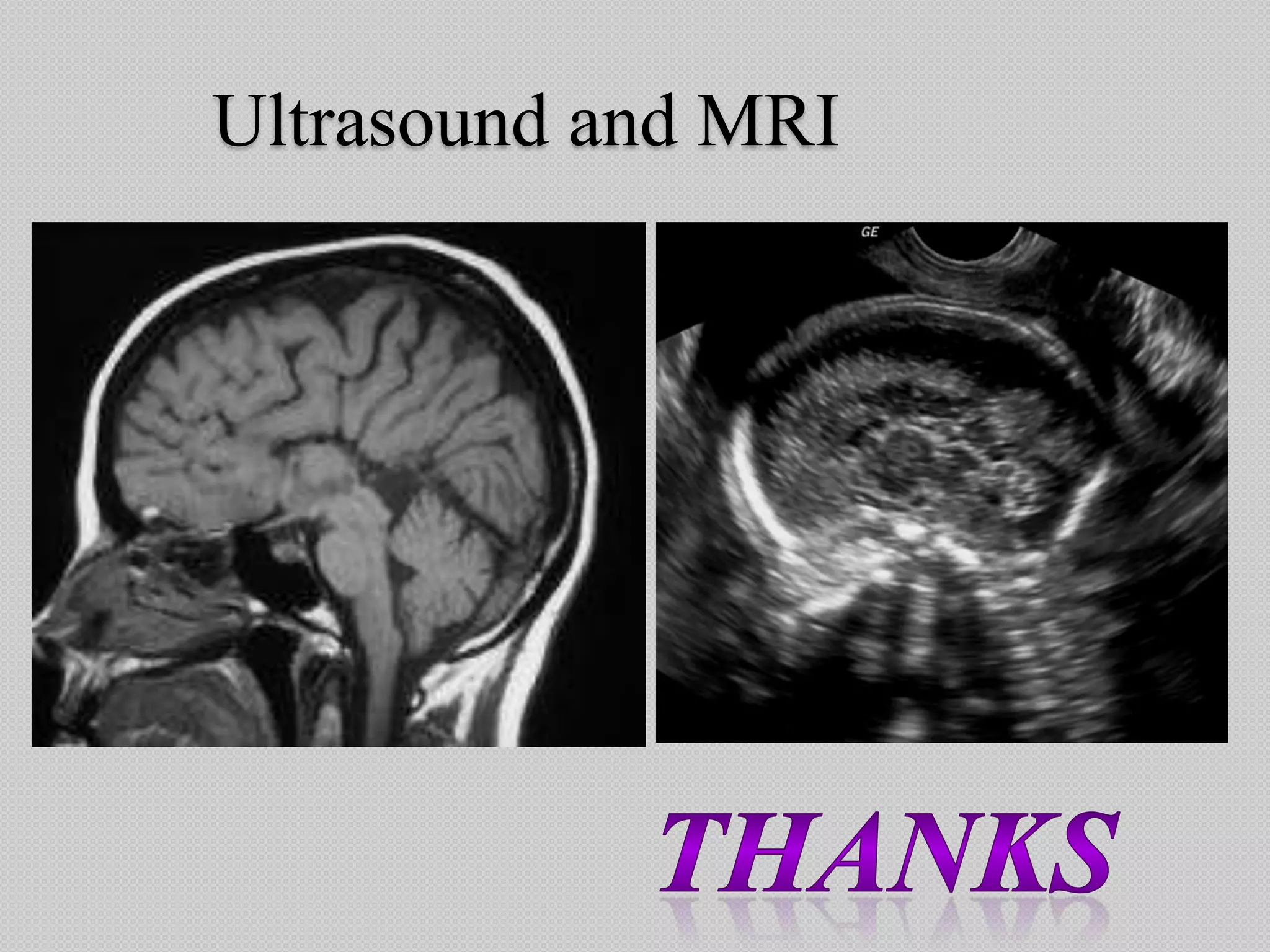
Abnormalities of corpus callosum
Dr.S.Lakshmy Ravi
Shri Lakshmi Clinic
Kaveripattinam
Callosal abnormality - incidence
Once thought to be a rare condition experts now
do not agree with this concept
National organisation for disorders of the corpus
callosum (NODCC) reports an incidence as high
as seven in 1000 children
Do we report such a high incidence
on antenatal ultrasound
An abnormality of corpus callosum is a frequently
missed malformation because imaging the entire corpus
callosum per se is not a part of our routine anomaly scan
Indirect markers of an evidence of abnormality of
callosum is easily obtained from routine axial views but
may sometimes be associated with pitfalls
Midline sagittal section of the brain is
generally not included in every anomaly
scan
Two structures of importance visualised in
the midsagittal view are the corpus
callosum and cerebellar vermis
Midsagittal view is inevitable in two CNS
anomalies
ACC DWM
The three standard views quoted in the anomaly
scan do not image the corpus callosum per se
 Transthalamic view
 Ventricular view
 Transcerebellar view
Axial views show only the cavum septum pellucidum
and not the corpus callosum
CAVUM SEPTUM PELLUCIDUM
The corpus callosum is in close anatomic and embryologic
relationship with underlying cavum septum pellucidum.
DIAGNOSIS OF CC ABNORMALITY
In the present scenario a suspicion of callossal
abnormality arises only by looking at the CSP.
Demonstration of the normal CSP is
considered sufficient in a routine anomaly scan
WHAT ARE THE PITFALLS IN IMAGING CSP
High riding third ventricle
Fused frontal horns mimic the presence of CSP
Lobar holoprosencephaly and septo-optic dysplasia
are associated with agenesis of corpus callosum and CSP
Mimickers of CSP in callosal agenesis
 High riding third ventricle
 Fusion of the frontal horns
 Presence of small interhemispheric cyst
Ideal to image the CSP along
with frontal horns
Corpus callosum
splenium
body
genu
rostrum
Etiology of ACC
 Chromosomal abnormalities
 Inherited genetic factors - genetic syndromes
 Metabolic syndromes
 Maternal phenylketonuria
 Prenatal infections (rubella) and injuries
 Prenatal toxic exposures (alcohol,valproate)
 Structural blockage by cysts or other
abnormalities.
Embryology
Derived from the massa commisuralis which is an
embryologic structure formed by the fusion of lateral
margins of the groove that separates the primitive
telencephalic vesicle.
Agenesis is caused by disruption to development of
fetal brain between 3rd and 12th week
Absence of the corpus callosum
 prosencephalic cleavage failure (holoprosencephaly),
 an abnormal commissural plate formation (agenesis of
the anterior commisures),
 an abnormality in the area of the commissural plate
destined to form the corpus callosum (agenesis of the
corpus callosum),
 failure of cortical axons to reach the corpus callosum
analogue (agenesis of the corpus callosum),
 a destructive lesion of the corpus callosum
Stages leading to the formation of the corpus callosum.
The light blue line represents a time-line.
Numbers on top of the blue line represent days of gestation.
The different stages are represented by the multicolor line above the light blue line.
Pink: prosencephalic cleavage (28 to 35 days);
dark blue: commisural plate formation (36 to 73 days);
green: corpus callosum formation (74 to 115 days);
gray: corpus callosum growth (after 115 days).
26%
5%
5%
4%
13%
26%
4% 13%
4%
corpus callossal abnormality
isolated
IUGR - TORCH
schizencephaly
interhemispheric cyst
lissencephaly
Other CNS malformation
Midfacial hypoplasia
ventriculomegaly - partial Acc
acrocallosal syndrome
Collection of 23 cases detected at our centre over a
period of four years
Agenesis of corpus callosum
 Axons form but are
unable to cross the
midline because of the
absence of massa
commissuralis.They
form aberrant
longitudinal fibre
bundles that run along
the medial hemispheric
walls.(Probst bundles)
 Axons and their parent
cell bodies fail to form in
the cerebral
cortex.Probst bundles are
not seen . This type is
associated with
lissencephaly and has a
poor prognosis.
Sonographic appearance of ACC is often a
diverse spectrum and are not strictly confined
Presence and absence of Probst bundles
Absence
of corpus
callosum
Failure of
axonal cross
over
Longtitutidinal
callossal
bundles
Probst bundles
indenting
lateral
ventricle
Absence of
cingulate
gyrus
Sulci extend
dorsally into
3rd ventricle
Radial
arrangement
of sulci
Roof of 3rd
ventricle
widens
Extends into
interhemispher
ic fissure
Interhemispher
ic cyst
Bodies of
lateral
ventricles
stay parallel
Dilation of
posterior and
occipital
horns
Tear drop
shaped
ventricles
colpocephaly
Architectual distortion in brain
Sonographic
markers of
ACC
Absence of the hypoechoic band of tissue in the midsagital plane
is the only diagnostic sign for definitive diagnosis for ACC
Abnormal appearance of CSP in axial plane should only
prompt us to look into the midsagittal and coronal planes
Normal course of pericallossal artery
ABNORMAL COURSE OF PERICALLOSAL ARTERY
Could be an isolated
malformation or may
be a part of spectrum
of anomalies
Abnormalities of corpus callosum
Suspicion of ACC and DWV
Suspicion confirmed
in the midsag plane
normal
Compare and contrast
20 wk fetus - probable diagnosis from midsag
view
Arnold - Chiari malformation
Ventriculomegaly
Agenesis of corpus callosum is associated
with 3% of cases with ventriculomegaly
Imaging the corpus callosum in the midsagital
plane should never be missed in evaluating cases
with ventriculomegaly
Ventriculomegaly- partial ACC
ACC with interhemispheric cyst
 Type I
- cyst is a diverticulum of ventricular system and thus
communicates with the ventricles
 Type II
- multiple cysts are present that do not communicate
with the ventricles often associated with subcortical
heterotopia
Twin gestation for routine anomaly scan
NORMAL CSP - FETUS A ABSENT CSP – FETUS B
ACC with multiple small interhemispheric cyst
Postnatal followup
Routine anomaly scan reported normal
presenting with a cystic lesion in 3rd trimester
ACC with large interhemispheric cyst
ACC in schizencephaly
2nd gravida - 15 week scan reported normal
presenting at 28 weeks
Impression
28 week live gestation with 5 weeks growth lag
Symmetric IUGR
Minimal unilateral ventriculomegaly
Inadequate imaging of CSP
? Intracerebral midline cyst
Single umbilical artery
Normal umbilical and MCA flow doppler
Midsagittal view showing absent corpus callosum and
dilated 3rd ventricle
ACC - sequelae of intrauterine infection
• Normal karyotype
Infection screening - High
titres of CMV and Rubella
Evaluation of fetus - symmetric IUGR
For chromosomal abnormality & intrauterine infection
Postnatal images
Primi,consanguinous marriage
for routine evaluation at 22 weeks
Impression
22 week live gestation with
HC falling below 5th percentile
Bilateral ventriculomegaly
Focal dilation of lateral ventricle ? communication with
interhemispheric cyst
Abnormal sulcation of fetal brain - underdeveloped brain
ACC IN LISSENCEPHALY
Lissencephaly
Increased subarachnoid
space in parasagital view
Acrocallosal syndrome
Involvement of acra - extremity
Callosal abnormality
Acrocallosal syndrome
 Rare congenital disorder involving absence or partial
formation of corpus callosum and polydactyly
 Described by Schinzel - Schinzel acrocallosal syndrome
 Acra - fingers and toes poly/syndactyly
 Autosomal recessive pattern of inheritance
 Each baby has a 25% risk of being affected
Primi, consanguinous marriage
Primi,consanguinous marriage,for routine
evaluation
Importance of midsag view in diagnosis of ACC
Spectrum of
callossal anomalies
in midsag view
In a nutshell
 Always image the CSP along with the
frontal horns in every anomaly scan
 Abnormal appearance of CSP in axial plan
should prompt us to look into the
midsagittal and coronal planes for a
definitive diagnosis
 In any suspected intracranial abnormality
please do image the corpus callosum in
midsagital plane
Ultrasound and MRI

Abnormalities of Corpus Callosum

  • 1.
    Abnormalities of corpuscallosum Dr.S.Lakshmy Ravi Shri Lakshmi Clinic Kaveripattinam
  • 2.
    Callosal abnormality -incidence Once thought to be a rare condition experts now do not agree with this concept National organisation for disorders of the corpus callosum (NODCC) reports an incidence as high as seven in 1000 children Do we report such a high incidence on antenatal ultrasound
  • 3.
    An abnormality ofcorpus callosum is a frequently missed malformation because imaging the entire corpus callosum per se is not a part of our routine anomaly scan Indirect markers of an evidence of abnormality of callosum is easily obtained from routine axial views but may sometimes be associated with pitfalls Midline sagittal section of the brain is generally not included in every anomaly scan
  • 4.
    Two structures ofimportance visualised in the midsagittal view are the corpus callosum and cerebellar vermis
  • 5.
    Midsagittal view isinevitable in two CNS anomalies ACC DWM
  • 6.
    The three standardviews quoted in the anomaly scan do not image the corpus callosum per se  Transthalamic view  Ventricular view  Transcerebellar view Axial views show only the cavum septum pellucidum and not the corpus callosum
  • 7.
    CAVUM SEPTUM PELLUCIDUM Thecorpus callosum is in close anatomic and embryologic relationship with underlying cavum septum pellucidum.
  • 8.
    DIAGNOSIS OF CCABNORMALITY In the present scenario a suspicion of callossal abnormality arises only by looking at the CSP. Demonstration of the normal CSP is considered sufficient in a routine anomaly scan WHAT ARE THE PITFALLS IN IMAGING CSP
  • 9.
  • 10.
  • 11.
    Fused frontal hornsmimic the presence of CSP Lobar holoprosencephaly and septo-optic dysplasia are associated with agenesis of corpus callosum and CSP
  • 12.
    Mimickers of CSPin callosal agenesis  High riding third ventricle  Fusion of the frontal horns  Presence of small interhemispheric cyst Ideal to image the CSP along with frontal horns
  • 13.
  • 14.
    Etiology of ACC Chromosomal abnormalities  Inherited genetic factors - genetic syndromes  Metabolic syndromes  Maternal phenylketonuria  Prenatal infections (rubella) and injuries  Prenatal toxic exposures (alcohol,valproate)  Structural blockage by cysts or other abnormalities.
  • 15.
    Embryology Derived from themassa commisuralis which is an embryologic structure formed by the fusion of lateral margins of the groove that separates the primitive telencephalic vesicle. Agenesis is caused by disruption to development of fetal brain between 3rd and 12th week
  • 16.
    Absence of thecorpus callosum  prosencephalic cleavage failure (holoprosencephaly),  an abnormal commissural plate formation (agenesis of the anterior commisures),  an abnormality in the area of the commissural plate destined to form the corpus callosum (agenesis of the corpus callosum),  failure of cortical axons to reach the corpus callosum analogue (agenesis of the corpus callosum),  a destructive lesion of the corpus callosum
  • 17.
    Stages leading tothe formation of the corpus callosum. The light blue line represents a time-line. Numbers on top of the blue line represent days of gestation. The different stages are represented by the multicolor line above the light blue line. Pink: prosencephalic cleavage (28 to 35 days); dark blue: commisural plate formation (36 to 73 days); green: corpus callosum formation (74 to 115 days); gray: corpus callosum growth (after 115 days).
  • 19.
    26% 5% 5% 4% 13% 26% 4% 13% 4% corpus callossalabnormality isolated IUGR - TORCH schizencephaly interhemispheric cyst lissencephaly Other CNS malformation Midfacial hypoplasia ventriculomegaly - partial Acc acrocallosal syndrome Collection of 23 cases detected at our centre over a period of four years
  • 20.
    Agenesis of corpuscallosum  Axons form but are unable to cross the midline because of the absence of massa commissuralis.They form aberrant longitudinal fibre bundles that run along the medial hemispheric walls.(Probst bundles)  Axons and their parent cell bodies fail to form in the cerebral cortex.Probst bundles are not seen . This type is associated with lissencephaly and has a poor prognosis. Sonographic appearance of ACC is often a diverse spectrum and are not strictly confined
  • 21.
    Presence and absenceof Probst bundles
  • 22.
    Absence of corpus callosum Failure of axonalcross over Longtitutidinal callossal bundles Probst bundles indenting lateral ventricle Absence of cingulate gyrus Sulci extend dorsally into 3rd ventricle Radial arrangement of sulci Roof of 3rd ventricle widens Extends into interhemispher ic fissure Interhemispher ic cyst Bodies of lateral ventricles stay parallel Dilation of posterior and occipital horns Tear drop shaped ventricles colpocephaly Architectual distortion in brain
  • 23.
  • 24.
    Absence of thehypoechoic band of tissue in the midsagital plane is the only diagnostic sign for definitive diagnosis for ACC Abnormal appearance of CSP in axial plane should only prompt us to look into the midsagittal and coronal planes
  • 25.
    Normal course ofpericallossal artery
  • 26.
    ABNORMAL COURSE OFPERICALLOSAL ARTERY
  • 27.
    Could be anisolated malformation or may be a part of spectrum of anomalies Abnormalities of corpus callosum
  • 28.
  • 29.
    Suspicion confirmed in themidsag plane normal
  • 30.
  • 31.
    20 wk fetus- probable diagnosis from midsag view
  • 32.
    Arnold - Chiarimalformation
  • 33.
    Ventriculomegaly Agenesis of corpuscallosum is associated with 3% of cases with ventriculomegaly Imaging the corpus callosum in the midsagital plane should never be missed in evaluating cases with ventriculomegaly
  • 34.
  • 35.
    ACC with interhemisphericcyst  Type I - cyst is a diverticulum of ventricular system and thus communicates with the ventricles  Type II - multiple cysts are present that do not communicate with the ventricles often associated with subcortical heterotopia
  • 36.
    Twin gestation forroutine anomaly scan
  • 37.
    NORMAL CSP -FETUS A ABSENT CSP – FETUS B
  • 38.
    ACC with multiplesmall interhemispheric cyst
  • 39.
  • 40.
    Routine anomaly scanreported normal presenting with a cystic lesion in 3rd trimester
  • 42.
    ACC with largeinterhemispheric cyst
  • 43.
  • 44.
    2nd gravida -15 week scan reported normal presenting at 28 weeks
  • 45.
    Impression 28 week livegestation with 5 weeks growth lag Symmetric IUGR Minimal unilateral ventriculomegaly Inadequate imaging of CSP ? Intracerebral midline cyst Single umbilical artery Normal umbilical and MCA flow doppler
  • 46.
    Midsagittal view showingabsent corpus callosum and dilated 3rd ventricle
  • 47.
    ACC - sequelaeof intrauterine infection • Normal karyotype Infection screening - High titres of CMV and Rubella Evaluation of fetus - symmetric IUGR For chromosomal abnormality & intrauterine infection
  • 48.
  • 49.
  • 51.
    Impression 22 week livegestation with HC falling below 5th percentile Bilateral ventriculomegaly Focal dilation of lateral ventricle ? communication with interhemispheric cyst Abnormal sulcation of fetal brain - underdeveloped brain
  • 52.
  • 53.
  • 54.
    Acrocallosal syndrome Involvement ofacra - extremity Callosal abnormality
  • 55.
    Acrocallosal syndrome  Rarecongenital disorder involving absence or partial formation of corpus callosum and polydactyly  Described by Schinzel - Schinzel acrocallosal syndrome  Acra - fingers and toes poly/syndactyly  Autosomal recessive pattern of inheritance  Each baby has a 25% risk of being affected
  • 56.
  • 58.
  • 61.
    Importance of midsagview in diagnosis of ACC
  • 62.
  • 63.
    In a nutshell Always image the CSP along with the frontal horns in every anomaly scan  Abnormal appearance of CSP in axial plan should prompt us to look into the midsagittal and coronal planes for a definitive diagnosis  In any suspected intracranial abnormality please do image the corpus callosum in midsagital plane
  • 64.