
Recommended
More Related Content
What's hot
What's hot (20)
Similar to Spinal cord Gross anatomy with Clinical Anatomy.pptx
Similar to Spinal cord Gross anatomy with Clinical Anatomy.pptx (20)
Recently uploaded
Recently uploaded (20)
Spinal cord Gross anatomy with Clinical Anatomy.pptx
- 1. CONTENTS 1. INTRODUCTION 2. GENERAL FEATURES 3. MENINGEAL COVERINGS 4. ENLARGEMENTS 5. EXTERNAL FEATURES 6. INTERNAL STRUCTURE 7. SPINAL NERVES 8. TRACTS OF SPINAL CORD 9. BLOOD SUPPLY 10. APPLIED ANATOMY
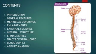
- 2. INTRODUCTION The spinal cord is the long cylindrical lower part of central nervous system. It is the main pathway for information connecting the brain and peripheral nervous system. It occupies upper two-thirds of vertebral canal. It is surrounded by the three meninges. It give rise to 31 pairs of spinal nerves . It retains the basic structural pattern.
- 3. GENERAL FEATURES DIMENSIONS : The spinal cord is 18 inches or 45 cm in an adult male and 42 cm in an adult female. The weight of spinal cord is 30 g. LOCATION : In an adult it extends from upper border of atlas vertebra to the lower border of L1 vertebra . In new-born , it extends up to L3 vertebra. Superiorly , it is continuous with the medulla oblongata . Inferiorly , it terminates as conus medullaris.
- 4. Segment or part of spinal cord to which a pair of dorsal nerve roots and a pair of ventral nerve roots is attached is called a spinal segment. As the spinal cord (45cm) is much shorter than the length of the vertebral column (65cm) , the spinal segments do not lie opposite to the corresponding vertebrae. A vertebral spine is always lower than the corresponding spinal segment . The level of spinal segment with their vertebral level is sown in the table below: Vertebral levels Spinal segments C1-C7 C1-C8 T1-T6 T1-T8 T7-T9 T9-T12 T10-T11 L1-L5 T12- L1 S1-S5 and Co1
- 5. General features of spinal cord
- 6. MENINGEAL COVERINGS The spinal cord is surrounded by three meninges . Outermost – Dura mater , middle one – Arachnoid mater and innermost- Pia mater The space between dura mater and arachnoid mater is called subdural space. The arachnoid and pia maters are separated by subarachnoid space which contains CSF. The CSF is withdrawn from the subarachnoid space during lumbar puncture. The spinal cord is enclosed only by the meningeal layer of dura mater . The space between the meningeal layer and endosteum of the vertebral canal is called epidural space, where epidural anaesthesia can be given.
- 7. Modifications of spinal pia mater 1. Ligamentum denticulatum: 21 pairs of teeth – like projections , fuse laterally with arachnoid and dura maters, Highest process attaches superior to foramen magnum Keeps the spinal cord in position 2. Linea splendens : Thickening seen at the anteromedian sulcus in the lower part of the spinal cord. 3. The filum terminale : 20 cm long After leaving through sacral hiatus ends by getting attached to the periosteun of dorsal surface of first segment of coccyx. It consists of two parts : Filum termnale internum – 15 cm ( upper part) Filum terminale externum – 5 cm ( lower part)
- 8. coverings and spaces in spinal cord
- 10. Enlargements 1. Cervical enlargement for supply of upper limb muscles : This extends from C4 to T1 spinal segments with maximum diameter of 38mm at level of C6. 2. Lumbar enlargement for supply of muscles of lower limb : It extends from level of L2 to S3 segments with maximum diameter of 35mm at the level of S1 segment. CAUDA EQUINA : Dorsal and ventral roots of right and left sides of L2 –L5 , S1-S5 and Co1 nerves lie almost vertically around filum terminale . These are called cauda equina as these resemble a horse’s tail.
- 11. External features Anteriorly , the spinal cord reveals a deep anterior median fissure lodging the anterior spinal artery. Posterior median sulcus is a thin longitudinal groove from which a septum runs in the depth of spinal cord. Each half is subdivided into – anterior , lateral and posterior regions by anterolateral and posterolateral sulci. Ventral or motor nerve roots emerge from the anterolateral sulcus. Dorsal or sensory nerve roots enter spinal cord from posterolateral sulcus.
- 12. External features with enlargement
- 13. Internal structure In a transverse section of a segment of spinal cord : White matter, i.e. nerve fibres , lies outside . Grey matter lies inside. In the centre of grey matter there is the central canal containing cerebrospinal fluid ( CSF). The canal is lined by single layer of ependymal cells. The grey matter is in the form of ‘H’ with a grey commissure joining the grey matter of right and left sides .
- 14. Grey matter comprises of one posterior horn and one anterior horn on each side in the entire extent of the cord . Only in T1-L2 and S2-S4 segments ,there is an additional lateral horn for the supply of the viscera. This horn is a part of autonomic nervous system. DORSAL HORN: Found at all spinal cord levels Comprised of sensory neurons VENTRAL HORN: Comprised of motor neurons That innervate skeletal muscle Shape and size of the horns differ in different segments due to functional reasons.
- 15. Shape of horns in different segments of spinal cord Segments of spinal cord Posterior horn Lateral horn Anterior horn Cervical , oval shape Slender Absent Narrow in 1-3 segments Broad in C4 –C8 segments for supply of upper limbs Thoracic , circular shape Slender Present for thoracolumbar outflow Slender in T2-T12 segments , broad in T1 segment Lumbar , circular shape Bulbous Present only in lumbar 1 and 2 segments Bulbous for supply of lower limbs Sacral , circular but smaller Thick Group of cells in sacral 2-4 segments for Bulbous for supply of lower limbs
- 16. Structure of the spinal cord
- 17. spinal nerves Spinal nerves arise in pairs . There are 31 pairs of spinal nerves as 8 cervical ,12 thoracic, 5 lumbar , 5 sacral and 1 coccygeal. Each spinal nerve arises by a series of six to eight dorsal and ventral nerve rootlets. These rootlets unite in or near the intervertebral foramen to form the spinal nerve. BRANCHES : Dorsal Ramus – It supplies the dorsal one-third of the body wall. They do not supply the limbs. Ventral Ramus-It supplies the ventral two-thirds of the body wall including the limbs.
- 18. TRACTS OF THE SPINAL CORD A collection of nerve fibres that connects two masses of grey matter within the central nervous system is called a tract . Tracts may be ascending (sensory) or descending (motor) . They are usually named after the masses of grey matter connected by them. Some tracts are called fasciculi or leminsci. Descending tracts are of two types : PYRAMIDAL or CORTICOSPINAL Tracts ( lateral and anterior) EXTRAPYRAMIDAL Tracts – rubrospinal , medial reticulospinal , lateral reticulospinal , olivospinal , vestibulospinal and tectospinal. Ascending tracts – lateral spinothalamic , anterior spinothalamic , fasciculus gracilis , fasciculus cuneatus , posterior spinocerebellar , anterior spinocerebral, spino – olivary and spinotectal.
- 22. Tracts of spinal cord
- 23. Blood supply The vertebral arteries are the main source of blood to the spinal cord . The following arteries branch from the vertebral arteries to directly supply the spinal cord : 1. One anterior spinal artery 2. Two posterior spinal arteries 3. Anterior and posterior radicular arteries 4. Arterial vasocorona – anastomose between the spinal arteries
- 24. Applied anatomy Cauda equina syndrome – Damage to cauda equina results into : Lower motor neuron type of paralysis due to compression of ventral nerve roots Root pain due to involvement of dorsal nerve roots Bladder and bowel movement is delayed Poliomyelitis – Viral disease involving anterior horn cells leading to flaccid paralysis of affected segments . It is a lower motor neuron paralysis If upper cervical segments affected , it may be fatal because of the involvement of C4 segment which supplies the diaphragm through phrenic nerve.
- 25. Applied anatomy Lumbar puncture – In a child , done at level of L4 Vertebra as spinal cord extends till L3 vertebra at birth. In an adult , done at level of L3 Vertebra as spinal cord ascends till level of L1 vertebra. Conus medullaris syndrome – Due to injury to S2-S4 segments of spinal cord Features are : Anaesthesia in perineum Involvement of bladder and bowel is early Sexual functions are affected
- 26. Applied anatomy Tabes dorsalis – Occurs during tertiary stage of syphills. Degenerative lesions of dorsal nerve roots and of posterior white columns Its feature is several pain in lower limbs and the lower limbs are mainly affected..
- 27. Applied anatomy