Downloaded 14 times


![6The Ecology of Breast Cancer
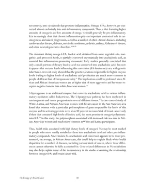
Breast cancer is an ancient disease. Its recorded history dates back to ancient Egypt
(3000-2500 BCE). Early documents describe what tumors looked like as they surfaced and
progressed.1,2
Recorded speculations about their origins appear much later. Hippocrates and
others espoused a humoral theory, thinking that imbalances among four bodily fluids—
blood, yellow bile, black bile, and phlegm—caused this to happen. Galen (130-c.200 CE)
subscribed to Hippocrates’ bodily humors theory, persuaded that he saw breast cancer more
often in melancholy (literally,“black bile”) women who were creative,kind,and considerate.
Some thought they saw cancer more generally in women who were anxious, depressed, or
grieving.3
For Galen and many who followed, breast cancer was a systemic disorder and not
confined to a single part of the body.
In the 17th
century, Italian physician Ramazzini saw that “tumors of this sort [breast cancer]
are found more often in nuns than in any other women. In my opinion, these tumors are not
due to amenorrhea, but rather to the celibate life led by these nuns.”4,5
Some theories pro-
posed that trauma or lymphatic or milk duct blockage was involved. But with the invention
of the microscope and emerging understanding of a cellular basis of anatomical structures,
cancer cells became visible, and breast cancer began to be seen as a more localized disease.
New anesthetic techniques aided a dramatic increase in surgery and, for decades, the radical
mastectomy, pioneered by William Halsted, dominated breast cancer treatment. Halsted
believed that removing enough tissue and precision to avoid spreading cancer cells during
surgery led to the best chances of cure.
Chapter 1
Toward a systems perspective of breast cancer](https://image.slidesharecdn.com/yzd484-150101095819-conversion-gate02/85/The-Ecology-of-Breast-Cancer-The-Promise-of-Prevention-and-the-Hope-for-Healing-13-320.jpg)
![Toward a systems perspective
of breast cancer
17
This committee is promoting new models for understanding the origins and treatment of
breast cancer.They emphasize the importance of a life-course approach, the timing of expo-
sures, and exposure to mixtures of risk factors. Multi-level, ecological frameworks are best
suited to this complex task.
References
1. Winchester D, Winchester D. Breast cancer: second edition. Hamilton, Ontario. BC Decker,
Inc. 2006.
2. Ekmektzoglou K, Xanthos T, German V, Zografos G. Breast cancer: from the earliest times
through to the end of the 20th
century. Eur J Obstet Gynecol Reprod Biol 2009; 145(1):3-8.
3. Berrios G. Melancholia and depression during the 19th century. A conceptual history. British
Journal of Psychiatry. 1988; 153: 298-304.
4. Mustacchi P. Ramazzini and Rigoni-Stern on parity and breast cancer. Clinical impression
and statistical corroboration. Arch Intern Med. 1961; 108:639-642.
5. Olson, James Stuart (2002). Bathsheba’s breast: women, cancer & history. Baltimore: The
Johns Hopkins University Press. pp. 32–33.
6. Beatson G. On treatment of inoperable cases of carcinoma of the mamma: suggestions for
a new method of treatment with illustrative cases. Lancet 1896;2:104–107.
7. Love R, Philips J. Oophorectomy for breast cancer: history revisited. JNCI J Natl Cancer
Inst. 2002; 94 (19): 1433-1434.
8. Stockwell S. Classics in oncology. George Thomas Beatson, M.D. (1848-1933). CA Cancer
J Clin. 1983; 33(2):105-121. Available at http://onlinelibrary.wiley.com/doi/10.3322/canj-
clin.33.2.105/pdf .
9. Allen E, Doisy E. An ovarian hormone: preliminary report on its localization, extraction and
partial purification and action in test animals. JAMA. 1923; 81:819 – 821.
10. Huggins C. Endocrine-induced regression of cancers. Science. 1967; 156(3778):1050-1054.
11. Hanahan D, Weinberg R. The hallmarks of cancer. Cell. 2000; 100:57-70.
12. Hanahan D, Weinberg R. Hallmarks of cancer: the next generation. Cell. 2011; 144:646-674.
13. Sonnenschein C, Soto A. The death of the cancer cell. Cancer Res. 2011; 71(13):4334-4337.
14. Sonnenschein C, Soto A. The aging of the 2000 and 2011 Hallmarks of Cancer reviews: A
critique. J Biosci. 2013; 38(3): 1–13.
15. Maffini M, Soto A, Calabro J, Ucci A, Sonnenschein C. The stroma as a crucial target in rat
mammary gland carcinogenesis.J. Cell Sci.2004; 117: 1495–1502.
16. Maffini M, Calabro J, Soto A, Sonnenschein C. Stromal regulation of neoplastic develop-
ment: Age-dependent normalization of neoplastic mammary cells by mammary stroma. Am.
J. Pathol. 2005; 167: 1405–1410.
17. Welch H, Black W. Using autopsy series to estimate the disease “reservoir” for ductal car-
cinoma in situ of the breast: how much more breast cancer can we find. Ann Intern Med.
1997; 127(11):1023-1028.
18. Nielsen M, Thomsen J, Primdahl S, Dyreborg U, Andersen J. Breast cancer and atypia among
young and middle-aged women: a study of 110 medicolegal autopsies. Br J Cancer. 1987;
56(6):814-819.
19. Esserman L, Thompson I. Overdiagnosis and overtreatment in cancer: an opportunity for
improvement. JAMA. 2013; ():-.doi:10.1001/jama.2013.108415. [Epub ahead of print]](https://image.slidesharecdn.com/yzd484-150101095819-conversion-gate02/85/The-Ecology-of-Breast-Cancer-The-Promise-of-Prevention-and-the-Hope-for-Healing-24-320.jpg)
![24The Ecology of Breast Cancer
framework discussed in chapter 1, we are learning that much of the available epidemiologic
research is limited to some extent by various features of study design that did not (and often,
could not) account for the complexity. For example, as noted in chapter 3, after decades of
research on diet and breast cancer, it became clear that much of that work was limited by
its failure to account for confounding or effect modification by exercise.18
That is, exercise
can independently influence both diet and breast cancer risk.Thus, it can be a confounder
of the relationship. Exercise can also influence biologic pathways that do link diet to breast
cancer—for example, inflammation and oxidative stress.Thus, exercise is a potential effect
modifier of any relationship between diet and breast cancer.This has practical importance
beyond complicating epidemiologic study design. It means that well-designed interventions
can be mutually reinforcing and have benefits that may exceed what would be predicted by
considering them individually.
As noted by the IOM committee report, more complex models “which attempt to depict
the multiplicity of factors that seem to have a role in breast cancer, help underline the bio-
logical complexity of the pathways along which those factors may be acting, the difficulty
of distinguishing truly causal effects from associations with intermediate factors, and the
challenges of designing, conducting, and interpreting studies that try to evaluate risk factors
for the various forms of this disease.19
Although these challenges share similarities across the
spectrum of risk factors evaluated in this report, they may be particularly acute for evaluat-
ing risk relationships from exposures to environmental chemicals.”
References
1. American Cancer Society. Breast Cancer Overview [Internet]. Atlanta: American Cancer
Society; c2012a-13 [updated 2012 Dec 5]. Available from: http://www.cancer.org/Cancer/
BreastCancer/OverviewGuide/breast-cancer-overview-key-statistics.
2. American Cancer Society. Global Cancer Facts & Figures. 2nd edition. Atlanta: American
Cancer Society; 2011.
3. American Cancer Society. Breast Cancer Overview [Internet]. Atlanta: American Cancer
Society; c2012a-13 [updated 2012 Dec 5]. Available from: http://www.cancer.org/Cancer/
BreastCancer/OverviewGuide/breast-cancer-overview-key-statistics.
4. Howlader N, Noone A, Krapcho M, Neyman N, et al. SEER Cancer Statistics Review,
1975-2009 (Vintage 2009 Populations) [internet]. Bethesda (MD): National Cancer Institute;
c2012-3 [updated 2012 Aug 20] Available from: http://seer.cancer.gov/csr/1975_2009_
pops09/.
5. Rossouw J, Anderson G, Prentice R, LaCroix A, et al. Risks and benefits of estrogen plus
progestin in healthy postmenopausal women: principal results from the Women’s Health
Initiative randomized controlled trial. JAMA. 2002; 288(3):321-333.
6. Kerlikowske K. Epidemiology of ductal carcinoma in situ. J Natl Cancer Inst Monogr. 2010;
2010(41):139-141.
7. National Cancer Institute. Surveillance Epidemiology and End Results. Available at: http://
seer.cancer.gov/csr/1975_2010/browse_csr.php.](https://image.slidesharecdn.com/yzd484-150101095819-conversion-gate02/85/The-Ecology-of-Breast-Cancer-The-Promise-of-Prevention-and-the-Hope-for-Healing-31-320.jpg)
![25 Breast cancer trends
and risk factors
8. Miller B, Feuer E, Hankey B. The increasing incidence of breast cancer since 1982: relevance
of early detection. Cancer Causes Control. 1991; 2(2):67-74.
9. Howlader N, Noone A, Krapcho M, Neyman N, et al. SEER Cancer Statistics Review,
1975-2009 (Vintage 2009 Populations) [internet]. Bethesda (MD): National Cancer Institute;
c2012-3 [updated 2012 Aug 20]. Available from: http://seer.cancer.gov/csr/1975_2009_
pops09/.
10. Amend K, Hicks D, Ambrosone CB. Breast cancer in African-American women: differences
in tumor biology from European-American women. Cancer Res. 2006;66(17):8327-8330.
11. For more detailed discussion of trends and tumor biology see Chapter 3 of “Breast Cancer
and the Environment: Prioritizing Prevention Report of the Interagency Breast Cancer and
Environmental Research Coordinating Committee,” available at http://www.niehs.nih.gov/
about/assets/docs/ibcercc_full.pdf. This report is the result of the 2008 Breast Cancer and
Environmental Research Act in which Congress required the Secretary of Health and Hu-
man Services to establish an Interagency Breast Cancer and Environmental Research Com-
mittee of federal and non-federal members to examine the current state of breast cancer and
the environment research and make recommendations for eliminating knowledge gaps.
12. National Cancer Institute. Surveillance Epidemiology and End Results. Available at: http://
seer.cancer.gov/csr/1975_2010/browse_csr.php.
13. American Cancer Society. Breast Cancer. Available at http://www.cancer.org/cancer/breast-
cancer/detailedguide/breast-cancer-risk-factors
14. Churpek J, Walsh T, Zheng Y, Casadei S, et al. Inherited mutations in breast cancer genes in
African American breast cancer patients revealed by targeted genomic capture and next-gen-
eration sequencing. J Clin Oncol 31, 2013 (suppl; abstr CRA1501). Available at: http://
meetinglibrary.asco.org/content/116465-132 .
15. Steingraber S. The falling age of puberty in US girls: what we know, what we need to
know. 2007. Available at http://www.breastcancerfund.org/media/publications/reports/
falling-age-of-puberty.html
16. IOM (Institute of Medicine). 2012. Breast cancer and the environment: A life course ap-
proach. Washington, DC: The National Academies Press. Available at http://www.iom.edu/
Reports/2011/Breast-Cancer-and-the-Environment-A-Life-Course-Approach.aspx
17. Printz C. Smoking studies produce new findings regarding breast and lung cancer link. Can-
cer. 2011; 117(13):2828-2829.
18. Holmes M, Chen W, Hankinson S, Willett W. Physical activity’s impact on the association of
fat and fiber intake with survival after breast cancer. Am J Epdemiol. 2009; 170(10):1250-
1256.
19. IOM (Institute of Medicine). 2012. Breast cancer and the environment: A life course ap-
proach. Washington, DC: The National Academies Press. Pg 181. Available at http://www.
iom.edu/Reports/2011/Breast-Cancer-and-the-Environment-A-Life-Course-Approach.aspx](https://image.slidesharecdn.com/yzd484-150101095819-conversion-gate02/85/The-Ecology-of-Breast-Cancer-The-Promise-of-Prevention-and-the-Hope-for-Healing-32-320.jpg)
![57 Diet, nutrition, and
breast cancer
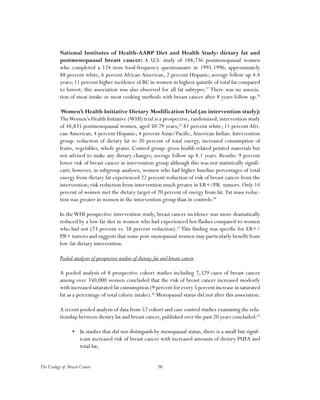
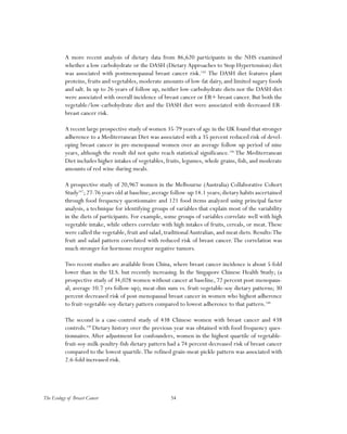
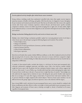
Another study of 516 post-menopausal women with breast cancer found that higher levels
of dietary fiber, fruits, and vegetables, and lower levels of dietary fat in the year prior to
diagnosis was associated with significantly lower risk of death from any cause over 7 years
of follow up.171
The CollaborativeWomen’s Longevity Study172
examined the relation between post-diagno-
sis dietary factors and survival in 4,441 women with invasive breast cancer.They were 20-79
years old at diagnosis and followed over a period of 7 years.The study used food-frequency
questionnaires and adjusted data for age, state of residence, menopausal status, smoking,
breast cancer stage, alcohol, and history of hormone replacement therapy.Women in the
highest compared to lowest levels of dietary saturated fat and trans fat had a significantly
higher risk of dying from any cause [for saturated fat (HR =1.41, 95 percent CI = 1.06-
1.87); for trans fat (HR = 1.78, 95 percent CI = 1.35-2.32]. Associations were similar,
though did not achieve statistical significance, for breast cancer-specific death.
Dietary soy prior to diagnosis and breast cancer prognosis
Two fairly large studies have looked at relationships between dietary soy prior to diagnosis
and course of the disease after diagnosis. In the population-based case control Long Island
Breast Cancer study, 1,508 women with breast cancer completed food frequency ques-
tionnaires reporting on their diets for the year prior to diagnosis.173
Over 6 years of follow
up, women with the highest intake of flavones, isoflavones, and anthocyanidins (in darkly
pigmented berries,red cabbage,eggplant) had reduced risk of death from any cause (37 per-
cent, 48 percent, and 36 percent reduction respectively) compared to those with the lowest
intake. Reductions in mortality were most marked among post-menopausal women. Breast
cancer specific mortality data were similar. Isoflavone intakes in this study ranged from very
low to 7.5 mg or more daily in the upper quintile.As previously noted, daily isoflavone in-
takes of 20 mg or more from traditional soy products are common among Asians.
In the Shanghai breast cancer study174
of 1,459 breast cancer patients, soy food intake was
assessed using a validated food frequency questionnaire at baseline. In an average follow-up
of 5.2 years, soy intake pre-diagnosis was unrelated to disease-free breast cancer survival
and this did not differ according to ER/PR status, tumor stage, age at diagnosis, body mass
index (BMI), or menopausal status. No information on tamoxifen use was provided.
These two studies are not comparable in that the Long Island study looked at risk of death
from breast cancer or other causes, whereas the Shanghai study used disease-free survival as
the outcome of interest.](https://image.slidesharecdn.com/yzd484-150101095819-conversion-gate02/85/The-Ecology-of-Breast-Cancer-The-Promise-of-Prevention-and-the-Hope-for-Healing-64-320.jpg)
![61 Diet, nutrition, and
breast cancer
References
1. Goodwin P, Ennis M, Pritchard K, Koo J, et al. Diet and breast cancer: evidence that ex-
tremes in diet are associated with poor survival. J Clin Oncol 2003; 21(13):2500-2507.
2. Michels K, Terry K, Eliassen A, Hankinson S, Willett W. Adult weight change and incidence
of premenopausal breast cancer. Int J Cancer 2011; Mar 16. Doi: 10.1002/ijc.26069. [Epub
ahead of print]
3. Hunter D, Spiegelman D, Adami H, Beeson L, et al. Cohort studies of fat intake and the risk
of breast cancer—a pooled analysis. N Engl J Med 1996;334:356–361.
4. Wu A, Pike M, Stram D. Meta-analysis: dietary fat intake, serum estrogen levels, and the risk
of breast cancer. J Natl Cancer Inst. 1999; 91(6):529–534.
5. Pierce J, Stefanick M, Flatt S, et al. Greater survival after breast cancer in physical-
ly active women with high vegetable-fruit intake regardless of obesity. J Clin Oncol
2007;25(17):2345–2351.
6. The role of soy isoflavones in menopausal health: report of the North American Menopause
Society/Wulf H. Utian Translational Science Symposium in Chicago, IL (Ocober 2010) No
authors listed. Menopause 2011 Jun 16 [Epub ahead of print]
7. Ju Y, Doerge D, Woodling K, Hartman J, et al. Dietary genistein negates the inhibitory
effect of letrozole on the growth of aromatase-expressing estrogen-dependant human breast
cancer cells (MCF-7Ca) in vivo. Carcinogenesis 2008; 29:2162–2168.
8. Ariazi J, Haag J, Lindstrom M, Gould M. Mammary glands of sexually immature rats are
more susceptible than those of mature rats to the carcinogenic, lethal, and mutagenic effects
of N-nitroso-N-methylurea. Mol Carcinog 2005;43:155–164.
9. MacLennan M, Ma D. Role of dietary FAs in mammary gland development and breast can-
cer. Breast Cancer Res. 2010; 12(5):211.
10. Frankel S, Gunnell D, Peters T, et al. Childhood energy intake and adult mortality from
cancer: The Boyd Orr cohort study. BMJ 1998;316:499-504.
11. Tretli S, Gaard M. Lifestyle changes during adolescence and risk of breast cancer: An ecolog-
ic study of the effect of World War II in Norway. Cancer Causes Control 1996;7:507-512.
12. Frazier A, Li L, Cho E, et al. Adolescent diet and risk of breast cancer. Cancer Causes Con-
trol 2004;15:73-82.
13. Land C. Studies of cancer and radiation dose among atomic bomb survivors: the example of
breast cancer. JAMA. 1995;274:402-407.
14. Ziegler R, Hoover R, Pike M, Hildesheim A, Nomura A, West D. Migration patterns and
breast cancer risk in Asian-American women. J Natl Cancer Inst 1993;85:1819–1827.
15. Cohn B, Wolff M, Cirillo P, Sholtz R. DDT and breast cancer in young women: new data on
the significance of age at exposure. Environ Health Perspect 2007; 115(10):1406-1414.
16. For a history of dietary trans fat see http://www.heart.org/HEARTORG/GettingHealthy/
FatsAndOils/Fats101/A-History-of-Trans-Fat_UCM_301463_Article.jsp.
17. Rose D, Boyar A, Wynder E. International comparisons of mortality rates for cancer of the
breast, ovary, prostate, and colon, and per capita food consumption. Cancer 1986;58:2363–
2371.
18. Hilakivi-Clarke L, Clarke R, Onojafe I, Raygada M, et al. A maternal diet high in n-6 poly-
unsaturated fats alters mammary gland development, puberty onset, and breast cancer risk
among female rat off spring. Proc Natl Acad Sci U S A 1997, 94:9372-9377.
19. Hilakivi-Clarke L, Clarke R, Lippman M: The influence of maternal diet on breast cancer
risk among female off spring. Nutrition 1999, 15:392-401.
20. Fay M, Freedman L, Clifford C, Midthune D. Effect of different types and amounts
of fat on the development of mammary tumors in rodents: a review. Cancer Res 1997;
57(18):3979-3988.](https://image.slidesharecdn.com/yzd484-150101095819-conversion-gate02/85/The-Ecology-of-Breast-Cancer-The-Promise-of-Prevention-and-the-Hope-for-Healing-68-320.jpg)
![63 Diet, nutrition, and
breast cancer
40. Carty C, Kooperberg C, Neuhouser M, Tinker L, et al. Low-fat dietary pattern and change in
body-composition traits in the Women’s Health Initiative Dietary Modification Trial. Am J
Clin Nutr 2011; 93(3):516-524.
41. Caan B, Aragaki A, Thomson C, Stefanick M. Vasomotor symptoms, adoption of a low-fat
dietary pattern, and risk of invasive breast cancer: a secondary analysis of the Women’s
Health Initiative randomized controlled dietary modification trial. J Clin Oncol 2009;
27(27):4500-4507.
42. Smith-Warner S, Spiegelman D, Adami H, Beeson WL, et al. Types of dietary fat and breast
cancer: a pooled analysis of cohort studies. Int J Cancer 2001;92:767–774.
43. Turner L. A meta-analysis of fat intake, reproduction, and breast cancer risk: An evolution-
ary perspective. Am J Hum Biol 2011 Jun 16. Doi 10.1002/ajhb.21176. [Epub ahead of
print]
44. MacLean C, Newberry S, Mojica W, Khanna P, et al. Effects ofomega-3 FAs on cancer risk: a
systematic review. JAMA 2006;295:403–415.
45. Zheng J, Hu X, Zhao Y, Yang J, Li D. Intake of fish and marine n-3 polyunsaturated FAs and
risk of breast cancer: meta-analysis of data from 21 independent prospective cohort studies.
BMJ. 2013; Jun 27;346:f3706. doi: 10.1136/bmj.f3706.
46. Gago-Dominguez M, Yuan J, Sun C, Lee H, Yu M. Opposing effects of dietary n-3 and n-6
fatty acids on mammary carcinogenesis: The Singapore Chinese Health Study. Br J Cancer.
2003;89:1686-1692.
47. Thiebaut A, Chajes V, Gerber M, Boutron-Ruault M, et al. Dietary intakes of omega-6 and
omega-3 polyunsaturated FAs and the risk of breast cancer. Int J Cancer. 2009; 124:924–931.
48. Harris R. Cyclooxygenase-2(cox-2) and the inflammogenesis of cancer. Subcell Biochem
2007; 42:93-126.
49. Stix G. A malignant flame. Sci American Jul 2007; 60-67.
50. Stein J, Schettler T, Rohrer B, Valenti M. Environmental Threats to Healthy Aging: with a
closer look at Alzheimer’s and Parkinson’s diseases. 2008. www.agehealthy.org Accessed
Jun 28, 2011.
51. Mathias R, Sergeant S, Ruczinski I, Torgerson D, et al. The impact of FADS genetic variants
on omega 6 polyunsaturated FA metabolism in African Americans. BMC Genet 2011; 12:50
http://www.biomedcentral.com/1471-2156/12/50
52. Stark A, Kleer C, Martin I, Awuah B, et al. African ancestry and higher prevalence of tri-
ple-negative breast cancer: findings from an international study. Cancer 2010; 116(21):4926-
4932.
53. Simopoulos A. Genetic variants in the metabolism of omega-6 and omega-3 FAs: their role
in the determination of nutritional requirements and chronic disease risk. Exp Biol Med
(Maywood). 2010; 235(7):785-795.
54. Wang J, John E, Ingles S. 5-lipoxygenase and 5-lipoxygenase-activating protein gene poly-
morphisms, dietary linoleic acid, and risk for breast cancer. Cancer Epidemiol Biomarkers
Prev 2008; 17(10):48-54.
55. Fung T, Schulze M, Manson J, Willett W, Hu F. Dietary patterns, meat intake, and the risk of
type 2 diabetes in women. Arch Intern Med 2004;164:2235–2240.
56. Hu F, Stampfer M, Manson J, et al. Dietary saturated fats and their food sources in relation
to the risk of coronary heart disease in women. Am J Clin Nutr 1999;70:1001–1008.
57. Friel S, Dangour A, Garnett T, Lock K. et al. Public health benefits of strategies to reduce
greenhouse-gas emissions: food and agriculture Lancet 2009; 374: 2016–2025.
58. O’Sullivan A, O’Sullivant K, Galvin K, Moloney A, et al. Grass silage versus maize silage
effects on retail packaged beef quality. J Anim Sci 2002; 80(6):1556-1563.](https://image.slidesharecdn.com/yzd484-150101095819-conversion-gate02/85/The-Ecology-of-Breast-Cancer-The-Promise-of-Prevention-and-the-Hope-for-Healing-70-320.jpg)
![64The Ecology of Breast Cancer
59. Daly C,Abbott A, Doyle P, Nader G, Larson S. A review of FA profiles and antioxidant
content in grass-fed and grain-fed beef. Nutr J. 2010; Mar 10;9:10. doi: 10.1186/1475-2891-
9-10.
60. Missmer S, Smith-Warner S, Spiegelman D, Yaun S, et al. Meat and dairy food consumption
and breast cancer: a pooled analysis. Int J Epidemiol 2002; 31(1):78-85.
61. Rogers I, Northstone K, Dunger D, Cooper A, et al. Diet throughout childhood and age at
menarche in a contemporary cohort of British girls. Public Health Nutr 2010; 13(12):2052-
2063.
62. Berkey C, Gardner J, Frazier A et al. Relation of childhood diet and body size to menarche
and adolescent growth in girls. Am J Epidemiol. 2000; 152, 446–452.
63. Rosell M, Appleby P, Key T. Height, age at menarche, body weight and body mass index in
life-long vegetarians. 2005; Public Health Nutr 8, 870–875.
64. Petridou E, Syrigou E, Toupadaki N et al. Determinants of age at menarche as early life
predictors of breast cancer risk.1996; Int J Cancer 68, 193–198.
65. Maclure M, Travis L, Willett W et al. A prospective cohort study of nutrient intake and age
at menarche. 1991; Am J Clin Nutr 54, 649–656.
66. Villamor E, Marin C, Mora-Plazas M, Baylin A. Vitamin D deficiency and age at menarche: a
prospective study. Am J Clin Nutr 2011; 94(4): 1020-1025.
67. Taylor V, Misra M, Mukherjee S. Is red meat intake a risk factor for breast cancer among
premenopausal women? Breast Cancer Res Treatment 2009; 117(1):1-8.
68. Fu Z, Deming S, Fair A, Shrubsole M, et al. Well-done meat intake and meat-derived mu-
tagen exposures in relation to breast cancer risk: the Nashville Breast Health Study. Breast
Cancer Res Treat 2011 May 3, [Epub ahead of print]
69. Kabat G, Cross A, Park Y, et al. Meat intake and meat preparation in relation to risk of
postmenopausal breast cancer in the NIH-AARP diet and health study. Int J Cancer
2009;124:2430–2435.
70. Cummings S, Tice J, Bauer S, Browner W, et al. Prevention of breast cancer in post-
menopausal women: approaches to estimating and reducing risk. J Natl Cancer Inst 2009;
101(6):384-398.
71. Tseng M, Olufade T, Evers K, Byrne C. Adolescent lifestyle factors and adult breast density
in U.S. Chinese immigrant women. Nutr Cancer 2011; 63(3):342-349.
72. Sellers T, Vachon C, Pankratz V, Janney C, Fredericksen Z. Association of childhood and ad-
olescent anthropometric factors, physical activity, and diet with adult mammographic breast
density. Am J Epidemiol 2007; 166(4):456-464.
73. Mishra G, dos Santos Silva I, McNaughton S, et al. Energy intake and dietary patterns in
childhood and throughout adulthood and mammographic density: results from a British
prospective cohort. Cancer Causes Contol 2011; 22(2):227-235.
74. Larsson S, Bergkvist L, Wolk A. Long-term meat intake and risk of breast cancer by oestro-
gen and progesterone receptor status in a cohort of Swedish women. Eur J Cancer (Epub
ahead of print 20 June 2009).
75. Wiley AS. 2011b. Milk intake and total dairy consumption: associations with early menarche
in NHANES 1999-2004 PLoS One 6:e14685. doi: 10.1371/journal.pone.0014685.
76. Cadogan J, Eastell R, Jones N, Barker M. Milk intake and bone mineral acquisition in adoles-
cent girls: randomized, controlled intervention trial. BMJ 1997;315(7118):1255-1260.
77. Berkey C, Colditz G, Rockett H, Frazier A, Willett W. Dairy consumption and female height
growth: prospective cohort study. Cancer Epidemiol Biomarkers Prev 2009; 18(6):1881-
1887.
78. Cho E, Spiegelman D, Hunter D, Chen W, et al. Pre-menopausal fat intake and risk of breast
cancer. J Natl Cancer Inst 2003; 95:1079–1085. .](https://image.slidesharecdn.com/yzd484-150101095819-conversion-gate02/85/The-Ecology-of-Breast-Cancer-The-Promise-of-Prevention-and-the-Hope-for-Healing-71-320.jpg)
![65 Diet, nutrition, and
breast cancer
79. Knekt P, Jarvinen R, Seppanen R, Pukkala E, Aromaa A: Intake of dairy products and the
risk of breast cancer.Br J Cancer 1996; 73: 687–691.
80. Michels K, Rosner B, Chumlea W, Colditz G, Willett WC. Preschool diet and adult risk of
breast cancer. Int J Cancer 2006; 118:749–754.
81. Hjartaker A, Laake P, Lund E. Childhood and adult milk consumption and risk of premeno-
pausal breast cancer in a cohort of 48,844 women—the Norwegian women and cancer
study. Int J Cancer 2001;93:888–893.
82. Pryor M, Slattery M, Robinson L, Egger M. Adolescent diet and breast cancer in Utah.
Cancer Res 1989;49:2161–2167.
83. Linos E, Willett W, Cho E, Frazier L. Adolescent diet in relation to breast cancer risk among
premenopausal women. Cancer Epidemiol Biomarkers Prev 2010; 19(3):689-696.
84. Qin L, Xu J, Tezuka H, Wang P, Hoshi K. Milk inhibits the regression of 7.12-dimethyl-
benz(a)anthracene-induced mammary tumors in ovariectomized rats. Nutr Cancer 2008;
60(4):505-510.
85. Nielsen T, Khan G, Davis J, Michels K, Hilakivi-Clarke L. Prepubertal exposure to cow’s
milk reduces susceptibility to carcinogen-induced mammary tumorigenesis in rats. Int J
Cancer. 2011; 128(1):12-20.
86. Kawaguchi H, Umekita Y, Souda M, Gejima K, et al. Effects of neonatally administered
high-dose diethylstilbestrol on the induction of mammary tumors induced by 7,12-dimethyl-
benz[a]anthracene in female rats. Vet Pathol 2009; 46(1):142-150.
87. Rock C. Carotenoids: biology and treatment. J Pharm Ther 1997; 75(3):185-197.
88. Haskell M. The challenge to reach nutritional adequacy for vitamin A: β-carotene bioavail-
ability and conversion—evidence in humans. Am J Clin Nutr 2012;96(5):1193S-1203S.
89. Zaineddin A, Vrieling A, Buck K, Becker S. Serum enterolactone and postmenopausal breast
cancer risk by estrogen, progesterone and herceptin 2 receptor status. Int J Cancer. 2012;
130(6):1401-1410.
90. Buck K, Vrieling A, Zaineddin A, Becker S, et al. Serum enterolactone and prognosis of
postmenopausal breast cancer. J Clin Oncol. 2011; 29(28):3730-3738.
91. Goldin B, Adlercreutz H, Dwyer J, Swenson L, et al. Effect of diet on excretion of estrogens
in pre- and postmenopausal women. Cancer Res. 1981; 41(9 Pt 2):3771-3773.
92. Gandini S, Merzenich H, Robertson C, Boyle P. Meta-analysis of studies on breast cancer
risk and diet: the role of fruit and vegetable consumption and the intake of associated mi-
cronutrients. Eur J Cancer 2000;36:636–646.
93. Smith-Warner SA, Spiegelman S-SY, Adami H-O, et al. Intake of fruits and vegetables and
risk of breast cancer. JAMA 2001;285:769–776.
94. Gonzalez C, Riboli E. Diet and cancer prevention: Contributions from the European
Prospective Investigation into Cancer and Nutrition (EPIC study). Eur J Cancer. 2010;
46(14):2555-2562.
95. Gandini S, Merzenich H, Robertson C, Boyle P. Meta-analysis of studies on breast cancer
risk and diet: the role of fruit and vegetable consumption and the intake of associated mi-
cronutrients. Eur J Cancer 2000;36:636–646.
96. Eliassen A, Hendrickson S, Brinton L, Buring J, et al. Circulating carotenoids and risk
of breast cancer: pooled analysis of eight prospective studies. J Natl Cancer Inst 2012;
104(24):1905-1916.
97. Reinwald S, Akabas S, Weaver C. Whole versus the piecemeal approach to evaluating soy. J
Nutr. 2010; 140(12):2335S-2343S.
98. Hilakivi-Clarke L, Andrade J, Helferich W. Is soy consumption good or bad for the breast? J
Nutr 2010; 140(12):2326S-2334S.](https://image.slidesharecdn.com/yzd484-150101095819-conversion-gate02/85/The-Ecology-of-Breast-Cancer-The-Promise-of-Prevention-and-the-Hope-for-Healing-72-320.jpg)
![68The Ecology of Breast Cancer
141. Hu F. Dietary pattern analysis: a new direction in nutritional epidemiology. Curr Opin Lipi-
dol 2002; 13: 3–9.
142. Flores R, Shi J, Fuhrman B, Xu X, et al. Fecal microbial determinants of fecal and systemic
estrogens and estrogen metabolites: a cross-sectional study. J Transl Med. 2012; Dec 21:
10-253.
143. Brennan S, Cantwell M, Cardwell C, Velentzis L, Woodside J. Dietary patterns and breast
cancer risk: a systematic review and meta-analysis. Am J Clin Nutr 2010; 91(5):1294-1302.
144. Fung T, Hu F, McCullough M, Newby P, et al. Diet quality is associated with the risk of es-
trogen receptor-negative breast cancer in postmenopausal women. J Nutr 2006; 136(2):466-
472.
145. Fung T, Hu F, Hankinson S, Willett W, Holmes M. Low-carbohydrate diets, dietary ap-
proaches to stop hypertension-style diets and the risk of postmenopausal breast cancer. Am
J Epidemiol 2011; 174(6):652-660.
146. Cade J, Taylor E, Burley V, Greenwood D. Does the Mediterranean dietary pattern or the
Healthy Diet Index influence the risk of breast cancer in a large British cohort of women?
Eur J Clin Nutr 2011; May 18 [Epub ahead of print]
147. Baglietto L, Krishnan K, Severi G, Hodge A, et al. Dietary patterns and risk of breast cancer.
Br J Cancer 2011; 104(3):524-531.
148. Butler L, Wu A, Wang R, Koh W, et al. A vegetable-fruit-soy dietary pattern protects against
breast cancer among postmenopausal Singapore Chinese women. Am J Clin Nutr 2010;
91(4):1013-1019.
149. Zhang C, Ho S, Fu J, Cheng S, et al. Dietary patterns and breast cancer risk among Chinese
women. Cancer Causes Control 2011; 22(1):115-124.
150. Cho Y, Kim J, Shin A, Park K, Ro J. Dietary patterns and breast cancer risk in Korean wom-
en. Nutr Cancer 2010;62(8):1161-1169.
151. Velentzis L, Keshtgar M, Woodside J, Leathem A, et al. Significant changes in dietary intake
and supplement use after breast cancer diagnosis in a UK multicentre study. Breast Cancer
Res Treat. 2011; 128(2):473-482.
152. Gregorio D, Emrich L, Graham S, Marshall J, Nemoto T. Dietary fat consumption and sur-
vival among women with breast cancer. J Natl Cancer Inst 1985; 75(1):37-41.
153. Newman S, Miller A, Howe G. A study of the effect of weight and dietary fat on breast
cancer survival time. Am J Epid 1986; 123(5):767-774.
154. Beasly J, Newcomb P, Trentham-Dietz A, Hampton J, et al. Post-diagnosis dietary factors
and survival after invasive breast cancer. Breast Cancer Res Treat. 2011; 128(1):229-236.
155. Nomura A, Marchand L, Kolonel L, Hankin J. The effect of dietary fat on breast cancer
survival among Caucasian and Japanese women in Hawaii. Breast Cancer Res Treat 1991;
18suppl 1:S135-141.
156. Kyogoku S, Hirohata T, Nomura Y, Shigematsu T, et al. Diet and prognosis of breast cancer.
Nutr Cancer 1992; 17(3):271-277.
157. Conroy S, Maskarinec G, Wilkens L, White K, et al. Obesity and breast cancer survival in
ethnically diverse postmenopausal women: the Multiethnic Cohort Study. Breast Cancer Res
Treat 2011; Apr 16 [Epub ahead of print]
158. Holmes M, Stampfer M, Colditz G, Rosner B, et al. Dietary factors and the survival of wom-
en with breast carcinoma. Cancer 1999; 86:826–835.
159. Hebert J, Hurley T, Ma Y. The effect of dietary exposures on recurrence and mortality in
early stage breast cancer. Breast Cancer Res Treat 1998; 51; 17–28.
160. Protani M, Coory M, Martin J. Effect of obesity on survival of women with breast cancer:
systematic review and meta-analysis. Breast Cancer Res Treat 2010; 123:627–635.
161. Jain M, Miller A, To T. Premorbid diet and the prognosis of women with breast cancer. J
Natl Cancer Inst 1994; 86(18):1390-1397.](https://image.slidesharecdn.com/yzd484-150101095819-conversion-gate02/85/The-Ecology-of-Breast-Cancer-The-Promise-of-Prevention-and-the-Hope-for-Healing-75-320.jpg)
![76The Ecology of Breast Cancer
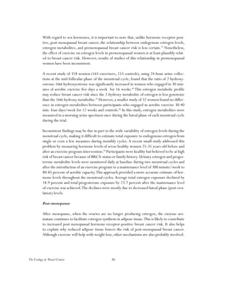
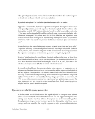
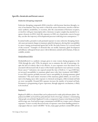
Table 4.2: Individual prospective cohort studies
Study
Study
Population
(number of
cases)
Follow-up
(years)
Levels of Physical
Activity Compared
Relative Risk (or Hazard Ratio) of Breast
Cancer in Physically Active Women
Compared with Inactive Women, RR or
hazard ratio HR (95 percent CI)
Pre-
menopausal
Post-
menopausal
Pre- and
post-
menopausal
combined
NIH-AARP Diet
and Health Study22,
23
182,862
(6,609 cases)
7
At least 20 min. physical
activity at least 5 times/
wk that caused increased
breathing, heart rate, or
sweating
vs.
inactive
0.92
(0.85-1.00)*
Nurses’ Health
Study 24
95,396
(4,782 cases)
20
27 or more
vs.
less than 3 MET hr/wk
0.88
(0.79-0.98)
French E3N
cohort 25
90,509
(3,424 cases)
11.4
22.3-33.8 MET hrs/wk
recreational activity
vs.
inactive
0.88
(0.79-0.98)
[protective
effect
persisted
regardless of
family history,
nulliparity,
HRT use,
BMI]
French E3N
cohor
90,509
(3,424 cases)
11.4
33.8 or more MET hrs/wk
recreational activity
vs.
inactive
0.81
(0.72-0.92)
EPIC 26 218,169
(3,423 cases)
6.4
Recreation:At least 42
vs.
less than 14 MET hrs/wk of
recreational activity
Household activity: > 90 vs <
23 Met hrs/wk
0.94
(0.76-1.15)
0.71 (0.55-0.90)
0.96
(0.85-1.08)
0.81 (0.70-0.93)
California Teachers
Study27
110,599
(2,649 cases)
6.6
5 or more hrs/wk moderate
physical activity
vs.
inactive
ER- tumors
0.53 (0.33-
0.85); ER+
tumors 0.98
(0.82-1.16)
Iowa Women’s
Health Study28
36,363
(2,548 cases)
15.3
High vs. low level of physical
activity
0.91
(0.82-1.01)
0.66 (0.46-0.94)
for ER+/PR-
tumors
National Breast
Cancer Screening
Study-Canada29 ##
40,318
(2,545 cases)
16.4
At least 1 hr/day vigorous
physical activity
vs.
inactive
0.87
(0.68-1.09)
1.00
(0.78-1.29)
0.93
(0.78-1.10)
Cancer Prevention
Study II (CPS II)30
72,608
(1,520 cases)
5
At least 42
vs.
less than 7 MET hrs/wk
physical activity
0.71
(0.49-1.02) Non-
recreational
activity not
associated with
BC risk](https://image.slidesharecdn.com/yzd484-150101095819-conversion-gate02/85/The-Ecology-of-Breast-Cancer-The-Promise-of-Prevention-and-the-Hope-for-Healing-83-320.jpg)
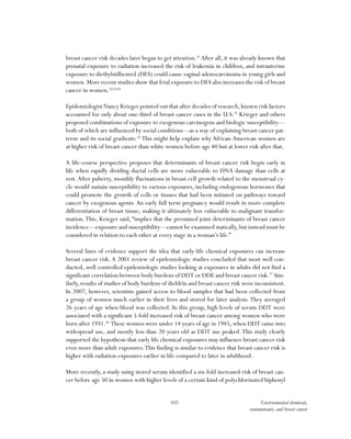
![83 Exercise, physical activity,
and breast cancer
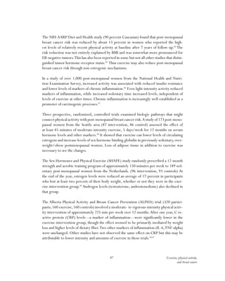
Study
Study
Population
(number of
participants)
Follow-up
(years)
Levels of
Physical
Activity
Compared
Relative Risk of Recurrence or Mortality in
Physically Active Women Compared with
Inactive Women, RR (95 percent CI)
Recurrence
All-cause
mortality
Breast-cancer
specific
mortality
Breast cancer
family registry62
4,153 cases; ages
< 35 - >60 yrs.
Median follow-up
7.8 yrs.
HR 0.77
(0.60-1.00) for
recreational
physical activity
of >38.2 vs 0
MET-h/wk within
last 3 yrs.; effect
mostly in ER+
tumors; beneficial
effects also at < 9
MET hrs/wk; No
significant effect
of earlier physical
activity levels
Population-based
survival study;
Norwegian
Counties Study 63
1,364 cases; ages
27-79 yrs. at
diagnosis
Mean follow-up
8.2 yrs.
Level of leisure
physical activity in
the year prior to
study entry
HR 1.47, (1.08–
1.99) for pre-
diagnostic BMI
> 30 compared
to BMI 18.5-25*;
Active compared
to inactive
women: HR 0.60,
(0.36–0.99)
Population-based
case control
study; So. CA64
717 cases; all pre-
menopausal
10.4 yrs.
Lifetime
recreational
exercise history;
from menarche
to one yr. before
diagnosis
No association
of exercise with
breast cancer
survival
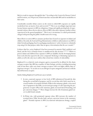
*effect stronger in pre/perimenopausal women. Women with BMI < 25 kg/m2 and age of diagnosis > 55 years had a 66 percent reduction in overall mortality
if they regularly exercised before diagnosis compared with sedentary women; HR = 0.34 (0.16–0.71). Women with the highest total cholesterol had a 29 percent
increase in mortality compared to women with the lowest cholesterol (HR = 1.29, [1.01–1.64]). Women with the highest blood pressure had a 41 percent increase
in mortality compared to women with the lowest BP. (HR = 1.41, [1.09–1.83]).
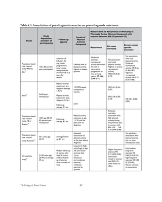
Table 4.4:Association of post-diagnosis exercise on outcomes
Study
Study
Population
(number of
participants)
Follow-up
(years)
Levels of
Physical
Activity
Compared
Relative Risk of Recurrence or Mortality in
Physically Active Women Compared with
Inactive Women, RR (95 percent CI)
Recurrence
All-cause
mortality
Breast-cancer
specific mortality
Nurse’s health
study65
3,846 cases;
average age at
diagnosis 58 yrs.
Median length
of follow-up 83
months, and
maximum length
of
follow-up 321
months.
Level of physical
activity after
diagnosis
Decreasing
risk associated
with increasing
amounts of
physical activity
(by quintile) RR
0.53 (0.39-0.71);
0.36 (0.26- 0.51);
0.28 (0.19- 0.41);
0.17 (0.11-0.27)](https://image.slidesharecdn.com/yzd484-150101095819-conversion-gate02/85/The-Ecology-of-Breast-Cancer-The-Promise-of-Prevention-and-the-Hope-for-Healing-90-320.jpg)
![91 Exercise, physical activity,
and breast cancer
36. Colditz G, Feskanich D, Chen W, Hunter D, Willett W. Physical activity and risk of breast
cancer in premenopausal women. Br J Cancer. 2003; 89(5):847-851.
37. Chang S, Ziegler R, Dunn B, et al. Association of energy intake and energy balance with
postmenopausal breast cancer in the prostate, lung, colorectal, and ovarian cancer screening
trial. Cancer Epidemiol Biomarkers Prev. 2006; 15(2):334-341.
38. Suzuki R, Iwasaki M, Kasuga Y, et al. Leisure-time physical activity and breast cancer risk by
hormone receptor status: effective life periods and exercise intensity. Cancer Causes Control.
2010; 21(11):1787-1798.
39. Moradi T, Adami H, Ekbom A, et al. Physical activity and risk for breast cancer a prospective
cohort study among Swedish twins. Int J Cancer. 2002; 100:76-81.
40. Pronk A, Ji B, Shu X, Chow W, et al. Physical activity and breast cancer risk in Chinese wom-
en. Br J Cancer 2011; 105(9):1443-1450.
41. Lynch B, Neilson H, Friedenreich C. Physical activity and breast cancer prevention. Recent
Results Cancer Res. 2011; 186:13-42.
42. Chandran U, Hirshfield K, Bandera E. The role of anthropometric and nutritional factors on
breast cancer risk in African-American women. Public Health Nutr. 2012; 15(4):738-748.
43. Schmitz K, Holtzman J, Courneya K, Masse L, et al. Controlled physical activity trials in
cancer survivors: A systematic review and meta-analysis. Cancer Epidemiol Biomarkers Prev.
2005;14:1588 – 1595.
44. Speck R, Courneya K, Masse L, Duval S, Schmitz K. An update of controlled physical ac-
tivity trials in cancer survivors: a systematic review and meta-analysis. J Cancer Surviv. 2010;
4(2):87-100.
45. Courneya K, Friedenreich C. Framework PEACE : an organizational model for examining
physical exercise across the cancer experience. Ann Behav Med. 2001;23:263 –272.
46. Mutrie N, Campbell A, Barry S, Hefferon K, et al. Five-year follow-up of participants in
a randomized controlled trial showing benefits from exercise for breast cancer survivors
during adjuvant treatment. Are there lasting effects? J Cancer Surviv. 2012; Jul 27. [Epub
ahead of print]
47. Markes M, Brockow T, Resch K. Exercise for women receiving adjuvant therapy for breast
cancer. Cochrane Database Syst Rev. 2006; Cot 18(4):CD005001.
48. McNeely M, Campbell K, Rowe B, Klassen T, et al. Effects of exercise on breast cancer
patients and survivors: a systematic review and meta-analysis. CMAJ. 2006; 175(1):34-41.
49. Speck R, Courneya K, Masse L, Duval S, Schmitz K. An update of controlled physical ac-
tivity trials in cancer survivors: a systematic review and meta-analysis. J Cancer Surviv. 2010;
4(2):87-100.
50. Dimeo F, Tilmann M, Bertz H, Kanz L, et al. Aerobic exercise in the rehabilitation of cancer
patients after high dose chemotherapy and autologous peripheral stem cell transplantation.
Cancer. 1997;79:1717–1722.
51. Gleeson M. Immune function in sport and exercise. J Appl Physiol 2007; 103(2):693-699.
52. Mutrie N, Campbell A, Barry S, Hefferon K, et al. Five-year follow-up of participants in
a randomized controlled trial showing benefits from exercise for breast cancer survivors
during adjuvant treatment. Are there lasting effects? J Cancer Surviv. 2012; Jul 27. [Epub
ahead of print]
53. Cohen L, Cole S, Sood A, Prinsloo S, et al Depressive symptoms and cortisol rhythmicity
predict survival in patients with renal cell carcinoma: role of inflammatory signaling. PLoS
One. Aug 1, 2012; 10.1371/journal.pone.0042324.
54. Lee M, Choi T, Ernst E. Tai chi for breast cancer patients: a systematic review. Breast Cancer
Res Treat 2010; 120(2):309-316.](https://image.slidesharecdn.com/yzd484-150101095819-conversion-gate02/85/The-Ecology-of-Breast-Cancer-The-Promise-of-Prevention-and-the-Hope-for-Healing-98-320.jpg)
![93 Exercise, physical activity,
and breast cancer
74. Kelesidis I, Kelesidis T, Mantzoros C. Adiponectin and cancer: a systematic review. Br J
Cancer. 2006; 94:1221-1225.
75. Tworoger S, Missmer S, Eliassen A, Barbieri R, et al. Physical activity and inactivity in rela-
tion to sex hormone, prolactin, and insulin-like growth factor concentrations in premeno-
pausal women – exercise and premenopausal hormones. Cancer Causes Control. 2007;
18(7):743-752.
76. Allen N, Appleby P, Kaaks R, Rinaldi S, et al. Lifestyle determinants of serum insulin-like
growth-factor-I (IGF-I), C-peptide and hormone binding protein levels in British women.
2003; Cancer Causes Control 14:65–74.
77. Eliakim A, Brasel J, Mohan S, Barstow T, et al. Physical fitness, endurance training, and the
growth hormone-insulin-like growth factor I system in adolescent females. J Clin Endocrinol
Metab 1996; 81:3986–3992.
78. Arikawa A, Kurzer M, Thomas W, Schmitz K. No effect of exercise on insulin-like growth
factor-1, insulin, and glucose in young women participating in a 16-week randomized con-
trolled trial. Cancer Epidemiol Biomarkers Prev 2010; 19(11):2987-2990.
79. Eliassen A, Spiegelman D, Xu X, Keefer L, et al. Urinary estrogens and estrogen metabolites
and subsequent risk of breast cancer among premenopausal women. Cancer Res 2012; Jan
16 [Epub ahead of print].
80. Smith A, Phipps W, Thomas W, Schmitz K, Kurzer M. The effects of aerobic exercise on
estrogen metabolism in healthy premenopausal women. Cancer Epidemiol Biomarkers Prev.
2013; 22(5):756-764.
81. Bradlow H, Hershcopf R, Martucci C, Fishman J. Estradiol 16alpha-hydroxylation in the
mouse correlates with mammary tumor incidence and presence of murine mammary tumor
virus: a possible model for the hormonal etiology of breast cancer in humans. Proc Natl
Acad Sci U S A 1985;82:6295–6299.
82. Campbell K, Westerlind K, Harber V, Bell G, et al. Effects of aerobic exercise training on
estrogen metabolism in premenopausal women: a randomized controlled trial. Cancer Epide-
miol Biomarkers Prev 2007;16:731–739.
83. Kossman D, Williams N, Domchek S, Kurzer M, et al. Exercise lowers estrogen and proges-
terone levels in premenopausal women at high risk of breast cancer. J Appl Physiol 2011;
111(6):1687-1693.
84. Peters T, Schatzkin A, Gierach G, Moore S, et al. Physical activity and postmenopausal breast
cancer risk in the NIH-AARP diet and health study. Cancer Epidemiol Biomarkers Prev.
2009; 18(1):289-296.
85. Friedenreich C, Cust A. Physical activity and breast cancer risk: impact of timing, type and
dose of activity, and population sub-group effects. Br J Sports Med 2008; 42:636–647.
86. Lynch B, Friedenreich C, Winkler E, Healy G, et al. Associations of objectively assessed
physical activity and sedentary time with biomarkers of breast cancer risk in postmenopausal
women: findings from NHANES (2003-2006). Breast Cancer Res Treat. 2011; 130(1):183-
194.
87. Hanahan D, Weinberg R. Hallmarks of cancer: the next generation. Cell 2011; 144(5):646-
674.
88. McTiernan A, Tworoger S, Ulrich C et al. Effect of exercise on serum estrogens in post-
menopausal women: a 12-month randomized clinical trial. 2004; Cancer Res 64:2923–2928
89. Monninkhof E, Peeters P, Schuit A. Design of the sex hormones and physical exercise
(SHAPE) study. 2007; BMC Public Health 7.
90. Stewart L, Earnest C, Blair S, Church T. Effects of different doses of physical activity on
C-reactive protein among women. Med Sci Sports Exerc 2010;42:70 1–7.](https://image.slidesharecdn.com/yzd484-150101095819-conversion-gate02/85/The-Ecology-of-Breast-Cancer-The-Promise-of-Prevention-and-the-Hope-for-Healing-100-320.jpg)
![94The Ecology of Breast Cancer
91. Jones S, Thomas G, Hesselsweet S, Alvarez-Reeves M, et al. Effect of exercise on markers
of inflammation in breast cancer survivors: the Yale Exercise and Survivorship Study. Cancer
Prev Res (Phila). 2102 Dec 4. [Epub ahead of print]
92. Irwin M, McTiernan A, Bernstein L, et al. Relationship of obesity and physical activity with
C-peptide, leptin, and insulin-like growth factors in breast cancer survivors. Cancer Epidemi-
ol Biomarkers Prev. 2005; 14(12): 2881–2888.
93. Abbenhardt C, McTiernan A, Alfano C, Wener M, et al. Effects of individual and combined
dietary weight loss and exercise interventions in postmenopausal women on adipnectin and
leptin levels. J Intern Med. 2013; Feb 25. doi: 10.1111/joim.12062. [Epub ahead of print]
94. Winzer B, Whiteman D, Reeves M, Paratz J. Physical activity and cancer prevention: a sys-
tematic review of clinical trials. Cancer Causes Control 2011; 22(6):811.826.](https://image.slidesharecdn.com/yzd484-150101095819-conversion-gate02/85/The-Ecology-of-Breast-Cancer-The-Promise-of-Prevention-and-the-Hope-for-Healing-101-320.jpg)

![121 Environmental chemicals,
contaminants, and breast cancer
75. Schernhammer E, Kroenke C, Laden F, Hankinson S. Night work and risk of breast cancer.
Epidemiology. 2006; 17:108–111.
76. Pesch B, Harth V, Rabstein S, Baisch C, et al. Night work and breast cancer—Results from
the German GENICA study. Scand J Work Environ Health. 2010; 36:134–141.
77. Diamanti-Kandarakis E, Bourguignon J, Guidice L, Hauser R, Prins G, et al. Endocrine-dis-
rupting chemicals: an Endocrine Society scientific statement. Endocr Rev. 2009; 30(4):293-
342.
78. Grandjean P, Bellinger D, Bergman A, Cordier S, et al. The Faroes statement: human health
effects of developmental exposure to chemicals in our environment. Basic Clin Pharmacol
Toxicol. 2008; 102(2):73-75.
79. Fenton S, Reed C, Newbold R. Perinatal environmental exposures affect mammary develop-
ment, function, and cancer risk in adulthood. Annu Rev Pharmacol Toxicol. 2012; 52:455-
479. (publicly available at http://www.ncbi.nlm.nih.gov/pubmed/22017681)
80. Hoover R, Hyer M, Pfeiffer R, Adam E, et al. Adverse health outcomes in women exposed
in utero to diethylstilbestrol. N Engl J Med. 2011; 365(14):1304-1314.
81. Boylan E, Calhoon R. Mammary tumorigenesis in the rat following prenatal exposure
to diethylstilbestrol and postnatal treatment with 7,12-dimethylbenz[a]anthracene, J.
Toxicol. Environ. Health. 1979; 5(6): 1059–1071.
82. Jenkins S, Betancourt A, Wang J, Lamartiniere C. Endocrine-active chemicals in mammary
cancer causation and prevention. J Steroid Biochem Mol Biol. 2012; 129(3-5):191-200.
83. Mendum T, Stoler E, VanBenschoten H, Warner J. Concentration of bisphenol A in thermal
paper. Green Chem Lett Rev 2011; 4: 81 86.
84. Liao C, Kannan K. Widespread occurrence of bisphenol A in paper and paper products:
implications for human exposure. Environ Sci Technol. 2011; 45(21):9372-9379.
85. Vandenberg L, Hunt P, Myers J, Vom Saal F. Human exposures to bisphenol A: mismatches
between data and assumptions. Rev Environ Health. 2013; 28(1):37-58.
86. Doerge D, Twaddle N, Woodling K, Fisher J. Pharmacokinetics of bisphenol A in neonatal
and adult rhesus monkeys. Toxicol Appl Pharmacol. 2010; 248(1):1-11.
87. For a general review see: Rubin B. Bisphenol A: an endocrine disruptor with widespread
exposure and multiple effects. J Steroid Biochem Mol Biol. 2011; 127(1-2):27-34.
88. See http://www.fda.gov/Food/NewsEvents/ConstituentUpdates/ucm360147.htm
89. Shelby, M. NIH Publ 08-5994. Natl. Toxicol. Program; Research Triangle Park, N.C: 2008.
NTP-CERHR monograph on the potential human reproductive and developmental effects
of bisphenol A.
90. Edlow A, Chen M, Smity N, Lu C, McElrath T. Fetal bisphenol A exposure: concentration
of conjugated and unconjugated bisphenol A in amniotic fluid in the second and third
trimesters. Reprod Toxicol 2012; 34(1):1-7.
91. Zhang J, Cooke G, Curran I, Goodyer C, Cao X. GC-MS analysis of bisphenol A in human
placental and fetal liver samples. J Chromatogr B Analyt Technol Biomed Life Sci. 2011;
879(2):209-214.
92. Nahar M, Liao C, Kannan K, Dolinoy D. Fetal liver bisphenol A concentrations and bio-
transformation gene expression reveal variable exposure and altered capacity for metabolism
in humans. J Biochem Molecular Toxicology. 2012; . doi: 10.1002/jbt.21459 [Epub before
print]
93. Gerona R, Woodruff T, Dickenson C, Pan J, et al. BPA, BPA glucuronide, and BPA sulfate
in mid-gestation umbilical cord serum in a northern California cohort. Environ Sci Technol.
August 13, 2013. [Epub ahead of print]
94. Wetherill Y, Akingbemi B, Kanno J, McLachlan J, et al. In vitro molecular mechanisms of
bisphenol A action. Reprod. Toxicol. 2007;24:178–198.](https://image.slidesharecdn.com/yzd484-150101095819-conversion-gate02/85/The-Ecology-of-Breast-Cancer-The-Promise-of-Prevention-and-the-Hope-for-Healing-128-320.jpg)
![122The Ecology of Breast Cancer
95. Munoz-de-Toro M, Markey C, Wadia P, Luque E, et al. Perinatal exposure to bisphenol-A
alters peripubertal mammary gland development in mice. Endocrinology. 2005; 146(9):4138–
4147.
96. Vandenberg L, Maffini M, Schaeberle C, Ucci A, et al. Perinatal exposure to the xenoestro-
gen bisphenol-A induces mammary intraductal hyperplasias in adult CD-1 mice. Reprod
Toxicol. 2008; 26:210–219.
97. Durando M, Kass L, Piva J, Sonnenschein C, et al. Prenatal bisphenol A exposure induces
preneoplastic lesions in the mammary gland in Wistar rats. Environ Health Perspect. 2007;
115(1):80–86.
98. Durando M, Kass L, Piva J, Sonnenschein C, et al. Prenatal bisphenol A exposure induc-
es preneoplastic lesions in the mammary gland in Wistar rats. Environ Health Perspect.
2007;115(1):80-86.
99. Murray T, Maffini M, Ucci A, Sonnenschein C, Soto A. Induction of mammary gland ductal
hyperplasias and carcinoma in situ following fetal bisphenol A exposure. Reprod Toxicol.
2007; 23(3):383–390.
100. Acevedo N, Davis B, Schaeberle C, Sonnenschein C, Soto A. Perinatally Administered
Bisphenol A Acts as a Mammary Gland Carcinogen in Rats. Environ Health Perspect. 2013.
http://dx.doi.org/10.1289/ehp.1306734 [Epub ahead of print]
101. Vandenberg L, Chahoud I, Heindel J, Padmanabhan V, et al. Urinary, circulating, and tissue
biomonitoring studies indicate widespread exposure to bisphenol A. Environ Health Per-
spect. 2010; 118(8):1055-1070.
102. Lamartiniere C, Jenkins S, Betancourt A, Wang J, Russo J. Exposure to the endocrine dis-
ruptor bisphenol A alters susceptibility for mammary cancer. Horm Mol Biol Clin Investig
2011;5(2):45-52.
103. Jenkins S, Raghuraman N, Eltoum I, Carpenter M, et al. Oral exposure to bisphenol A in-
creases dimethylbenzanthracene-induced mammary cancer in rats. Environ Health Perspect.
2009; 117(6):910-915.
104. Betancourt A, Eltoum I, Desmond R, Russo J, Lamartiniere C. In utero exposure to bisphe-
nol A shifts the window of susceptibility for mammary carcinogenesis in the rat. Environ
Health Perspect. 2010; 118(11):1614-1619.
105. Weber Lozada K, Keri R. Bisphenol A increases mammary cancer risk in two distinct mouse
models of breast cancer. Biol Reprod. 2011; 85(3):490—497.
106. Tharp A, Maffini M, Hunt P, VandeVoort C, et al. Bisphenol A alters the development of
the rhesus monkey mammary gland. Proc Natl Acad USA. 2012; 109(21):8190-8195.
107. Weyandt J, Ellsworth R, Hooke J, Shriver C, Ellsworth D. Environmental chemicals and
breast cancder risk—a structural chemistry perspective. Curr Med Chem. 2008; 15(26):2680-
2701.
108. Rastogi S, Schouten A, de Kruijf N, Weijland J. Contents of methyl-, ethyl-, propyl-, butyl-
and benzylparaben in cosmetic products. Contact Dermatitis. 1995;32: 28-30.
109. Byford J, Shaw L, Drew M, Pope G, et al. Oestrogenic activity of parabens in MCF7 human
breast cancer cells. J. Steroid Biochem. Mol. Biol.,2002;80:49-60.
110. Gomez E, Pillon A, Fenet H, Rosain D, et al. Estrogenic activity of cosmetic components in
reporter cell lines: parabens, UV screens, and musks. J. Toxicol. Environ. Health A, 2005;68:
239-251.
111. Pugazhendhi D, Sadler A, Darbre P, Comparison of the global gene expression profiles pro-
duced by methylparaben, n-butylparaben and 17β-oestradiol in MCF7 human breast cancer
cells. J. Appl. Toxicol., 2007; 27: 67-77.
112. Zhang Z, Sun L, Hu Y, Jiao J, Hu J. Inverse antagonist activities of parabens on human oes-
trogen-related receptor γ (ERRγ): in vitro and in silico studies. Toxicol Appl Pharmacol. 2013;
270(1):16-22.](https://image.slidesharecdn.com/yzd484-150101095819-conversion-gate02/85/The-Ecology-of-Breast-Cancer-The-Promise-of-Prevention-and-the-Hope-for-Healing-129-320.jpg)
![124The Ecology of Breast Cancer
135. Centers for Disease Control and Prevention. National report on human exposure to envi-
ronmental chemicals. Available at http://www.cdc.gov/exposurereport/
136. Macon M, Villanueva L, Tatum-Gibbs K, Zehr R, et al. Prenatal perfluorooctanoic acid ex-
posure in CD-1 mice: low dose developmental effects and internal dosimetry. 2011;Toxicol.
Sci. 122:134–145.
137. White S, Calafat A, Kuklenyik L, Villanueva R, et al. Gestational PFOA exposure of mice is
associated with altered mammary gland development in dams and female offspring. Toxicol
Sci. 2007; 96(1):133–144.
138. Bonefeld-Jorgensen E, Long M, Bossi R, Ayotte P, et al. Perfluorinated compounds are
related to breast cancer risk in Greenlandic Inuit: a case control study. Environ Health.
2011;10: 88. doi: 10.1186/1476-069X-10-88.
139. Vieira V, Hoffman K, Shin HM, Weinberg J, et al. Perfluorooctanoic acid exposure and
cancer outcomes in a contaminated community: a geographical analysis. Environ Health
Perspect. 2013; 121(3):318-323.
140. The Inventory of Sources and Environmental Releases of Dioxin-Like Compounds in the
United States: The Year 2000 Update (External Review Draft, March 2005; EPA/600/p-
03/002A). US EPA. National Center for Environmental Assessment. Available at: http://
www.epa.gov/ncea/pdfs/dioxin/2k-update/
141. Schecter A, Birnbaum L, Ryan J, Constable J. Dioxins: an overview. Environ Res. 2006;
101(3):419-428.
142. Matthews J, Gustafsson J. Estrogen receptor and aryl hydrocarbon receptor signaling path-
ways. Nucl Recept Signal. 2006; e016. Epub 2006 Jul 7.
143. Jenkins S, Rowell C, Wang J, Lamartiniere C. Prenatal TCDD exposure predisposes for
mammary cancer in rats. Repro Toxicol 2007; 23(3):391-396.
144. Wang T, Gavin H, Arlt V, Lawrence B, et al. Aryl hydrocarbon receptor activation during
pregnancy, and in adult nilliparous mice, delays the subsequent development of DMBA-in-
duced mammary tumors. Int J Cancer. 2011; 128(7):1509-1523.
145. Manz A, Berger J, Dwyer J, Flesch-Janys, et al. Cancer mortality among workers in chemical
plant contaminated with dioxin. Lancet. 1991; 338(8773):959-964.
146. Pesatori A, Consonni D, Rubagotti M, Grillo P, Bertazzi P. Cancer incidence in the popu-
lation exposed to dioxin after the “Seveso accident”: twenty years of follow-up. Environ
Health 2009; Sept 15:8:39.
147. Singletary K, McNary M. Effect of moderate ethanol consumption on mammary gland
structural development and DNA synthesis in the female rat. Alcohol. 1992; 9(2):95-101.
148. Rosenberg L, Slone D, Shapiro S, Kaufman D, et al. Breast cancer and alcoholic-beverage
consumption. Lancet. 1982; 1(8266):267-270.
149. Talamini R, La Vecchia C, Decarli A, et al. Social factors, diet and breast cancer in a north-
ern Italian population. Br J Cancer. 1984; 49:723–729.
150. Hiatt R, Klatsky A, Armstrong M. Alcohol and breast cancer. Prev Med. 1988; 17(6):683-
685.
151. IARC Monographs on the Evaluation of carcinogenic risks to humans. Alcohol Consump-
tion and Ethyl Carbamate, Vol.96.Lyon, France: International Agency for Research on
Cancer, 2010.
152. Seitz H, Pelucchi C, Bagnardi V, La Vecchia C. Epidemiology and pathophysiology of alco-
hol and breast cancer: Update 2012. Alcohol Alcohol. 2012; 47(3):204-212.
153. Klatsky A. Alcohol and cardiovascular diseases. Expert Rev Cardiovasc Ther. 2009;7(5):499–
506.
154. Brooks P, Zakari S. Moderate alcohol consumption and breast cancer in women: from
epidemiology to mechanisms and interventions. Alcohol Clin Exp Res. 2012; Oct 16. doi:
10.1111/j.1530-0277.2012.01888.x. [Epub ahead of print]](https://image.slidesharecdn.com/yzd484-150101095819-conversion-gate02/85/The-Ecology-of-Breast-Cancer-The-Promise-of-Prevention-and-the-Hope-for-Healing-131-320.jpg)
![128The Ecology of Breast Cancer
and plants exposed to ultraviolet rays from sunlight make forms of vitamin D from existing
precursors.*
It has diverse, essential biologic functions.4
Vitamin D deficiency causing abnormal calcium metabolism and rickets became a major
public health problem at the beginning of the industrial revolution when children began
to spend increasing amounts of time in sunless environments.The importance of sunlight
and consequences of its absence was confirmed.A search for food that would help prevent
rickets identified cod liver oil, the flesh of some fatty fish, and to a lesser extent, some
mushrooms and eggs that contain naturally-occurring vitamin D. In the United States many
dairy products and cereals are now fortified with vitamin D. It is also available as a dietary
supplement.
Vitamin D obtained from sun exposure, food, and supplements is biologically inert and
must undergo metabolic transformation to the active form. The liver converts vitamin D to
25-hydroxyvitamin D [25(OH)D], also known as calcidiol. A second step yields the phys-
iologically active 1,25-dihydroxyvitamin D [1,25(OH)2D], known as calcitriol. This con-
version occurs primarily in the kidney and to a lesser extent in other tissues, including the
breast. Calcitriol binds to vitamin D receptors (VDRs) and initiates biologic effects. Some
VDRs are present in the cell nucleus and, when occupied by vitamin D, interact with DNA
to modulate gene expression. OtherVDRs are present in cell membranes and when activat-
ed, initiate a different cascade of events.Vitamin D receptors are present in most body cells,
including the small intestine, colon, brain, heart, skin, prostate, gonads, breast, lympho-
cytes, osteoblasts, and β-islet pancreatic cells.
Historically, the role of vitamin D in calcium metabolism and bone health has received most
attention,but in recent years it has become clear that vitamin D has multiple functions in the
regulation of cellular growth and differentiation more generally. Inadequate vitamin D levels
have been linked to a range of acute and chronic illnesses, including some cancers, immune
disorders, infectious diseases, diabetes, neurocognitive disorders, and overall mortality.5
In support of the idea that inadequate levels of vitamin D might be linked to cancer, the
authors of a paper published in 1980 proposed that lower levels of vitamin D at higher lat-
* Here the term vitamin D refers to vitamin D2 (ergocalciferol) and vitamin D3 (cholecalciferol) Both
are produced by photolysis from naturally occurring precursors with light in the UVB spectrum (280–
320 nm). Vitamin D2 is produced from ergosterol, a compound found only in plants and fungi. Vitamin
D3 is produced from 7-dehydrocholesterol (7-DHC), found in high concentration in the skin of animals,
including humans, and some plants.](https://image.slidesharecdn.com/yzd484-150101095819-conversion-gate02/85/The-Ecology-of-Breast-Cancer-The-Promise-of-Prevention-and-the-Hope-for-Healing-135-320.jpg)
![138The Ecology of Breast Cancer
With regard specifically to cancer, in 2001 the InternationalAgency for Research on Cancer
(IARC) classified ELF-EMF as possibly carcinogenic to humans, based on an association
between higher levels of exposure to EMFs from proximity to high voltage power lines and
increased risk of childhood leukemia. In 2011, IARC classified RF-EMFs (cell phones and
related technology) as possibly carcinogenic to humans, based on an increased risk of glio-
ma, a malignant brain tumor, associated with wireless phone use.76
Investigators have also
examined the possibility that exposure to ELF-EMF might be associated with an increased
risk of breast cancer.
Studying the health impacts of ELF-EMF exposure is challenging. Most importantly, expo-
sure assessments are difficult. At a basic level, it is not always obvious which aspect of the
EMF is most biologically important. ELF-EMF exposures have both electric field and mag-
netic field components. Most epidemiologic studies of ELF and breast cancer have focused
on associated magnetic fields. But it may be that electric field exposures also matter.77
More-
over, investigators often use estimates of average exposures, but peak exposures or even rate
of change may be equally or more important.And, since ELF-EMFs are not perceptible and
vary substantially with everyday circumstances, they must either be directly measured or
estimated using proxies based on conditions that influence exposure—e.g. occupation or
electric blanket use.Thus, epidemiologic studies are often limited by imprecise exposure
assessments, subject to exposure misclassification, and are likely to be biased toward finding
no association, even if one truly exists.
Decreased melatonin production associated with higher exposures is one proposed mecha-
nism by which ELF-EMF could influence breast cancer risk.As previously noted melatonin
is a powerful anti-oxidant and has various other properties that are likely to reduce breast
cancer risk. Laboratory studies show that melatonin can inhibit proliferation of ER+ breast
cancer cells. Studies in cell cultures show that ELF-EMFs can interfere with this effect.78,79
Other cell culture studies show that the magnetic field associated with ELF-EMF at typical
environmental levels can not only interfere with the suppressive action of melatonin but also
tamoxifen, a pharmaceutical estrogen antagonist commonly used in the treatment of ER+
breast cancer.80,81
Thus, ELF-EMFs could plausibly promote ER+ breast cancer.
Results from melatonin studies in various laboratory animal species and humans are incon-
sistent. Some show that ELF-EMFs reduce melatonin production and activity while others
do not.82
The reasons for these inconsistencies are not entirely clear but differences in study
design, including variable exposure patterns and timing of melatonin measurements, are
likely to be at least partly responsible.
A 2001 meta-analysis of 15 case-control and 21 cohort studies found a 12 percent increased
risk of breast cancer associated with higher ELF-EMF exposures in women [relative risk
1.12 (95 percent CI: 1.09, 1.15)] and a 37 percent increased risk in men [relative risk of](https://image.slidesharecdn.com/yzd484-150101095819-conversion-gate02/85/The-Ecology-of-Breast-Cancer-The-Promise-of-Prevention-and-the-Hope-for-Healing-145-320.jpg)
![139 The electromagnetic spectrum
and breast cancer
1.37 (95 percent CI: 1.11, 1.71)].83
The findings in men may be particularly instructive
since men do not have many other known risk factors and breast cancer in men is much less
common than in women.In the 19 studies of men included in this meta-analysis (5 case-con-
trol; 14 cohort), nine used job title or job-exposure matrix to estimate ELF-EMF exposure
while the remainder used job title and various other estimates of exposure. Some degree of
exposure misclassification is almost inevitable, potentially biasing the results toward finding
no association, even if one exists.Thus, the 37 percent increase in relative risk may actually
be an under-estimate.
A more recent meta-analysis of 15 studies published between 2000 and 2009 found no sig-
nificant association between ELF-EMF exposure and female breast cancer risk (OR =0.988,
95 percent CI: 0.898–1.088), including subgroup analyses by exposure modes, menopausal
status, or estrogen receptor status.84
Subgroup exposure modes included occupational vs.
residential exposures and electric blanket exposures specifically.
No studies of RF-EMF and breast cancer have been published. However, IARC’s recognition
of the possible link of RF-EMF to brain cancer has raised concerns about other cancer risks
associated with widespread cell phone use and its accompanying infrastructure.Even though
RF-EMF is not ionizing radiation, some studies show evidence of genotoxicity associated
with experimental RF exposures similar to those from cell phones while others do not.85,86,87
This has sparked considerable debate inasmuch as exposure to RF-EMF is widespread.Ac-
cording to a UN report, about six billion people throughout the world now have access to
cell phones.88,89
Anecdotal reports of breast cancer in young women who carried their cell phones in their
bras have helped to reveal just how widespread the practice is today.90
Inasmuch as solid
tumors like breast and brain cancer have long latency periods, it will be many years before
definitive studies resolve uncertainties about the safety of cell phones and related technolo-
gies.To the extent that RF-EMF exposures raise cancer risks even modestly,the public health
consequences will be large because of such widespread exposures.
The best ways to reduce RF-EMF exposures from cell phones include:
• keep conversations on cell phones as short and infrequent as possible; use a land
line or send texts instead;
• do not put it against your body. Put it in your purse, your backpack, or your case;
• do not keep your cell phone in your bra or pocket;
• always try to keep it a few inches away from your body.The strength of the antenna
signal decreases quickly with increasing distance from the source;
• do not call in vehicles (car, bus, train). If your mobile does not have an external
antenna,the radiation levels go up in moving vehicles. This is because each time the](https://image.slidesharecdn.com/yzd484-150101095819-conversion-gate02/85/The-Ecology-of-Breast-Cancer-The-Promise-of-Prevention-and-the-Hope-for-Healing-146-320.jpg)
![141 The electromagnetic spectrum
and breast cancer
17. Krishnan A, Swami S, Feldman D. The potential therapeutic benefits of vitamin D in the
treatment of estrogen receptor positive breast cancer. Steroids. 2012;77(11):1107-1112.
18. Shin M, Holmes M, Hankinson S, Wu K, et al. Intake of dairy products, calcium and vitamin
D and risk of breast cancer. J Natl Cancer Inst. 2002;94:1301–1311.
19. Su X, Colditz G, Collins L, Baer H, et al. Adolescent intakes of vitamin D and calcium and
incidence of proliferative benign breast disease. Breast Cancer Res Treat 2012; May 24.
[Epub ahead of print]
20. Kabat G, Jones J, Olson N, Negassa A, et al. A multi-center prospective cohort study of
benign breast disease and risk of subsequent breast cancer. Cancer Causes Control 2010;
21(6): 821-828.
21. John E, Schwartz G, Dreon D, Koo J. Vitamin D and breast cancer risk: The NHANES I
Epidemiologic Follow-Up Study, 1971–1975 to 1992. Cancer Epidemiol Biomarkers Prev.
1999;8:399–406.
22. McCullough M, Rodriguez C, Diver W, Feigelson H, et al. Dairy, calcium, vitamin D intake
and postmenopausal breast cancer risk in the Cancer Prevention Study II Nutrition Cohort.
Cancer Epidemiol Biomarkers Prev. 2005;14:2898–2904.
23. Garland C, Gorham E, Mohr S, Grant W, et al. Vitamin D and prevention of breast cancer:
pooled analysis. J Steroid Biochem Mol Biol 2007; 103(3-5):708-711.
24. Yin L, Grandi N, Raum E, Haug U, et al. Meta-analysis: serum vitamin D and breast cancer
risk. Eur J Cancer 2010; 46(12):2196-2205.
25. Bauer S, Hankinson S, Bertone-Johnson E, Ding E. Plasma vitamin d levels, menopause,
and risk of breast cancer: dose-response meta-analysis of prospective studies. Medicine
(Baltimore). 2013; 123-131.
26. Chen P, Hu P, Xie D, Qin Y, et al. Meta-analysis of vitamin D, calcium and the prevention of
breast cancer. Breast Cancer Res Treat. 2010; 121(2):469-477.
27. Engel P, Fagherazzi G, Boutten A, Dupre T, et al. Serum 25(OH)vitamin D and risk of
breast cancer: a nested case-control study from the French E3N cohort. Cancer Epidemiol
Biomarkers Prev 2010; 19(9):2341-2350.
28. Kuhn T, Kaaks R, Becker S, Eomois P, et al. Plasma 25-hydroxyvitamin D and the risk of
breast cancer in the European prospective investigation into cancer and nutrition: a nested
case-control study. Int J Cancer. 2013; 133(7):1689-1700.
29. Yao S, Zirpoli G, Bovbjerg D, Jandorf L, et al. Variants in the vitamin D pathway, serum
levels of vitamin D, and estrogen receptor negative breast cancer among African-American
women: a case-control study. Breast Cancer Res 2012; 14(2):R58 [Epub ahead of print]
30. Yao S, Sucheston L, Millen A, Johnson C, et al. Pretreatment serum concentrations of 25-hy-
droxyvitamin D and breast cancer prognostic characteristics: a case-control and a case-series
study. PLoS One. 2011; 6(*2):e17251.
31. Chlebowski R, Johnson K, Kooperberg C, Pettinger M, et al. Calcium plus vitamin D supple-
mentation and the risk of breast cancer. J Natl Cancer Inst 2008; 100(22):1581-1591.
32. Bolland M, Grey A, Gamble G, Reid I. Calcium and vitamin D supplements and health
outcomes: a reanalysis of the Women’s Health Initiative (WHI) limited-access data set. Am J
Clin Nutr. 2011; 94(4):1144-1149.
33. Lappe J, Travers-Gustafson D, Davies K, Recker R, Heaney R. Vitamin D and calcium
supplementation reduces cancer risk: results of a randomized trial. Am J Clin Nutr. 2007;
85(6):1586-1591.
34. Anderson L, Cotterchio M, Kirsh V, Knight J. Ultraviolet sunlight exposure during adoles-
cence and adulthood and breast cancer risk: a population-based case-control study among
Ontario women. Am J Epidemiol 2011 Jun 9 [Epub ahead of print]
35. Anderson L, Cotterchio M, Vieth R, Knight J. Vitamin D and calcium intakes and breast
cancer risk in pre- and postmenopausal women. Am J Clin Nutr 2010; 91(6):1699-1707.](https://image.slidesharecdn.com/yzd484-150101095819-conversion-gate02/85/The-Ecology-of-Breast-Cancer-The-Promise-of-Prevention-and-the-Hope-for-Healing-148-320.jpg)
![142The Ecology of Breast Cancer
36. Knight J, Lesosky M, Barnett H, Raboud J, Vieth R. Vitamin D and reduced risk of breast
cancer: a population-based case-control study. Cancer Epidemiol Biomarkers Prev 2007;
16(3):422-429.
37. Poole E, Shu X, Caan B, Flatt S, et al. Postdiagnosis supplement use and breast cancer prog-
nosis in the After Breast Cancer Pooling Project. Breast Cancer Res Treat. 2013; 139(2):529-
537.
38. IOM (Institute of Medicine). 2011. Dietary Reference Intakes for Calcium and Vitamin D.
Washington, DC: The National Academies Press.
39. Centers for Disease Control and Prevention. NCHS Data Brief. Vitamin D status: United
States; 2001-2006. Available at http://www.cdc.gov/nchs/data/databriefs/db59.htm .
40. Turer C, Lin H, Flores G. Prevalence of vitamin D deficiency among overweight and obese
US children. Pediatrics. 2013; 131(1):e152-161.
41. Endocrine Society Clinical Practice Guidelines. Available at http://www.guideline.gov/con-
tent.aspx?id=34761
42. ACOG Committee on Obstetric Practice. ACOG Committee Opinion No. 495: Vitamin D:
screening and supplementation during pregnancy. Obstet Gynecol. 2011;118 (1):197-198.
43. Perrine C, Sharma A, Jeffers M, Serdula M, Scanlon K. Adherence to vitamin D recommen-
dations among US infants. Pediatrics. 2010; 125(4):627-632.
44. IARC Monographs on the Evaluation of Carcinogenic Risks to Humans. Vol.98. Painting,
Firefighting, Shift Work. Lyon, France. 2010.
45. Hardeland R. Antioxidative protection by melatonin: multiplicity of mechanisms from radi-
cal detoxification to radical avoidance. Endocrine. 2005; 27 (2): 119–130.
46. Proietti S, Cucina A, Reiter R, Bizzarri M. Molecular mechanisms of melatonin’s inhibitory
actions on breast cancers. Cell Mol Life Sci. 2013; 70(12):2139-2157.
47. Hill S, Blask D, Xiang S, Yuan L, et al. Melatonin and associated signaling pathways that
control normal breast epithelium and breast cancer. J Mammary Gland Biol Neoplasia.
2011; 16(3):235-245.
48. Liu R, Fu A, Hoffman A, Zheng T, Zhu Y. Melatonin enhances DNA repair capacity possi-
bly by affecting genes involved in DNA damage responsive pathways. BMC Cell Biol. 2013;
Jan 7:14:1. Doi 10.1186/1471-2121-14-1.
49. Sanchez-Barcelo E, Cos S, Fernandez R et al. Melatonin and mammary cancer: a short
review. Endocr Relat Cancer. 2003; 10:153–159.
50. Flynn-Evans E, Stevens R, Tabandeh H, Schernhammer E, Lockley S. Total visual blindness
is protective against breast cancer. Cancer Causes Control. 2009; 20(9):1753-1756.
51. Lewy A, Newsome D. Different types of melatonin circadian secretory rhythms in some
blind subjects. J Clin Endocrinol Metab 1983;56:1103–1107.
52. Klerman E, Zeitzer J, Duffy J, Khalsa S, Czeisler C. Absence of an increase in the duration
of the circadian melatonin secretory episode in totally blind human subjects. J Clin Endocri-
nol Metab. 2001; 86(7):3166-3170.
53. Ward M, Berry D, Power C, Hypponen E. Working patterns and vitamin D status in mid-
life: a cross-sectional study of the 1958 British birth cohort. Occup Environ Med. 2011;
68(12):902-907.
54. Ijaz S, Verbeek J, Seidler A, Lindbohm M, et al. Night-shift work and breast cancer—a
systematic review and meta-analysis. Scand J Work Environ Health. 2013; 2013 Jun 26. pii:
3371. doi: 10.5271/sjweh.3371. [Epub ahead of print]
55. Pinheiro S, Schernhammer E, Tworoger S, Michels K. A prospective study on habitual dura-
tion of sleep and incidence of breast cancer in a large cohort of women. Cancer Res 2006;
66:5521–5525.](https://image.slidesharecdn.com/yzd484-150101095819-conversion-gate02/85/The-Ecology-of-Breast-Cancer-The-Promise-of-Prevention-and-the-Hope-for-Healing-149-320.jpg)
![143 The electromagnetic spectrum
and breast cancer
56. Wu A, Stanczyk F, Wang R, Koh W-P, et al. Sleep duration, spot urinary 6-sulfatoxymel-
atonin levels and risk of breast cancer among Chinese women in Singapore. Int J Cancer
132:891–896.
57. Girschik J, Heyworth J, Fritschi L. Self-reported sleep duration, sleep quality, and breast
cancer risk in a population based case-control study. Am J Epidemiol [epub ahead of print]
doi:10.1093/aje/kws42.
58. McElroy J, Newcomb P, Titus-Ernstoff L, Trentham-Dietz A, et al. Duration of sleep and
breast cancer risk in a large population-based case-control study. J Sleep Res. 2006; 15:241–
249.
59. Kakizaki M, Kuriyama S, Sone T, Ohmori-Matsuda K, et al. Sleep duration and the risk of
breast cancer: the Ohsaki Cohort Study. Br J Cancer 2008; 99:1502–1505.
60. Verkasalo P, Lillberg K, Stevens R, Hublin C, et al. Sleep duration and breast cancer: a pro-
spective cohort study. Cancer Res 2005; 65:9595–9600.
61. Girschik J, Fritschi L, Erren T, Heyworth J. Quantitative exposure metrics for sleep distur-
bance and their association with breast cancer risk. Cancer Causes Control. 2013; 24(5):919-
928.
62. Erren T. Sleep duration and cancer risk: time to use a “sleep-years” index? Cancer Causs
Control.2012; 23(9):1399-1403.
63. US EPA. Radiation: Non-ionizing and ionizing. http://www.epa.gov/rpdweb00/under-
stand/#nonionizing
64. Simon N, Silverstone S. Radiation as a cause of breast cancer. Bull NY Acad Med. 1976;
52(7):741-751.
65. Mackenzie I. Breast cancer following multiple fluoroscopies. Br J Cancer. 1965; 19(1):1-8.
66. Adams E, Brues A. Breast cancer in female radium dial workers first employed before 1930.
J Occup Med 1980; 22(9):583-587.
67. Baverstock K, Papworth D, Vennart J. Risks of radiation at low dose rates. Lancet 1981;
1(8217):430-433.
68. President’s Cancer Panel. Reducing Environmental Cancer Risk: what we can do now. 2008-
2009. U.S. Dept of Health and Human Services; National Institutes of Health; Nation-
al Cancer Institute
69. IOM (Institute of Medicine). 2012. Breast cancer and the environment: A life course ap-
proach. Washington, DC: The National Academies Press.
70. US EPA. Radiation: Non-ionizing and ionizing. http://www.epa.gov/rpdweb00/under-
stand/#nonionizing
71. FCC Begins Review of its RF Safety Rules. Microwave News. March 29, 2013. Available at:
http://microwavenews.com/short-takes-archive/fcc-updating-rf-rules .
72. Cell Phone Carriers and the FCC: Cozy and Colluding. Microwave News. April 2, 2013.
Available at: http://microwavenews.com/news-center/cell-phone-carriers-fcc-cozy .
73. GAO to FCC:”Reassess” RF limits for cell phones. Microwave News. August 7, 2012.
Available at: http://microwavenews.com/short-takes-archive/gao-fcc-reassess-rf-limits-cell-
phones .
74. BioInitiative Working Group, Cindy Sage and David O. Carpenter, Editors. BioInitiative
Report: A Rationale for Biologically-based Public Exposure Standards for Electromagnetic
Radiation at www.bioinitiative.org, December 31, 2012.
75. L. Verschaeve. Evaluations of International Expert Group Reports on the Biological Effects
of Radiofrequency Fields. (2012). In Wireless Communications and Networks – Recent
Advances, Dr. Ali Eksim (Ed.), ISBN: 978-953-51-0189-5, InTech, Available from: http://
www.intechopen.com/books/wireless-communications-and-networks-recent-advances/
evaluations-of-international-expert-group-reports-on-the-biological-effects-of-radiofrequen-
cy-fields .](https://image.slidesharecdn.com/yzd484-150101095819-conversion-gate02/85/The-Ecology-of-Breast-Cancer-The-Promise-of-Prevention-and-the-Hope-for-Healing-150-320.jpg)

![151 Stress, social support,
and breast cancer
• A later meta-analysis used qualitative and quantitative data from 27 studies (10 ret-
rospective case-control, 4 prospective case-control, 9 limited prospective cohort
[participants waiting for biopsy results], and 4 prospective cohort studies).37
The
categories of stressful life events generally (OR 1.77, 95 percent CI 1.31–2.40),
death of spouse (OR 1.37,95 percent CI 1.10–1.71) and death of relative or friend
(OR 1.35, 95 percent CI 1.09–1.68) were associated with a modestly increased
risk of breast cancer. But after controlling for publication bias, death of a spouse
was the only stressful event that remained significantly associated with increased
risk.
At least some of the inconsistency in findings is likely to be due to differences in study design
and variability in the measures of stress. For example, marital separation and divorce may be
more stressful than bereavement after the death of a spouse.38
Stress may have more marked
effects on immune function when it is associated with depression.39
Incorporating these and
other more precise details into study design can be challenging.
Another limit of many studies follows from the latency period, perhaps as long as 15-20
years, between breast tumor initiation and when it becomes clinically apparent.Thus, stud-
ies that examine the influence of stressful events within the five years immediately before
diagnosis are more likely measuring their impacts on tumor promotion than as an initial
contributing cause.
Many studies also fail to consider the context in which major stressful events occur—an
important component of the maladaptive stress model. For example, a limited prospective
study of 514 women requiring follow up after a suspicious finding on mammography found
no relationship between a major stressful event within the past two years and the likelihood
of having breast cancer.40
However, further analysis showed that a major stressful event in
combination with lack of intimate emotional support was strongly associated with increased
risk. Models that integrate stressful events with the capacity and resources to respond better
accommodate the biology of stress than those addressing single variables independently.
The timing and duration of stress also appear to be important.A prospective study of 1213
women, averaging 43 years old at baseline, followed up14-16 years later, found that ma-
ternal death in childhood or lifelong depression with periods of severe exacerbation were
independently strongly associated with increased risk of breast cancer.41
In this study, recent
stressful events were not associated with increased risk.
Another limited prospective study in Finland found that women with breast cancer were
somewhat more likely to have reported more severe losses and cumulative stresses in child-
hood and adolescence than women with benign breast disease.42](https://image.slidesharecdn.com/yzd484-150101095819-conversion-gate02/85/The-Ecology-of-Breast-Cancer-The-Promise-of-Prevention-and-the-Hope-for-Healing-158-320.jpg)
![159 Stress, social support,
and breast cancer
• An analysis of the association of social networks and survival in 4,530 women, av-
erage 64 years old, who were participants in theWomen’s Health Initiative study,
found that in those with high levels of social support, being married was related to
lower all-cause mortality.77
In contrast, among women with high social burdens,
those with a higher number of first-degree relatives, including siblings, parents,
and children, had higher all-cause and breast cancer-specific mortality.The authors
concluded that social relationships may have both beneficial and adverse influences
on breast cancer survival, depending on the context of women’s relationships.
References
1. Lerner M. Choices in Healing: Integrating the best of conventional and complementary
approaches to cancer. Cambridge; MIT Press, 1994.
2. Berrios G. Melancholia and depression during the 19th century. A conceptual history. British
Journal of Psychiatry. 1988; 153: 298-304.
3. LeShan L, Worthington R. Some recurrent life history patterns observed in patients with ma-
lignant disease. J Nerv Ment Dis. 1956; 124(5):460-465.
4. Bahnson C. Psychophysiological complementarity in malignancies: past work and future
vistas. Ann NY Acad Sci. 1969; 164(2):319-334.
5. Selye H. Stress and disease. Science. 1955; 122: 625-631.
6. Selye H, Fortier C. Adaptive reaction to stress. Psychosom Med. 1950; 12(3):149-157.
7. Sterling P, Eyer J. Biological basis of stress-related mortality. Soc Sci Med 1981;15E:3–42.
8. Sterling P, Eyer J. Biological basis of stress-related mortality. Soc Sci Med1981;15E:3–42.
9. McEwen B, Stellar E. Stress and the individual. Mechanisms leading to disease. Archives of
Internal Medicine 1993;153:2093–2101.
10. Karatsoreos I, McEwen B. Psychobiological allostasis: resistance, resilience and vulnerability.
Trends Cogn. Sci. 2011;15: 576–584.
11. Sapolsky R, Romero L, Munck A. How do glucocorticoids influence stress responses?
Integrating permissive, suppressive, stimulatory, and preparative actions. Endocr Rev. 2000;
21(1):55-89. Also,for links to lectures by Robert Sapolsky and others at a symposium on
stress and human health sponsored by the American Association for Advancement of Sci-
ence see http://www.aaas.org/programs/centers/pe/abelson/content_2008/abelson2008.
xml
12. Mattei J, Demissie S, Falcon L, Ordovas J, Tucker K. Allostatic load is associated with chron-
ic conditions in the Boston Puerto Rican Health Study. Soc Sci Med. 2010; 70(12):1988-1996.
13. Bahreinian S, Ball G, Vander Leek T, Colman I, et al. Allostatic load biomarkers and asthma
in adolescents. Am J Respir Crit Car Med. 2012; Sept 6. [Epub ahead of print]
14. Parente V, Hale L, Palermo T. Association between breast cancer and allostatic load by race:
National Health and Nutrition Examination Survey 1999-2008. Psychooncology. 2012; Jan
31. Doi: 10.1002/pon.3044. [Epub ahead of print]
15. Franklin T, Saab B, Mansuy I. Neural mechanisms of stress resilience and vulnerability.
Neuron. 2012; 75(5):747-761.
16. Franklin T, Saab B, Mansuy I. Neural mechanisms of stress resilience and vulnerability.
Neuron. 2012; 75(5):747-761.
17. Sapolsky R. The influence of social hierarchy on primate health. Science. 2005;
308(5722):648-652.](https://image.slidesharecdn.com/yzd484-150101095819-conversion-gate02/85/The-Ecology-of-Breast-Cancer-The-Promise-of-Prevention-and-the-Hope-for-Healing-166-320.jpg)
![160The Ecology of Breast Cancer
18. Levy S, Herberman R, Lippman M, et al. Correlation of stress factors with sustained depres-
sion of natural killer cell activity and predicted prognosis in patients with breast cancer. J
Clin Oncol. 1987; 5:348-353.
19. Glaser R, Kiecolt-Glaser J. Stress-induced immune dysfunction: implications for health. Nat.
Rev. Immunol. 2005; 5: 243–251.
20. McEwen B, Stellar E. Stress and the individual. Mechanisms leading to disease. Archives of
Internal Medicine 1993;153:2093–2101.
21. Reiche E, Nunes S, Morimoto H. Stress, depression, the immune system, and cancer. Lancet
Oncol 5: 617–625.
22. Roberti M, Mordoh J, Levy E. Biological role of NK cells and immunotherapeutic approach-
es in breast cancer. Front Immunol 2012; 3:375. Doi. 10.3389/fimmu.2012.00375. Epub
2012 Dec 12.
23. Dhabhar F. Enhancing versus suppressive effects of stress on immune function: implica-
tions for immunoprotection and immunopathology. NeuroImmunoModulation. 2009; 16(5):
300–317.
24. Tilan J, Kitlinska J. Sympathetic neurotransmitters and tumor angiogenesis—link between
stress and cancer progression. J Oncol 2010; 2010:539706. doi: 10.1155/2010/539706. Epub
2010 May 20.
25. Powell N, Tarr A, Sheridan J. Psychosocial stress and inflammation in cancer. Brain Behav
Immun 2012; Jul 9 [Epub ahead of print]
26. Hara M, Kovacs J, Whalen E, Rajagopal S, et al. A stress response pathway regulates DNA
damage through beta-2- adrenoreceptors and beta-arrestin-1. Nature. 2011;477(7364):349-
353.
27. Flint M, Baum A, Episcopo B, Knickelbein K, et al. Chronic exposure to stress hormones
promotes transformation and tumorigenicity of 3T3 mouse fibroblasts. Stress. 2013;
16(1):114-121.
28. Gidron Y, Russ K, Tissarchondou H, Warner J: The relation between psychological factors
and DNA-damage: a critical review. Biol Psychol. 2006; 72:291-304.
29. Joergensen A, Broedbaek K, Weimann A, Semba R, et al. Association between urinary excre-
tion of cortisol and markers of oxidatively damaged DNA and RNA in humans. PLoS One.
2011; 6(6):e20795.
30. Thompson M, Jensen R, Obermiller P, Page D, Holt J. Decreased expression of BRCA1 ac-
celerates growth and is often present during sporadic breast cancer progression. Nat Genet
1995;9:444–450.
31. Antonova L, Mueller C. Hydrocortisone down-regulates the tumor suppressor gene BRCA1
in mammary cells: a possible molecular link between stress and breast cancer. Genes Chro-
mosomes Cancer 2008;47:341–352.
32. Ritter H, Antonova L, Mueller C. The unliganded glucocorticoid receptor positively regulates
the tumor suppressor gene BRCA1 through GABP beta. Mol Cancer Res. 2012; 10(4):558-
569.
33. Sephton S, Sapolsky R, Kraemer H, Spiegel D. Diurnal cortisol rhythm as a predictor of
breast cancer survival. J Natl Cancer Inst. 2000; 92(12):994-1000.
34. Reviewed in: Reiche E, Nunes S, Morimoto H. Stress, depression, the immune system, and
cancer. Lancet Oncol 5: 617–625.
35. Kolata G. Is there a link between stress and cancer? In: The New York Times. November 29
2005. http://www.nytimes.com/2005/11/29/health/29canc.html .
36. Petticrew M, Fraser J, Regan M. Adverse life-events and risk of breast cancer: a meta-analy-
sis. Br J Health Psych 1999;4 (1):1–17.
37. Duijts S, Zeegers M, Borne B. The association between stressful life events and breast cancer
risk: a meta-analysis. Int J Cancer. 2003; 107(6):1023-1029.](https://image.slidesharecdn.com/yzd484-150101095819-conversion-gate02/85/The-Ecology-of-Breast-Cancer-The-Promise-of-Prevention-and-the-Hope-for-Healing-167-320.jpg)
![161 Stress, social support,
and breast cancer
38. Bloom B, Asher S, White S. Marital disruption as a stressor: a review and analysis. Psychol
Bull1987;85:867–94.
39. Schleifer S, Keller S, Bartlett J, et al. Immunity in young adults with major depressive disor-
der. Am J Psychiatry1996;153:477–82.
40. Price M, Tennant C, Butow P, Smith R, et al. The role of psychosocial factors in the devel-
opment of breast carcinoma: Part II. Life event stressors, social support, defense style, and
emotional control and their interactions. Cancer. 2001; 91(4):686-697.
41. Jacobs J, Bovasso G. Early and chronic stress and their relation to breast cancer. Psychol
Med 2000; 30(3):669-678.
42. Eskelinen M, Ollonen P: Life stress due to losses and deficit in childhood and adolescence
as breast cancer risk factor: a prospective case–control study in Kuopio, Finland. Anticancer
Res 2010, 30:4303-4308.
43. Ferlic M, Goldman A, Kennedy B. Group counseling in adult patients with advanced cancer.
Cancer 1979; 43: 760-766.
44. Lane D, Liss-Levinson W. Education and counseling for cancer patients—Lifting the shroud
of silence. Patient Counseling and Health Education. 1980;2(4): 154–160.
45. Spiegel D, Bloom J. Group therapy and hypnosis reduce metastatic breast carcinoma pain.
Psychosom Med. 1983; 45(4):333-339.
46. Fawzy F, Fawzy N, Arndt L, Pasnau R. Critical review of psychosocial interventions in can-
cer care. Arch Gen Psychiatry. 1995; 52(2):100-113.
47. Newell S, Sanson-Fisher R, Savolainen N. Systematic review of psychological therapies for
cancer patients: overview and recommendations for future research. J Natl Cancer Inst.
2002; 94(8):558-584.
48. Galway K, Black A, Cantwell M, Cardwell C, et al. Psychosocial interventions to improve
quality of life and emotional wellbeing for recently diagnosed cancer patients. Cochrane
Database Syst Rev. 2012; Nov 14; 11:CD007064.
49. Antoni M. Psychosocial intervention effects on adaptation, disease course, and biobehavioral
processes in cancer. Brain Behav Immun. 2012; May 22. [Epub ahead of print]
50. Funch D, Marshall J. The role of stress, social support and age in survival from breast can-
cer. J Psychosom Res. 1983; 27(1):77-83.
51. Ramirez A, Craig T, Watson J, Fentiman I, et al. Stress and relapse of breast cancer. Br Med J
1989;298:291-293.
52. Morgenstern H, Gellert G, Walter S, Ostfeld A, Siegel B. The impact of a psychosocial
support program on survival with breast cancer: the importance of selection bias in program
evaluation. J Chronic Dis. 1984; 37(4):273-282.
53. Gellert G, Maxwell R, Siegel B. Survival of breast cancer patients receiving adjunctive psy-
chosocial support therapy: a 10-year follow-up study. J Clin Oncol. 1993; 11(1):66–69.
54. Spiegel D, Bloom J, Kraemer H, Gottheil E. Effect of psychosocial treatment on survival of
patients with metastatic breast cancer. Lancet. 1989;2(8668):888-891.
55. Spiegel D, Butler L, Giese-Davis J, Koopman C, et al. Effects of supportive-expressive
group therapy on survival of patients with metastatic breast cancer: a randomized prospec-
tive trial. Cancer. 2007; 110(5):1130–1138.
56. Kissane D, Grabsch B, Clarke D, et al. Supportive–expressive group therapy for women with
metastatic breast cancer: survival and psychosocial outcome from a randomized controlled
trial. Psycho-Oncology 2007; 16:277–286.
57. Goodwin P, Leszcz, M, Ennis M, Koopmans J, et al. The effect of group psychosocial sup-
port on survival in metastatic breast cancer. N. Engl. J. Med. 2001; 345: 1719–1726.
58. Andersen B, Yang H, Farrar W, Golden-Kreutz D, et al. Psychologic intervention improves
survival for breast cancer patients: A randomized clinical trial. Cancer. 2008; 113: 3450–3458.](https://image.slidesharecdn.com/yzd484-150101095819-conversion-gate02/85/The-Ecology-of-Breast-Cancer-The-Promise-of-Prevention-and-the-Hope-for-Healing-168-320.jpg)
![162The Ecology of Breast Cancer
59. Andersen B, Thornton L, Shapiro C, Farrar W, et al. Biobehavioral, immune and health
benefits following recurrence for psychological intervention participants. Clin. Cancer Res.
2010; 16: 3270–3278.
60. Block K, Gyllenhaal C, Tripathy D, Freels S, et al. Survival impact of integrative cancer care
in advanced metastatic breast cancer. Breast J. 2009; 15(4):357-366.
61. Antoni M. Psychosocial intervention effects on adaptation, disease course, and biobehavioral
processes in cancer. Brain Behav Immun. 2012; May 22. [Epub ahead of print]
62. Available from: http://www.mentalhealth.org.uk/publications. Mental Health Foundation—
Be Mindful Report, United Kingdom, 2010.
63. See http://www.youtube.com/watch?v=3nwwKbM_vJc
64. Zainal N, Booth S, Huppert F. The efficacy of mindfulness-based stress reduction on mental
health of breast cancer patients: a meta-analysis. Psychooncology. 2012 Sep 7. doi: 1002/
pon.3171. [Epub ahead of print]
65. Cramer H, Lauche R, Paul A, Dobos G. Mindfulness-based stress reduction for breast can-
cer-a systematic review and meta-analysis. Curr Oncol. 2012; 19(5):e343-352. doi: 10.3747/
co.19.1016.
66. Phillips K, Antoni M, Lechner S, Blomberg B, et al. Stress management intervention reduces
serum cortisol and increases relaxation. Psychosom Med. 2008; 70(9):1044-1049.
67. Antoni M, Lechner S, Diaz A, Vargas S, et al. Cognitive behavioral stress management
effects on psychosocial and physiologica adaptation in women undergoing treatment for
breast cancer. Brain Behav Immun. 2009; 23(5):580-591.
68. Hsiao F, Jow G, Kuo W, Chang K, et al. The effects of psychotherapy on psychological
well-being and diurnal cortisol patterns in breast cancer survivors. Psychother Psychosom.
2012; 81(3):173-182.
69. Antoni M, Lutgendorf S, Blomberg B, Carver C, et al. Cognitive –behavioral stress man-
agement reverses anziety-related leukocyte transcriptional dynamics. Biol Psychiatry. 2012;
71(4):366-372.
70. Chou A, Stewart S, Wild R, Bloom J. Social support and survival in young women with
breast carcinoma. Psychooncology. 2012;21(2):125-133.
71. Bloom J, Stewart S, Johnston M, Banks P, Fobair P. Sources of support and the physical and
mental well-being of young women with breast cancer.Soc Sci Med 2001;53(11):1513–1524.
72. Pinquart M, Duberstein P. Associations of social networks with cancer mortality: a me-
ta-analysis. Crit Rev Oncol Hematol 2010; 75(2):122-137. This paper is publicly available at
http://www.ncbi.nlm.nih.gov/pubmed/19604706
73. Chou A, Stewart S, Wild R, Bloom J. Social support and survival in young women with
breast carcinoma. Psychooncology. 2012; 21(2):125-133.
74. Kroenke C, Kubzansky L, Schernhammer E, Holmes M, Kawachi I. Social networks, social
support, and survival after breast cancer diagnosis. J Clin Oncol. 2006; 24(7):1105–1111.
75. Kroenke C, Quesenberry C, Kwan M, Sweeney C, et al. Social networks, social support, and
burden in relationships, and mortality after breast cancer diagnosis in the Life After Breast
Cancer Epidemiology (LACE) Study. Breast Cancer Res Treat. 2013; 137(1):261-271.
76. Beasley J, Newcomb P, Trentham-Dietz A, Hampton J, et al. Social networks and survival
after breast cancer diagnosis. J Cancer Surviv. 2010; 4(4):372-380.
77. Kroenke C, Michael Y, Tindle H, Gage E, et al. Social networks, social support and burden
in relationships, and mortality after breast cancer diagnosis. Breast Cancer Res Treat. 2012;
133(1):375-385.](https://image.slidesharecdn.com/yzd484-150101095819-conversion-gate02/85/The-Ecology-of-Breast-Cancer-The-Promise-of-Prevention-and-the-Hope-for-Healing-169-320.jpg)
![168The Ecology of Breast Cancer
tions,governments,businesses,schools,and health care institutions in more comprehensive,
multi-level approaches.
These summaries of specific risk factors are based on more detailed material from previous
chapters. Supporting references are not repeated here.This is not intended to be medical
advice since individuals’ needs, health status, and circumstances vary. But, these summaries
can offer general guidance, based on what is known about the associations of each variable
with breast cancer onset and prognosis after diagnosis and initial treatment.
Diet, nutrition, and the food environment (Chapter 3)
These conclusions are based on the findings of many studies examining links between diet
and breast cancer reviewed in Chapter 3. In addition to serving as a guide for individuals and
families, health care professionals, food-service providers, local, state, and federal govern-
ment officials, and other policy makers should be able to identify opportunities they have
to use this information to help improve diets and nutritional status in people of all ages. In
general, following these guidelines is likely to improve health in other ways as well, although
some individuals may have health conditions for which they are not appropriate.
• Beginning in childhood, emphasize consumption of fruits and vegetables.Yellow
and orange fruits and vegetables and leafy greens are particularly beneficial as they
contain higher levels of carotenoids. Many studies show foods containing higher
levels of carotenoids and associated substances to be beneficial for health general-
BOX 8.1:The biologic terrain
“Biologic terrain” is a concept that comes from Antoine Béchamp, Claude Bernard, and Louis Pasteur.
Bernard described the milieu intérieur—the internal physiologic environment and its relevance to health
and disease. Pasteur formulated the germ theory of disease and emphasized the invasion of the body by
external “germs” as an explanation for illnesses. Béchamp argued that germs could not invade a host and
create disease without internal host susceptibility.
It is widely claimed that, on his death bed, Pasteur said,“Bernard [or Béchamp] was right.The pathogen
is nothing.The terrain is everything” (“Le microbe n’est rien,le terrain est tout”). Pasteur had come to realize
how critical the internal terrain is to the susceptibility to infectious diseases.This concept can be broad-
ened beyond the biologic terrain of individuals to include the eco-social terrain in which people live.
That, too, helps shape the milieu intérieur. It is also applicable to most non-infectious as well as infectious
diseases.The eco-social terrain is a major determinant of who gets sick or remains healthy.](https://image.slidesharecdn.com/yzd484-150101095819-conversion-gate02/85/The-Ecology-of-Breast-Cancer-The-Promise-of-Prevention-and-the-Hope-for-Healing-175-320.jpg)
![192The Ecology of Breast Cancer
16. Gillies C, Abrams K, Lambert P, Cooper N, et al. Pharmacological and lifestyle interventions
to prevent or delay type 2 diabetes in people with impaired glucose tolerance: systematic
review and meta-analysis. BMJ. 2007; 334(7588):299.
17. American Academy of Pediatrics. See http://www2.aap.org/breastfeeding/.
18. Ip S, Chung M, Raman G, et al. 2007. Breastfeeding and Maternal and Infant Health
Outcomes in Developed Countries. 153. Rockville, MD, Agency for Healthcare Research
and Quality. Evidence Report/Technology Assessment. 2007. Available at : http://www.
ncbi.nlm.nih.gov/books/NBK38337/.
19. Kwan M, Kushi L, Weltzien E, Tam E, et al. Alcohol consumption and breast cancer
recurrence and survival among women with early-stage breast cancer: the life after cancer
epidemiology study. J Clin Oncol 2010; 28(29):4410-4416.
20. Newcomb P, Kampman E, Trentham-Dietz A, Egan K, et al. Alcohol consumption before
and after breast cancer diagnosis: Associations with survival from breast cancer, cardiovascu-
lar disease, and other causes. J Clin Oncol 2013;Apr 8 [Epub ahead of print]
21. Katz R, Edelson M. The Cancer Fighting Kitchen: Nourishing big-flavor recipes for cancer
treatment and recovery. 2009. New York: Ten Speed Press.
22. A list is available at http://www.cancer.org/cancer/bookstore/acs-cooking-nutrition-books
23. Stein J, Schettler T, Rohrer B, Valenti M. Environmental threats to healthy aging: with a
closer look at Alzheimer’s and Parkinson’s diseases. 2008. Available at www.agehealthy.org/
pdf/GBPSRSEHN_HealthyAging1017.pdf
24. American Institute for Cancer Research. http://www.aicr.org/
25. American Heart Association. http://www.heart.org/HEARTORG/GettingHealthy/Di-
et-and-Lifestyle-Recommendations_UCM_305855_Article.jsp
26. Salas-Salavado J, Martinez-Gonzalez M, Bullo M, Ros E. The role of diet in the prevention
of type 2 diabetes. Nutr Metab Cardiovasc Dis. 2011; 21(suppl 2):B32-46.
27. Putnam J, Allshouse J, Kantor L. US per capita food supply trends: more calories, refined
carbohydrates, and fats. FoodReview[serial online]. 2002;25(3):2–15. Available at: http://
www.ers.usda.gov/publications/FoodReview/DEC2002/frvol25i3.pdf.
28. American Soybean Association. 2011. SoyStats: A Reference Guide to Important Soy-
bean Facts and Figures: US Fats & Edible Oils Consumption 2010. http://www.soystats.
com/2011/Default-frames.htm
29. Centers for Disease Control and Prevention. Office of Surveillance, epidemiology, and lab-
oratory services. Prevalence and trends data. Fruits and vegetables. Available at http://apps.
nccd.cdc.gov/BRFSS/display.asp
30. CDC. MMWR. State-specific trends in fruit and vegetable consumption among adults:
US—2000-2009. Available at http://www.cdc.gov/mmwr/preview/mmwrhtml/mm5935a1.
htm.
31. Daniel C, Cross A, Koebnick C, Sinha R. Trends in meat consumption in the USA. Public
Health Nutr. 2011; 14(4):575-583.
32. http://www.ers.usda.gov/topics/crops/sugar-sweeteners/background.aspx#production
33. Wallinga D. Agricultural policy and childhood obesity: A food systems and public health
commentary. Health Affairs. 2010; 29(3): 404–409.
34. Federal Trade Commission. Marketing Food to Children and Adolescents: A Review of In-
dustry Expenditures, Activities, and Self-Regulation. 2008. Available at http://www.ftc.gov/
opa/2008/07/foodmkting.shtm .
35. Castonguay J, Kunkel D, Wright P, Duff C. Healthy characters? An investigation of market-
ing practices in childrens’s food advertising. J Nutr Educ Behav. 2013 Jul 13. pii: S1499-
4046(13)00112-7. doi: 10.1016/j.jneb.2013.03.007. [Epub ahead of print]
36. Lynch B, Neilson H, Friedenreich C. Physical activity and breast cancer prevention. Recent
Results Cancer Res. 2011; 186:13-42.](https://image.slidesharecdn.com/yzd484-150101095819-conversion-gate02/85/The-Ecology-of-Breast-Cancer-The-Promise-of-Prevention-and-the-Hope-for-Healing-199-320.jpg)
![193 Designing for breast cancer
prevention and improved outcomes
37. World Cancer Research Fund / American Institute for Cancer Research. Food, Nutrition,
Physical Activity, and the Prevention of Cancer: a Global Perspective. Washington DC:
AICR, 2007
38. Garber C, Blissmer B, Deschenes M, Franklin B, et al. American College of Sports Medicine
position stand. Quantity and quality of exercise for developing and maintaining cardiore-
spiratory, musculoskeletal, and neuromotor fitness in apparently healthy adults: guidance for
prescribing exercise. Med Sci Sports Exerc. 2011; 43(7):1334-1359.
39. Schmitz K, Courneya K, Matthews C, Demark-Wahnefried W, et al. American College of
Sports Medicine roundtable on exercise guidelines for cancer survivors. Med Sci Sports
Exerc 2010; 42(7):1409-1426.
40. Centers for Disease Control and Prevention. MMWR. School health guidelines to promote
healthy eating and physical activity. http://www.cdc.gov/mmwr/preview/mmwrhtml/
rr6005a1.htm?s_cid=rr6005a1_w
41. Centers for Disease Control and Prevention. MMWR. Adult Participation in Aerobic and
Muscle-Strengthening Physical Activities — United States, 2011. http://www.cdc.gov/
mmwr/preview/mmwrhtml/mm6217a2.htm
42. Tucker J, Welk G, Beyler N. Physical activity in US: adults compliance with the physical
activity guidelines for Americans. Am J Prev Med. 2011; 40(4):454-461.
43. Singh A, Uijtdewilligen L, Twisk J, van Mechelen W, Chinapaw M. Physical activity and
performance at school: a systematic review of the literature including methodological quality
assessment. Arch Pediatr Adolesc Med. 2012; 166(1):49-55.
44. Foresight Tackling obesities: future choices—project report. London: The Stationery Of-
fice, 2007. http://www.foresight.gov.uk/Obesity/obesity_final/Index.html.
45. McCormack G, Shiell A. In search of causality: a systematic review of the relationship be-
tween the built environment and physical activity among adults. Int J Behav Nutr Phys Act
2011; 8:125.
46. IOM (Institute of Medicine). 2013. Educating the student body: Taking physical activity and
physical education to school.Washington, DC: The National Academies Press.
47. Centers for Disease Control and Prevention. School health guidelines to promote healthy
eating and physical activity. Available at http://www.cdc.gov/healthyyouth/npao/strategies.
htm.
48. Thompson W, Berry D, Hu J. A church-based intervention to change attitudes about
physical activity among Black adolescent girls: a feasibility study. Public Health Nurs. 2013;
30(3):221-230.
49. Walkers for Wellness. See http://www.preventioninstitute.org/component/sbxmapper/
article/324.html
50. Bauman A, Reis R, Sallis J, Wells J, et al. Correlates of physical activity: why are some peo-
ple physically active and others are not? Lancet. 2012; 380(9838):258-271.
51. Williams L. Commitment to sport and exercise: re-examining the literature for a practical
and parsimonious model. J Prev Med Public Health. 2013; 46(suppl 1): S35-S42. Available
at http://www.ncbi.nlm.nih.gov/pmc/articles/PMC3567317/ .
52. Aaltonen S, Rottensteiner M, Kaprio J, Kujala U. Motives for physical activity among
active and inactive persons in their mid-30s. Scand J Med Sci Sports. 2013; doi: 10.1111/
sms.12040. [Epub ahead of print].
53. Ding D, Sallis J, Kerr J, Lee S, Rosenberg D. Neighborhood environment and physical activi-
ty among youth: a review. Am J Prev Med. 2011; 41:442–455.
54. See, for example: Centers for Disease Control and Prevention. Healthy Aging Research
Network. Available at http://www.prc-han.org/](https://image.slidesharecdn.com/yzd484-150101095819-conversion-gate02/85/The-Ecology-of-Breast-Cancer-The-Promise-of-Prevention-and-the-Hope-for-Healing-200-320.jpg)
![195 Designing for breast cancer
prevention and improved outcomes
77. Willett W, Stampfer M. Current evidence on healthy eating. Annu Rev Public Health. 2013;
34:77-95.
78. Bonde J, Hansen J, Kolstad H, Mikkelsen S, et al. Work at night and breast cancer—report
on evidence-based options for preventive actions. Scand J Work Environ Health. 2012;
38(4):380-390.
79. Hopkins G, Carroll K. Relationship between amount and type of dietary fat in promotion
of mammary carcinogenesis induced by 7,12-dimethylbenz[a]anthracene. J Natl Cancer Inst.
1979; 62(4):1009-1012.
80. Moral R, Escrich R, Solanas M, Vela E, et al. Diets high in corn oil or extra-virgin olive oil
provided from weaning advance sexual maturation and differentially modify susceptibility to
mammary carcinogenesis in female rats. Nutr Cancer. 2011; 63(3):410-420.
81. Manna S, Janarthan M, Ghosh B, Rana B, et al. Fish oil regulates cell proliferation, protect
DNA damages and decrease HER-2/neu and c-Myc protein expression in rat mammary
carcinogenesis. Clin Nutr. 2010; 29(4):531-537.
82. Olivo S, Hilakivi-Clarke L. Opposing effects of prepubertal low- and high-fat n-3 polyun-
saturated fatty acid diets on rat mammary tumorigenesis. Carcinogenesis. 2005; 26(9):1563-
1572.
83. Rogers A, Hafer L, Iskander Y, Yang S. Black tea and mammary gland carcinogenesis by
7,12-dimethylbenz[a]anthracene in rats fed control or high fat diets. Carcinogenesis. 1998;
19(7):1269-1273.
84. Jacobson E, James K, Newmark H, Carroll K. Effects of dietary fat, calcium, and vitamin
D on growth and mammary tumorigenesis induced by 7,12-dimethylbenz(a)anthracene in
female Sprague-Dawley rats. Cancer Res. 1989; 49(22):6300-6303.
85. deAssis S, Warri A, Cruz M, Laja O, et al. High-fat or ethinyl-oestradiol intake during preg-
nancy increases mammary cancer risk in several generations of offspring. Nat Commun.
2012; 3:1053. doi: 10.1038/ncomms2058.
86. Block K, Gyllenhaal C, Tripathy D, Freels S, et al. Survival impact of integrative cancer care
in advanced metastatic breast cancer. Breast J. 2009; 15(4):357-366.
87. Buchanan N, Roland K, Rodriguez J, Miller J, Fairley T. Opportunities for public health
communication, intervention, and future research on breast cancer in younger women. J
Womens Health (Larchmt). 2013; 22(4):293-298.
88. Block K, Gyllenhaal C, Tripathy D, Freels S, et al. Survival impact of integrative cancer care
in advanced metastatic breast cancer. Breast J. 2009; 15(4):357-366.
89. Dobos G, Voiss P, Schwidde I,Choi KE, et al. Integrative oncology for breast can-
cer patients: introduction of an expert-based model. BMC Cancer. 2012; 12:539. doi:
10.1186/1471-2407-12-539
90. Lu W, Ott M, Kennedy S, Mathav M, et al. Integrative tumor board: a case report and dis-
cussion from Dana-Farber Cancer Institute. Integr Cancer Ther. 2009; 8(3):235-241.
91. See for example http://ww5.komen.org/BreastCancer/GailAssessmentModel.html and
http://www.cancer.gov/bcrisktool/
92. IOM (Institute of Medicine). 2012. Breast cancer and the environment: A life course
approach. Washington, DC: The National Academies Press. Available at: http://www.iom.
edu/Reports/2011/Breast-Cancer-and-the-Environment-A-Life-Course-Approach.aspx
93. HHS Interagency Breast Cancer and the Environment Research Coordinating Committee.
Breast cancer and the environment: Prioritizing prevention. Available at: http://www.niehs.
nih.gov/about/boards/ibcercc/](https://image.slidesharecdn.com/yzd484-150101095819-conversion-gate02/85/The-Ecology-of-Breast-Cancer-The-Promise-of-Prevention-and-the-Hope-for-Healing-202-320.jpg)
![199
“The findings…highlight the influence of insulin resistance on breast cancer
progression. In the era of treatment selectivity and molecular-targeted anti-
cancer drugs, the accumulating evidence of common pathways linking breast
cancer and impaired glucose intolerance or diabetes is increasingly pointing the
way forward.The time has come to overcome the conventional tunnel vision
that results in two diseases being treated by separate clinicians, and to move
towards a comprehensive approach that ideally integrates oncologists, inter-
nists, nutritionists, and other health care professionals in an attempt to improve
breast cancer prognosis in a significant proportion of patients.”27
A number of plausible biologic pathways link obesity, insulin resistance, and diabetes to
increased breast cancer risk, particularly post-menopausal, and less favorable outcomes af-
ter diagnosis and treatment regardless of menopausal status.These biologic pathways are
favorably influenced by adoption of healthy dietary patterns, weight control, and regular
exercise, and their benefits are demonstrated in epidemiologic studies.28
They should be
routinely included in the daily lives of individuals and encouraged via public health policy
decisions.
References
1. Strasser B. Physical activity in obesity and metabolic syndrome. Ann NY Acad Sci. 2013;
1281:141-159.
2. Boule N, Haddad E, Kenney G, et al. Effects of exercise on glycemic control and body
mass in type 2 diabetes mellitus: a meta-analysis of controlled clinical trials. JAMA. 2001;
286:1218–1227.
3. Boule N, Weisnagel S, Lakka T ,et al. Effects of exercise training on glucose homeostasis:
the HERITAGE Family Study. Diabetes Care. 2005; 28:108–114.
4. Andersen C, Fernandez M. Dietary strategies to reduce metabolic syndrome. Rev Endocr
Metab Disord. 2013; Aug 13 [Epub ahead of print]
5. Weiss R, Bremer A, Lustig R. What is metabolic syndrome, and why are children getting it?
Ann NY Acad Sci. 2013; 1281:123-140.
6. Frassetto L,Schloetter M, Mietus-Synder M, Morris R, Sebastian A. Metabolic and physi-
ologic improvements from consuming a Paleolithic, hunter-gatherer type diet. Eur J Clin
Nutr. 2009; 63(8):947-955.
7. Alonso-Magdalena P, Quesada I, Nadal A. Endocrine disruptors in the etiology of type 2
diabetes mellitus. Nat Rev Endocrinol. 2011; 7(6):346-353.
8. Lee D. Persistent organic pollutants and obesity-related metabolic dysfunction: focusing on
type 2 diabetes. Epidemiol Health. 2012;34:e2012002. doi: 10.4178/epih/e2012002. Epub
2012 Jan 27.
9. Ligibel J, Srickler H. Obesity and its impact on breast cancer. Am Soc Clin Oncol Educ
Book. 2013; 2013:52-59.
10. Cavaghan M, Ehrmann D, Polansky K. Interactions between insulin resistance and insulin
secretion in the development of glucose intolerance. J Clin Invest. 2000; 106(3):329-333.
11. Mahler R, Adler M. Clinical review 102: Type 2 diabetes mellitus: update on diagnosis,
pathophysiology, and treatment. J Clin Endocrinol Metab. 1999; 84(4):1165-1171.](https://image.slidesharecdn.com/yzd484-150101095819-conversion-gate02/85/The-Ecology-of-Breast-Cancer-The-Promise-of-Prevention-and-the-Hope-for-Healing-206-320.jpg)
![200
12. Kaaks R. Nutrition, hormones, and breast cancer: is insulin the missing link? Cancer Causes
Control. 1996; 7(6):605-625.
13. Poretsky L, Cataldo N, Rosenwaks Z, Giudice L. The insulin-related ovarian regulatory
system in health and disease. Endocrin Rev 1999 20(4):535-538.
14. Gatenby R. The potential role of transformation-induced metabolic changes in tumor-host
interaction. Cancer Res 1995; 55:4151–4156.
15. Sieri S, Muti P, Claudia A, Berrino F, et al. Prospective study on the role of glucose metabo-
lism in breast cancer occurrence. Int J Cancer 2011; doi: 10.1002/ijc.26071. [Epub ahead of
print]
16. Stattin, P, Bjor, O, Ferrari, P, Lukanova, A, Lenner, P, Lindahl, B, Hallmans, G, Kaaks, R.
Prospective study of hyperglycemia and cancer risk. Diabetes Care 2007; 30: 561-567.
17. Rapp, K, Schroeder, J, Klenk, J, Ulmer, H, Concin, H, Diem, G, Oberaigner, W, Weiland, S.
Fasting blood glucose and cancer risk in a cohort of more than 140,000 adults in Austria.
Diabetologia 2006; 49: 945-952.
18. Bjorge, T, Lukanova, A, Jonsson, H, Tretli, S, Ulmer, H, Manjer, J, Stocks, T, Selmer, R, Na-
gel, G, Almquist, M, Concin, H, Hallmans, G et al. Metabolic syndrome and breast cancer
in the mecan (metabolic syndrome and cancer) project. Cancer Epidemiol Biomarkers Prev
2010; 19: 1737-1745.
19. Muti, P, Quattrin, T, Grant, BJ, Krogh, V, Micheli, A, Schunemann, HJ, Ram, M, Freuden-
heim, JL, Sieri, S, Trevisan, M, Berrino, F. Fasting glucose is a risk factor for breast cancer: a
prospective study. Cancer Epidemiol Biomarkers Prev 2002; 11: 1361-1368.
20. Mink P, Shahar E, Rosamond W, Alberg A, Folsom A. Serum insulin and glucose levels
and breast cancer incidence: the atherosclerosis risk in communities study. Am J Epidemiol
2002; 156: 349-352.
21. Kabat, GC, Kim, M, Caan, BJ, Chlebowski, RT, Gunter, MJ, Ho, GY, Rodriguez, BL, Shika-
ny, JM, Strickler, HD, Vitolins, MZ, Rohan, TE. Repeated measures of serum glucose and
insulin in relation to postmenopausal breast cancer. Int J Cancer 2009; 125: 2704-2710.
22. Manjer, J, Kaaks, R, Riboli, E, Berglund, G. Risk of breast cancer in relation to anthropom-
etry, blood pressure, blood lipids and glucose metabolism: a prospective study within the
Malmo Preventive Project. Eur J Cancer Prev 2001; 10: 33-42.
23. Peairs K, Barone B, Snyder C, Yeh H, et al. Diabetes mellitus and breast cancer outcomes: a
systematic review and meta-analysis. J Clin Oncol 2011; 29(1):40-46.
24. Erickson K, Patterson R, Flatt S, Natarajan L, et al. Clinically defined type 2 diabetes melli-
tus and prognosis in early-stage breast cancer. J Clin Oncol 2011; 29(1):54-60.
25. Du W, Simon M. Racial disparities in treatment and survival of women with stage I-III
breast cancer at a large academic medical center in metropolitan Detroit. Breast Cancer Res
Treat 2005; 91(3):243-248.
26. Duggan C, Irwin M, Xiao L, Henderson K, et al. Associations of insulin resistance and
adiponectin with mortality in women with breast cancer. J Clin Oncol 2011; 29(1):32-39.
27. DeCensi A, Gennari A. Insulin breast cancer connection: confirmatory data set the stage for
better care. J Clin Oncol 2011; 29(1):7-10.
28. Ligibel J, Srickler H. Obesity and its impact on breast cancer. Am Soc Clin Oncol Educ
Book. 2013; 2013:52-59.](https://image.slidesharecdn.com/yzd484-150101095819-conversion-gate02/85/The-Ecology-of-Breast-Cancer-The-Promise-of-Prevention-and-the-Hope-for-Healing-207-320.jpg)

This chapter discusses the history of perspectives on breast cancer and argues that an ecological or systems perspective is most appropriate. Breast cancer has been documented since ancient Egypt, but its causes were not well understood. Early theories focused on humoral imbalances or localized factors. Later, hormones and the host environment were recognized as important influences. Currently, some view cancer as a disease of aberrant cells while others argue it is a tissue-based disease involving changes in the cellular environment. An ecological perspective acknowledges cancer as a multi-factorial, multi-stage process resulting from interactions between genetic and environmental factors at multiple levels.
![Obesity Final Paper [402119]](https://cdn.slidesharecdn.com/ss_thumbnails/6a3503c0-ed46-49fd-ab98-456b356bdeee-160113180021-thumbnail.jpg?width=640&height=640&fit=bounds)


