Radiation hazard and radiation protection
•Download as PPTX, PDF•
8 likes•2,514 views

Effects of radiation on health and how to prevent them

Recommended
More Related Content
What's hot
What's hot (20)
Similar to Radiation hazard and radiation protection
Similar to Radiation hazard and radiation protection (20)
More from Dr. Soe Moe Htoo
More from Dr. Soe Moe Htoo (11)
Recently uploaded
Recently uploaded (19)
Radiation hazard and radiation protection
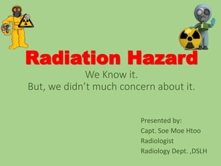
- 1. Radiation Hazard We Know it. But, we didn’t much concern about it. Presented by: Capt. Soe Moe Htoo Radiologist Radiology Dept. ,DSLH
- 2. OBJECTIVE • To become familiar with the mechanisms & biological effects following exposure to ionizing radiation. • To be aware of the risks of ionizing radiation . • To know main safety issues of protection.
- 3. RADIATION • Radiation is the energy that comes from a source and travels through some material or space as waves or photons.
- 4. Ionizing Radiation • Radiation that can cause specific changes is called ionizing radiation (>10ev) • Has enough energy to break chemical bonds. • Interaction with matter produce ions. Non-Ionizing Radiation • Radiation that does not have enough energy to break chemical bonds but can vibrate atom. • Low-energy radiation e.g. sound waves, visible light, microwaves • Cannot produce ions.
- 6. UNITS OF RADIATION Roentgen (R) • Radiaton exposure in a volume of air. Rem • It is the unit of effective dose. • Used only in radiation protection. • The SI unit is Sievert • 1 Sv= 100 rem • 1 mSv = 0.001 Sv = 0.1 rem
- 7. Linear Energy Transfer (LET) • The average energy deposited per unit length of track. • Measured in kiloelectron volts per micron • Low mass, increased travel distance (gamma rays, X rays). • Large mass, decreased travel distance (alpha particles, protons, low energy neutrons). • Causes dense ionization along its path with a high probability of interacting directly with DNA
- 10. Outcomes after cell exposure DAMAGE TO DNA DAMAGE REPAIRED CELL DEATH (APOPTOSIS) TRANSFORMED CELL Viable Cell Non-Viable Cell Cancer?
- 11. Reactions Within Body • Reactions produce 1. free electrons e- 2. ions H- and OH- 3. free radicals H* and OH*
- 12. Free Radicals • A free radical is an atom or molecule that has an unpaired electron in its valence shell. • Pair with electrons from other atoms, including those that make up the DNA molecule.
- 13. Direct Action • Causes damage directly to DNA or other important molecules in the cell. • More likely when the beam of charged particles consist of alpha particles, protons, or electrons
- 14. Indirect Action • Causes damage by interacting with the cellular medium producing free radicals which then damage the DNA molecule. • More likely when x-rays or gamma-rays compose the beam.
- 17. Radiosensitivity • Actively reproducing cells are more radiosensitive than mature cells. • During mitosis, the cell is in a stressed state and shows an increase in damage caused by radiation.
- 18. Radiosensitivity HIGH MEDIUM LOW Bone marrow Spleen Thymus Lymph nodes Gonads Eye lens Lymphocytes Skin Mesodermal origins (Liver, heart, lungs, …) Muscle Bones Nervous system
- 19. Dose-Response Relationships • Two effects of radiation exposure: • Deterministic (threshold) • Stochastic: cancer
- 20. Non-Stochastic (Deterministic) Effects • Occurs above threshold dose • Severity increases with dose • Alopecia (hair loss) • Cataracts • Erythema • Radiation Sickness • Temporary Sterility Stochastic (Probabilistic) Effects • Occurs by chance • Probability increases with dose • Carcinogenesis • Mutagenesis • Teratogenesis
- 21. Effects in eye • Eye lens is highly RS. • Coagulation of proteins occur with doses greater than 2 Gy. • There are 2 basic effects: Effect Sv single exposure Sv per years for many years Detectable opacities 0.5 – 2.0 > 0.1 Visual impairment (Catarct) 5.0 > 0.15
- 22. Threshold Doses for Deterministic Effects • Cataracts of the lens of the eye 2-10 Gy • Permanent sterility • males 3.5-6 Gy • females 2.5-6 Gy • Temporary sterility • males 0.15 Gy • females 0.6 Gy
- 23. Symptoms of Acute Radiation Sickness • Hemopoietic: 3-8 Gy LD50/60 radiation damages precursors to red/white blood cells & platelets: eg. Chernobyl personnel (203 exhibited symptoms, 13 died)
- 24. Gastrointestinal : >10 Gy • radiation depopulates GI epithelium (crypt cells) • abdominal pain/fever, diarrhea, dehydration • death 3 to 10 days (no record of human survivors above 10 Gy) examples include Chernobyl firefighters Cerebrovascular : > 100 Gy • death in minutes to hours
- 26. RADIATION PROTECTION • Based on two components. • A) JUSTIFICATION. • B) OPTIMIZATION.
- 27. JUSTIFICATION • Applications of ionising radiation are only justified when they provide a net benefit with minimization of risks of radiation for people.
- 28. GUIDELINES FOR REFERRING PHYSICIANS: 1) Repeating investigations which have already been done 2) Investigation when results are unlikely to affect patient management 3) Investigating too often: i.e. before the disease could have progressed or resolved 4) Doing the wrong investigation 5) Failing to provide appropriate clinical information & questions that imaging Investigation should answer.
- 29. 0.01 mSv 0.5 CXR 0.02mSv 1 CXR 0.07 mSv 3.5 CXR 1.3mSv 65 CXR 0.7mSv 35 CXR 1.0mSv 50 CXR 2.5mSv 125 CXR 3.0mSv 150 CXR 7.0mSv 350 CXR
- 31. 4 mSv 200 CXR 15 mSv 750 CXR
- 32. THYROID SCAN 1.0 mSv 50 CXR RENAL SCAN 1.0 mSv 50 CXR LUNG PERFUSION SCAN 1.0 mSv 50 CXR BONE SCAN 4.0 mSv 200 CXR FDG PET (HEAD) 5.0 mSv 250 CXR CARDIAC GATED 6.0 mSv 300 CXR
- 33. ALARA PRINCIPLE OPTIMIZATION • Once a practice is justified, the exposure to ionising radiation should be kept as low as reasonably achievable (ALARA).
- 34. FUNDAMENTAL PRINCIPLES OF RADIATION PROTECTION 1. Distance. 2. Exposure time. 3. Barriers & Shielding.
- 35. DISTANCE INVERSE SQUARE LAW: • Intensity of radiation is inversely proportional to the square of the distance from the source of radiation. • For Example: If the dose is 9 R at 3 feet, stepping back to a distance of 6 feet will cause the dose to decrease to 2.25 R.
- 36. EXPOSURE TIME • The amount of radiation received is proportional to the length of the exposure time. • Minimized by conducting procedures as quickly as possible. • For Example, using short bursts of fluoroscopy. • Employing image intensifiers & Intensifying screens. • Using high kVp , low mAs techniques.
- 37. BARRIERS & SHIELDING • Most commonly used protective material lead. • High density and high atomic number • Lead equivalent: thickness of lead which provide the same degree of protection as the material. ROOM SHIELDING: • Should be located as far as away from areas of high occupancy and general traffic. • Wall on which primary beam falls should not be less than 35 cm thick brick or equivalent. • Shielding of 1.7mm lead (23 cm brick) in front of doors & windows of x-ray room.
- 38. X-RAY CONTROL ROOM: • Walls & viewing windows of control booth should have lead equivalent of 1.5 mm. • Distance between control panel & X-ray unit / chest stand should be minimum 3 meters.
- 39. PATIENT WAITING ROOM: • Provided outside X-ray room a proper warning signal when unit is in use. • Warning devices may include audible and visual signs.
- 40. LEAD APRON • Typically thickness of 0.5 mm lead equivalent is used. • Weight ranges from 2.5 to 7 kg. • Should cover much of red bone marrow & breast.
- 41. LEAD GLOVES • Lead salts or metallic lead are added to rubber or plastic. • Lead equivalent of these is about ¼ mm.
- 42. LEAD GLASS • Made by adding lead salts to silicates , in the manufacturing of glass. • It is acceptably transparent and a better protective material. • Contains 60% of lead by weight.
- 43. GONADAL SHIELDING • Must be 0.5 mm of lead. • Must be used when gonads will lie within 5 cm of the collimated area. • Separate male vs. female shielding available. MALE GONADAL SHIELD OVARIAN PROTECTION
- 44. THYROID COLLER
- 45. • Have standard projections for specific indications. • Additional views - on a case-by-case basis • Use PA projections, where practical, for chest and spine radiographs. • Avoid repeating exposures. • Use safe exposure factors – high KVp and low mAs technique. • Never stand in the primary beam. • Always wear protective apparel when not behind a protective barrier. • Always wear a radiation monitor and position it outside the protective apron at collar level. • The person holding the patient must wear protective apron and if possible gloves. • Always collimate to smallest field size appropriate to examination.
- 46. Summary • Ionizing radiation use should be only used when benefit outweighs possible risks. • Every examination should be Justified. • Optimized protocols for lowering patient dose without affecting accurate diagnosis should be done. • Use all kinds of radiation protection during work...It's your life.!
- 48. THANK YOU… HAVE A NICE DAY!