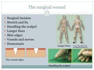
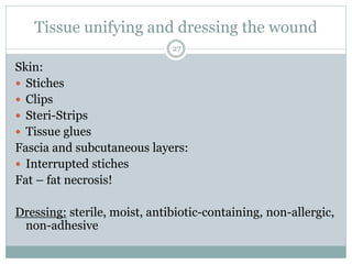
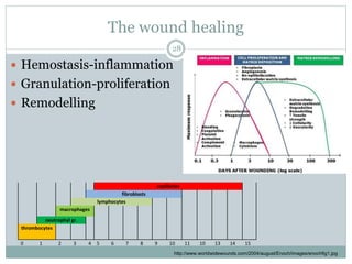
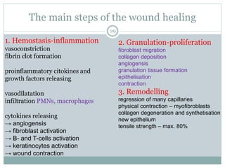
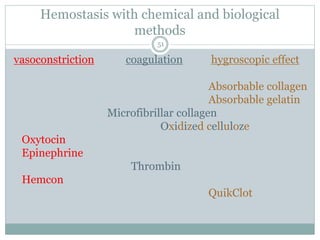
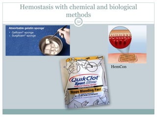
Call Girls Cuttack Just Call 9907093804 Top Class Call Girl Service Available
Wound lecture 1.ppt
1. GYÖRGYI SZABÓ
A S S I S T A N T P R O F E S S O R
DEPARTMENT OF SURGICAL
RESEARCH AND TECHNIQUES
Classification and management of
wound, principle of wound healing,
haemorrhage and bleeding control
Basic Surgical Techniques, Faculty of Medicine, 3rd year
2021/13 Academic Year, Second Semester
1
3. What is a wound?
It is a circumscribed injury which is caused by an external
force and it can involve any tissue or organ.
surgical, traumatic
It can be mild, severe, or even lethal.
Simple wound
Compound wound
Acute
Chronic
3
4. Parts of the wound
Wound edge Wound
corner
Surface of
the wound
Base of the wound
Cross section of a simple wound
Skin surface
Subcutaneus tissue
Superficial fascia
Muscle layer
Base of the wound
Wound edge
Surface of
the wound
Wound
cavity
4
5. The ABCDE in the injured assessment
The mnemonic ABCDE is used to remember the order
of assessment with the purpose to treat first that kills
first.
A: Airway and C-spine stabilization
B: Breathing
C: Circulation
D: Disability
E: Environment and Exposure
5
6. Wound management - anamnesis
When and where was the wound occured?
Alcohol and drug consumption
What did caused the wound?
The circumstances of the injury
Other diseases eg. diabetes mellitus, tumour,
atherosclesosis, allergy
The state of patient’s vaccination against Tetanus
Prevention of rabies
The applied first-aid
6
7. Classification of the accidental wounds
1. Based on the origine
I. Mechanical:
1. Abraded wound (vulnus abrasum)
2. Puncured wound (v. punctum)
3. Incised wound (v. scissum)
4. Cut wound (v. caesum)
5. Crush wound (v. contusum)
6. Torn wound (v. lacerum)
7. Bite wound (v. morsum)
8. Shot wound (v. sclopetarium)
II. Chemical:
1. Acid
2. Base
III. Wounds caused by radiation
IV. Wounds caused by thermal forces:
1. Burning
2. Freezing
V. Special
7
8. 1.) Abraded wound
(v. abrasum)
2.) Punctured wound
(v. punctum)
Superficial part of the epidermal
layer
Good wound healing
Sharp-pointed object
Seems negligible
BUT
Anaerobic infection
Injury of big vessels and nerves
Mechanical wounds
8
14. 1.) Acid 2.) Base
in small concentration – irritate
in large concentration –
coagulation necrosis
colliquative necrosis
Chemical wounds
14
15. Symptoms and severity
depend on:
Amount of radiation
Length of exposure
Body part that was exposed
Symptoms may occur immediately,
after a few days, or even as long
as months.
What part of the body is
most sensitive during
radiation sickness?
bone marrow
gastrointestinal tract
Wounds caused by radiation
15
16. 1.) Burning 2.) Freezing
a – normal skin
1 - 1st degree – superficial injury
(epidermis)
2 – 2nd degree –partial or deep partial
thickness (epidermis+superficial or deep
dermis)
3 – 3rd degree – full thickness (epidermis
+ entire dermis)
4 – 4th degree – (skin + subcutaneous
tissue + muscle and bone)
Treatment:
Cooling – cold water and clean covering
Wounds caused by thermal forces
16
Metabolic change! - toxemia mild, moderate, severe (redness,
bullas, necrosis)
rewarm – not only the frozen area
but the whole body
18. Classification of the wounds
2. According to the bacterial contamination
Clean wound
Clean-contaminated wound
Contaminated wound
Heavily contaminated wound
18
19. Superficial
Partial thickness
Full thickness
Deep wound
Classification of the wounds
2. Depending on the depth of injury
+ bone, opened cavities, organs…etc.
19
source: http://www.funscrape.com/Search/1/skin+layers.html
20. Wound management - history
Ancient Egypt – lint (fibrous base-wound site closure), animal grease
(barrier) and honey (antibiotic)
„closing the wound preserved the soul”
Greeks – acute wound= „fresh” wound; chronic wound = „non-healing”
wound
maintaining wound-site moisture
Ambroise Paré – hot oil oil of roses and turpentine, ligature of
arteries instead of cauterization
Lister pretreated surgical gauze – Robert Wood Johnson 1870s;
gauze and wound dressings treated with iodide
20
32. Complications of wound healing
I. Early complications
Seroma
Hematoma
Wound disruptin
Superficial wound infection
Deep wound infection
Mixed wound infection
32
33. 1.) Seroma 2.) Hematoma
Filled with serous fluid, lymph
or blood
Fluctuation, swelling, redness,
tenderness, subfebrility
TREATMENT:
Sterile punture and
compression
Suction drain
Early complications of wound healing
33
Bleeding, short drainage time,
anticoagulant
Risk of infection
Swelling, fluctuation, pain,
redness
TREATMENT
Sterile puncture
Surgical exploration
34. 3.) Wound disruption
A. partial – dehiscenece
B. complete - disruption
Surgical error
Increased intraabdominal
pressure
Wound infection
Hypoproteinaemia
TREATMENT:
U-shaped sutures
Early complications of wound healing
34
35. 1.) Diffuse 2.) Localized
Located below the skin
TREATMENT
Resting position
Antibiotic
Dermatological consultation
Anywhere
TREATMENT
Surgical exploration
Drainage
X-ray examination
Early complications of wound healing
Superficial wound infection
35
e.g. erysipelas e.g. abscess
36. 1.) Diffuse 2.) Localized
TREATMENT
Surgical exploration
Open therapy
H2O2 and antibiotics
e.g. anaerobic necrosis
Inside the tissues or body cavities
TREATMENT
surgical exploration
drainage
Early complications of wound healing
Deep wound infection
36
37. Mixed wound infection
e.g. gangrene
necrotic tissues
putrid and anaerobic
infection
a severe clinical picture
TREATMENT
aggresive surgical
debridement
effective and specified
(antibiotic) therapy
37
Complications of wound healing
I. Early complications
38. Complications of wound healing
II. Late complications
Hyperthrophic scar
Keloid formation
Necrosis
Inflammatory infiltration
Abscesses
Foreign body containing abscesses
38
39. Hypertrophic scar Keloid
Develop in areas of thick
chorium
Non-hyalinic collagen
fibres and fibroblasts
Confine to the incision
line
TREATMENT
Regress spontaneously
(1-2 yrs)
Late complications
39
Mostly African and Asian
population
Well-defined edge
Emerging, tough structure
Overproliferation of collagen
fibers in the subcutaneous tissue
Subjective complains
TREATMENT
Postoperative radiation
Corticosteroid + local anaesthetic
injection
41. Anatomical Diffuse
Arterial – bright red,
pulsate
Venous – dark red,
continuous
Capillary – can become
serious
Parenchymal
Bleeding
41
42. Bleeding
Severity of bleeding – the volume of the lost blood and
time
42
source: http://lifeinthefastlane.com/2012/03/trauma-tribulation-025/
43. The direction of hemorrage
External
Internal
In a luminar organ (hematuria, hemoptoe, melena)
In body cavities (intracranial, hemothorax, hemascos,
hemopericardium, hemarthros)
Among the tissues (hematoma, suffusion)
43
44. Bleeding
Preoperative hemorrhage
Prehospital care! – maintenance of the airways, ventillation and circulation
bandages, direct pressure, turniquets
Intraoperative hemorrhage
anatomical and/or diffuse
depending on the surgeon, the surgery, position,
the size of the vessel, pressure in the vessel
ANESTHESIA!
Postoperative bleeding
ineffective local hemostasis, undetected hemostatic defect, consumptive
coagulopathy or fibrinolysis
44
45. Local General
Hematoma, suffusion,
ecchymosis
Compression in the pleural
cavity, in pericardium, in the
skull
Functional disturbancies – e.g.
hyperperistalsis
Pale skin, cyanosis, decreased
BP. and tachycardia, difficulty
in breeding, sweeting,
decreased body temperature,
unconsciousness, cardiac and
laboratory standstill, laboratory
disorders, signs of shock
Signs of the bleeding
45
46. Surgical hemostasis
Aim – to prevent the flow of blood from the incised or
transected vessels
Mechanical methods
Thermal methods
Chemical and biological methods
46
47. Surgical hemostasis
Mechanical methods
Digital pressure – direct pressure,
e.g. Pringle maneuver
Tourniquet
Ligation
Suturing
Preventive hemostasis
Clips
Bone wax
other
47
48. Thermal methods
Low temperature
Hypothermia – eg. stomach bleeding
Cryosurgery
dehidratation and denaturation of fatty tissue
decreases the cell metabolism
vasoconstriction
48
49. Thermal methods
High temperature
Electrosurgery – electrocauterization
Monopolar diathermy
Bipolar diathermy
Laser surgery
coagulation and vaporization
for fine tissues
49
50. Thermal methods
High temperature
Electrocoagulation
Electrofulguration (A)
Electrodessication
Electrosection
50
51. Hemostasis with chemical and biological
methods
vasoconstriction coagulation hygroscopic effect
Absorbable collagen
Absorbable gelatin
Microfibrillar collagen
Oxidized celluloze
Oxytocin
Epinephrine
Thrombin
Hemcon
QuikClot
51
A kialakult lőcsatorna első rétegében elhalt szövetek és idegen anyagok helyezkednek el (pl.: ruhadarabok). Körülötte kialakul az elsődleges necroticus zóna, amelyben a lökéshullám és a hőhatás miatt elhalt szöveteket észlelünk. E körül a necrobioticus zóna helyezkedik el, amelyben vérzést, érkárosodást, trombusokat észlelünk, majd körkőrösen a molekuláris rázkódtatás zónája, amely éles határ nélkül megy át az ép szövetekbe. A lőcsatornát mindig fel kell tárni!
ulcers on people’s backs, chests, mouth, etc. are common when one is exposed to a vast amount of radiation
Wound exudate
» View large image.
If a wound produces a large amount of fluid (exudate)1, the healing process can be slowed down. Increased exudate levels can be a symptom of infection and increased oedema. And the more a wound exudes, the higher the risk of maceration of the surrounding skin2 – and exudate leakage. Exudate leakage is not only clinically challenging to treat – it also prevents some patients from participating in social activities, or from going out altogether. Problems often associated with exuding wounds are [1-2] :
a negative impact on the patient’s quality of life
very time consuming treatment
complications such as infection, wound pain and maceration that slows the healing process
The composition and amount of wound exudate1 varies depending on the stage of the healing process. In the inflammatory phase of wound healing, exudate levels are usually high, which may be a sign of wound infection. Unlike acute wound exudate, chronic wound exudate contains enzymes (proteases) that break down proteins and newly formed cells. To break the vicious cycle in a non-healing wound, the exudate must be led away from the wound with an absorbent dressing. Controlling wound exudateControl of exudate, removal of unhealthy tissue by debridement and management of bacterial load are all part of good wound bed preparation. A good wound dressing keeps the wound moist and absorbs exudate, locking it inside the dressing to prevent maceration. Offering excellent absorption and retention, Biatain foam dressings are proven to deliver superior exudate management.[3-4] With longer wear time (up to seven days), minimal risk of leakage and maceration, your patients’ wounds will heal faster.[1,5-7]
Tertiary Intention
When a wound is intentionally kept open to allow edema or infection to resolve or to permit removal of exudate, the wound heals by tertiary intention, or delayed primary intention. These wounds result in more scarring than wounds that heal by primary intention but less than wounds that heal by secondary intention. (Johnstone, Farley,& Hendry, 2005)
Breast reduction – dehiscence – 2weeks
Diffuse – erysipelas – caused by hemolytic streptococci
Localized – joint abscess
This patient developed an abscess over the metacarpophalangeal joint of the thumb after an accidental cut with a kitchen knife.
See Also
Scar location related to patient and parent satisfaction with ...
A 15-year-old Hispanic girl with bump
12-year-old boy admitted for febrile illness
What is your diagnosis?
Source: Jeray KJ
Answer
The answer is methicillin-resistant Staphylococcus aureus.
Surgeons at one hospital system found that MRSA may become a more common bacteriologic flora of hand infections, and if so, surgeons must adjust their choices for empiric antibiotics.
After noticing a marked increase in the number of community-acquired methicillin-resistant Staphylococcus aureus (MRSA) cases, surgeons at the Greenville Hospital System in South Carolina conducted a retrospective chart review of all orthopedic consultations done for hand infections from November 2003 to October 2005.
Localised – massive empyema (see the article)
Figure 1: Diffuse terminal ileal ulceration and angiomal formtion with oozing hemorrhage from these ulcers.
Mentions: In the absence of a primary ulcer, Dieulafoy's lesion was seen in the terminal ileum with oozing hemorrhage from this lesion 10 cm away from the ileocecal valve (Figure 1). The bleeding could not be controlled by endoscopic hemostasis using thermal coagulation or any other endoscopic intervention. A decision was made accordingly for urgent explorative laparotomy to save her life. An ileocolectomy (emergency limited segmental resection for a known bleeding source) was also done on our patient (Figure 2). Histology revealed this to be of the Dieulafoy type of lesion in the distal ileum (Figure 3).
Conjunctival suffusion with subconjunctival hemorrhage (ou), which was suggestive of leptospirosis, developed on the second hospitalization day.
The Pringle maneuver. The portal triad is occluded by guiding the posterior blade of the clamp through the foramen of Winslow with the aid of the left index finger.
-6. kép: amennyiben vérnyomásmérő áll rendelkezésre, úgy pneumatikus vértelenség felhelyezése. 280 Hgmm-re felfújjuk a vérnyomásmérő mandzsettát, így a sebalapot megtekinthetjük. Betadines vagy Octeniseptes fedőkötés, steril pólya, korrekt nyomókötés
7-8.kép: a nyomókötés felhelyezése után a vértelenség felengedése. Amennyiben erős vérzést észlelünk, ismételten felfújjuk a mandzsettát és revideáljuk a kötést. (az erős vérzés forrása csak technikai hiba lehet az elsősegélynyújtó részéről).
Timed spot freeze technique used to treat a malignancy (possibly a small basal cell cancer), demonstrating freeze ball formation and the 5-mm treatment margins necessary to achieve a temperature of −50ºC (−58 ºF) and, thus, the required depth of 4 to 5 mm.
Cryosurgery is a method of superfreezing tissue in order to destroy it. The technique is used to treat tumors, control pain, and control bleeding.
Information
The cold is introduced through a probe which has liquid nitrogen circulating through it. To destroy diseased tissue, the tissue is cooled to below -20 degrees Celsius. Other procedures that control pain or bleeding are cooled to a lesser degree to prevent tissue damage.
Electrocoagulation: A fine wire probe or other delivery mechanism is used to transmit radio waves to tissues near the probe. Molecules within the tissue are caused to vibrate which lead to a rapid increase of the temperature, causing coagulation of the proteins within the tissue, effectively killing the tissue. At higher powered applications, full desiccation of tissue is possible.
Two forms of electrosurgery: (A) Electrodesiccation with an active electrode tip touching the skin and showing penetration of planned tissue damage. (B) Fulguration with sparking from electrode to tissue. Treatment area is more superficial than in desiccation.Added by BiomedGuy
AboutEdit
Fulguration, also called electrofulguration, is the destruction of tissue by means of a high-frequency electric current applied with a needlelike electrode. In fulguration, the electrode is held away from the skin to produce a sparking at the skin surface and more shallow tissue destruction Fulguration is especially useful in treating superficial epidermal lesions, such as a superficial basal cell carcinoma of the trunk.[1]
LinksEdit
ReferenceEdit
↑ http://www.aafp.org/afp/2002/1001/p1259.html | Electrosurgery for the Skin | BARRY L. HAINER, M.D.,RICHARD B. USATINE, M.D., | Am Fam
HemCon Medical Technologies, Inc. began with funding from the United States Army and access to research by Dr. Kenton Gregory, Dr. Bill Wiesmann, the Oregon Medical Laser Center, and Providence Health Systems. The result was the HemCon® Bandage, which was designed to control life threatening bleeding.
Related Links: History & MissionManagement TeamBoard of DirectorsProductsEducational ResourcesDistribution and SalesProduct TrainingSuccess StoriesMedia RoomCustomer FeedbackCareers
The bandage was ushered through the FDA clearance process in a near-record 48 hours, and was soon deployed on the battlefield. Since then, the bandage has been used extensively and is credited with helping save over 100 lives with no adverse events reported. In 2005, the Army Surgeon General mandated that any soldier serving in Iraq or Afghanistan will carry at least one HemCon Bandage. This commitment by the U.S. Army is a testament to the efficacy and value of the HemCon Bandage. HemCon is rapidly changing from a military provider to a broad-based supplier of medical technology. With new products, strong partnerships in distribution, and a world class development team, we will continue to be unwavering in our commitment to innovate in all that we strive to achieve.