Downloaded 30 times
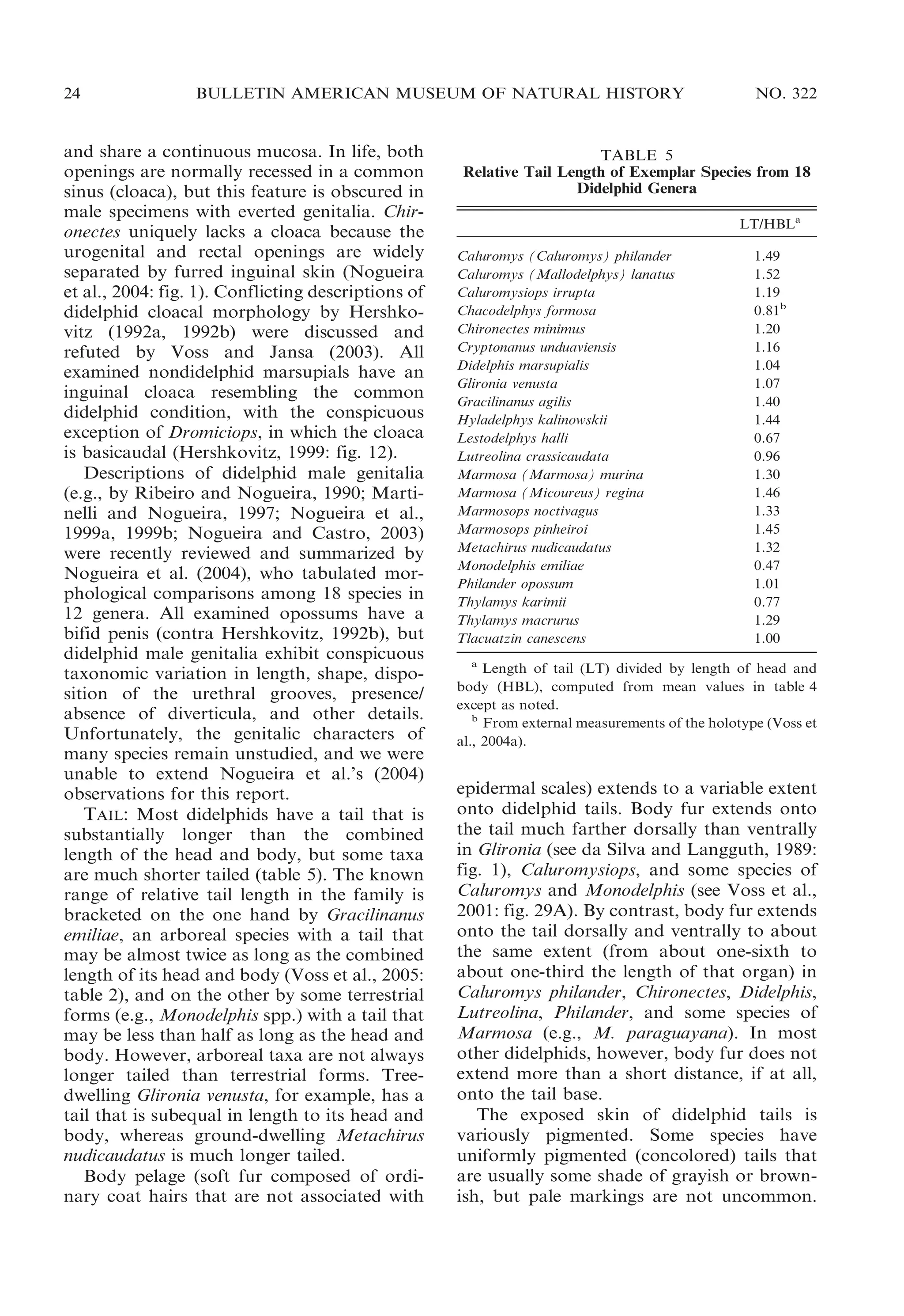
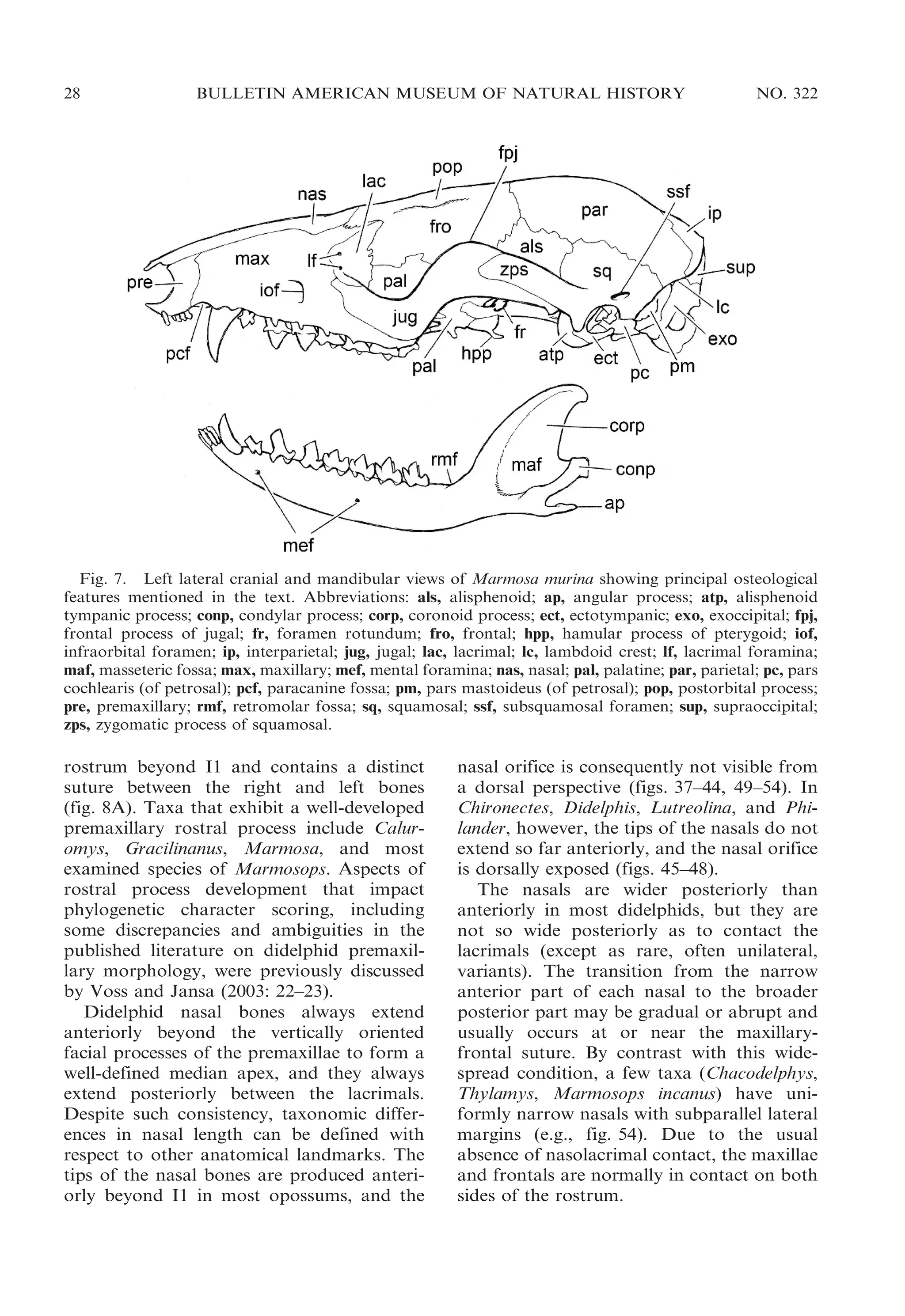
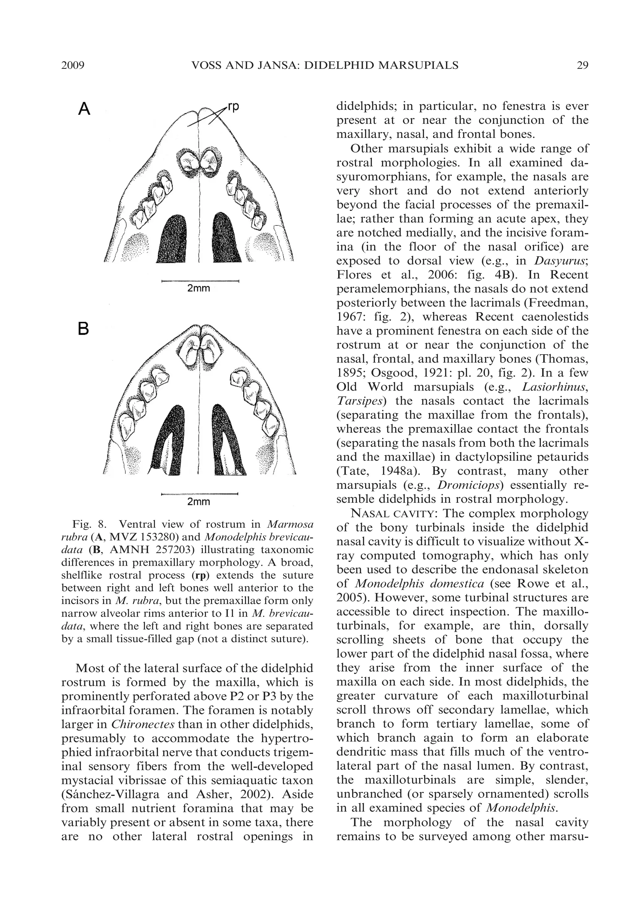
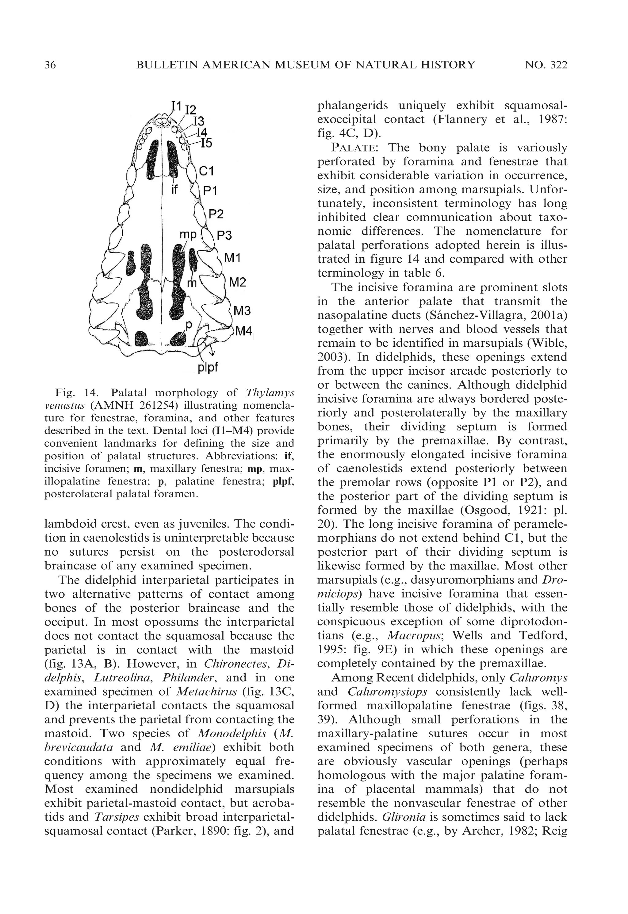
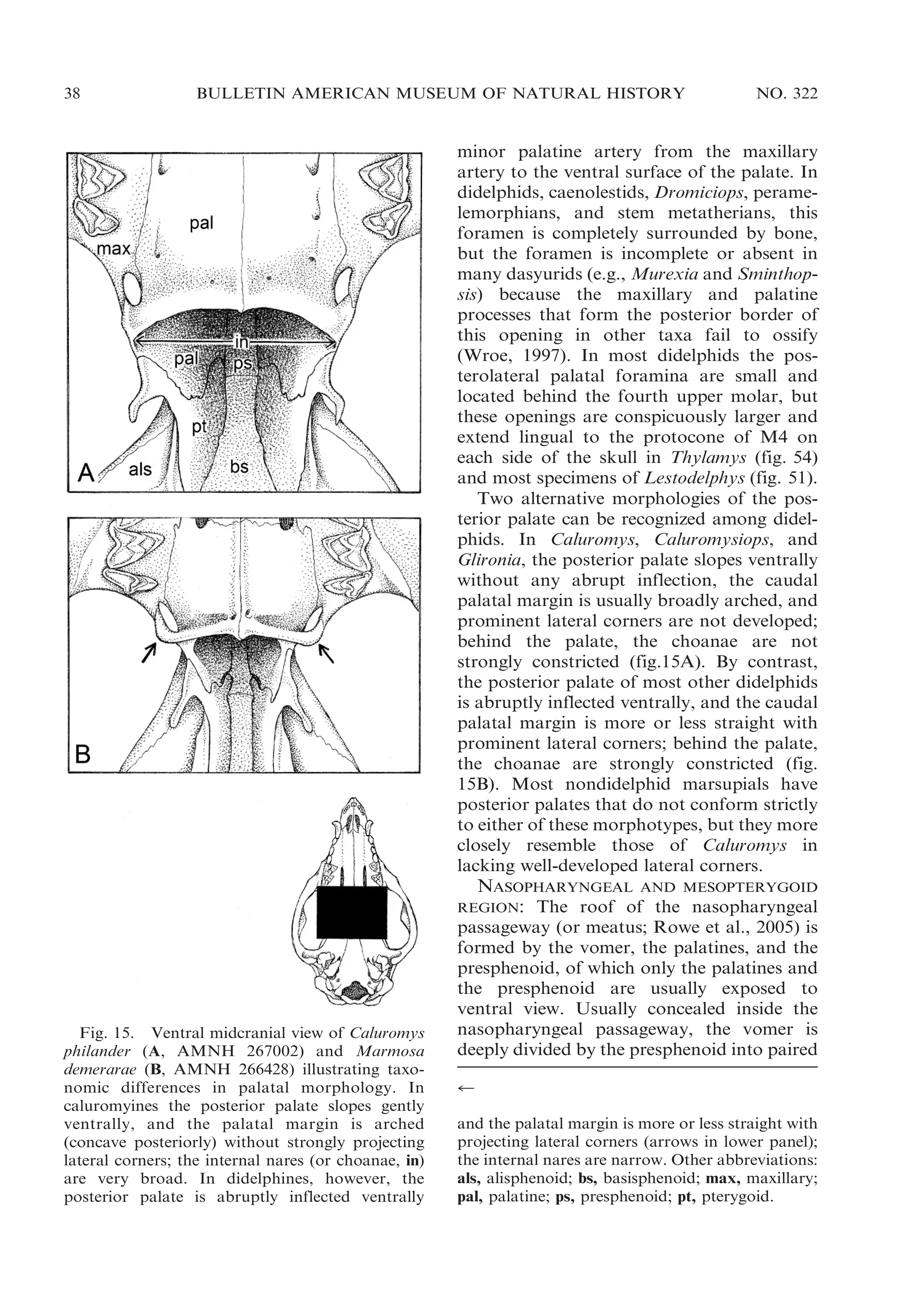
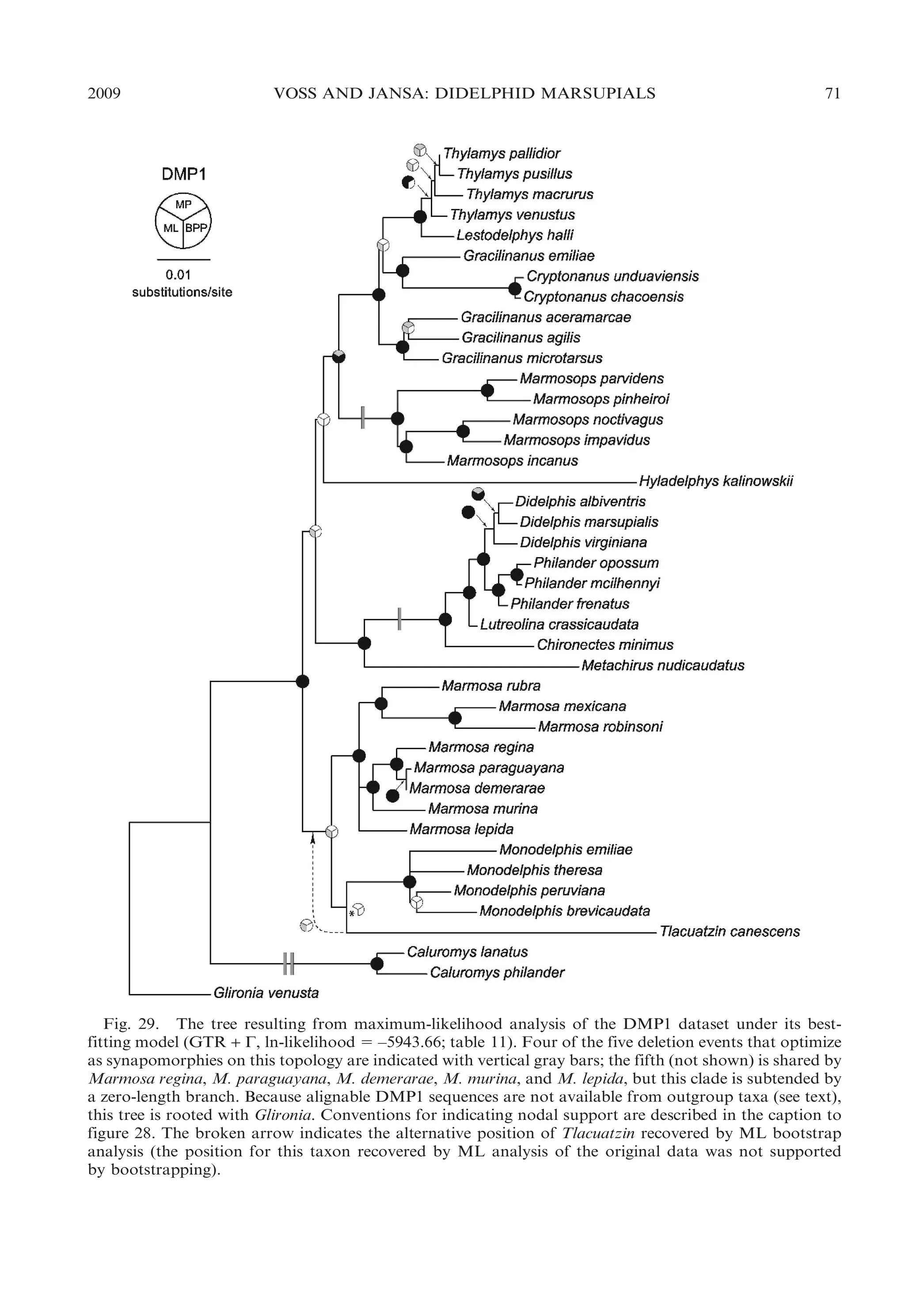
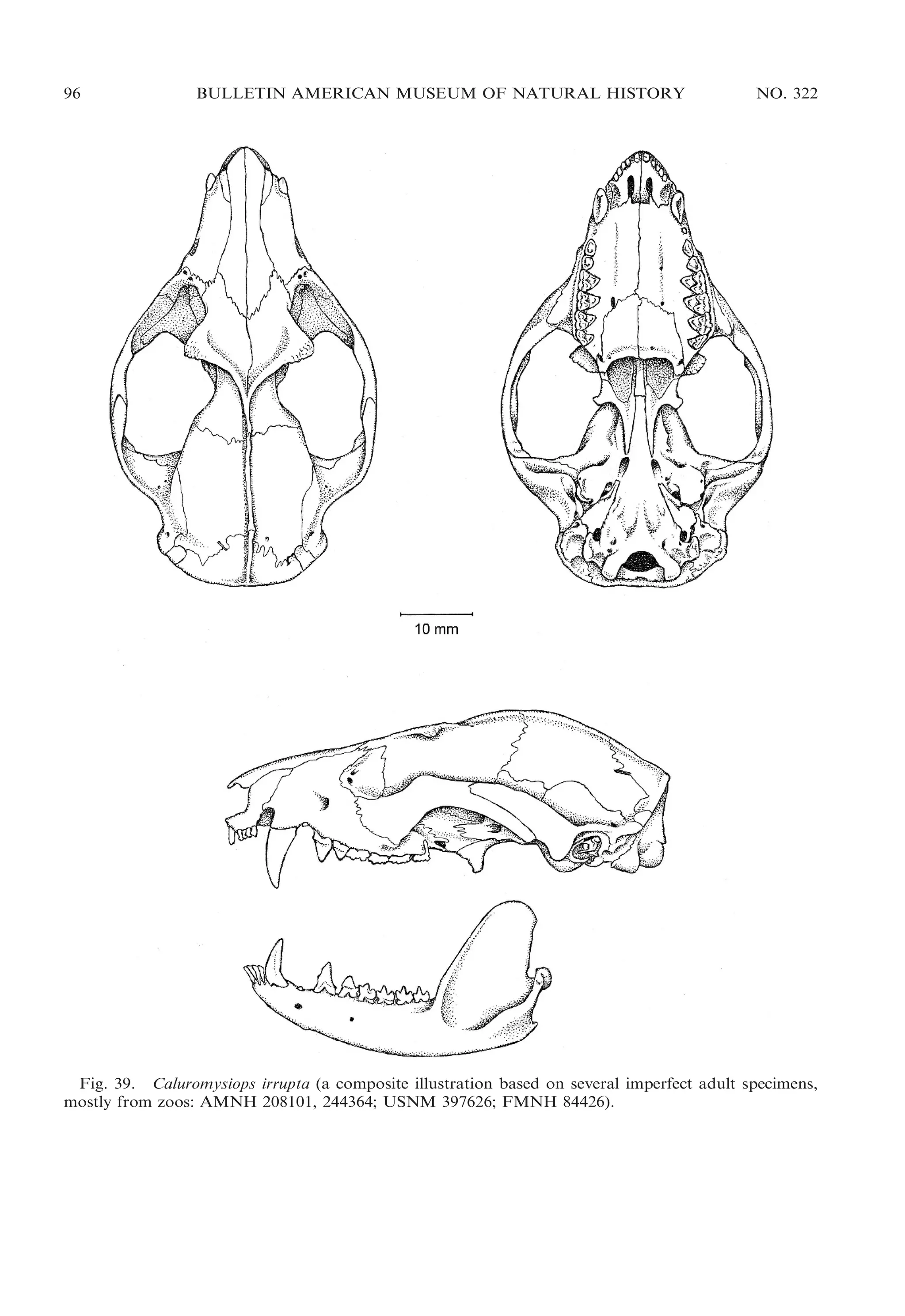
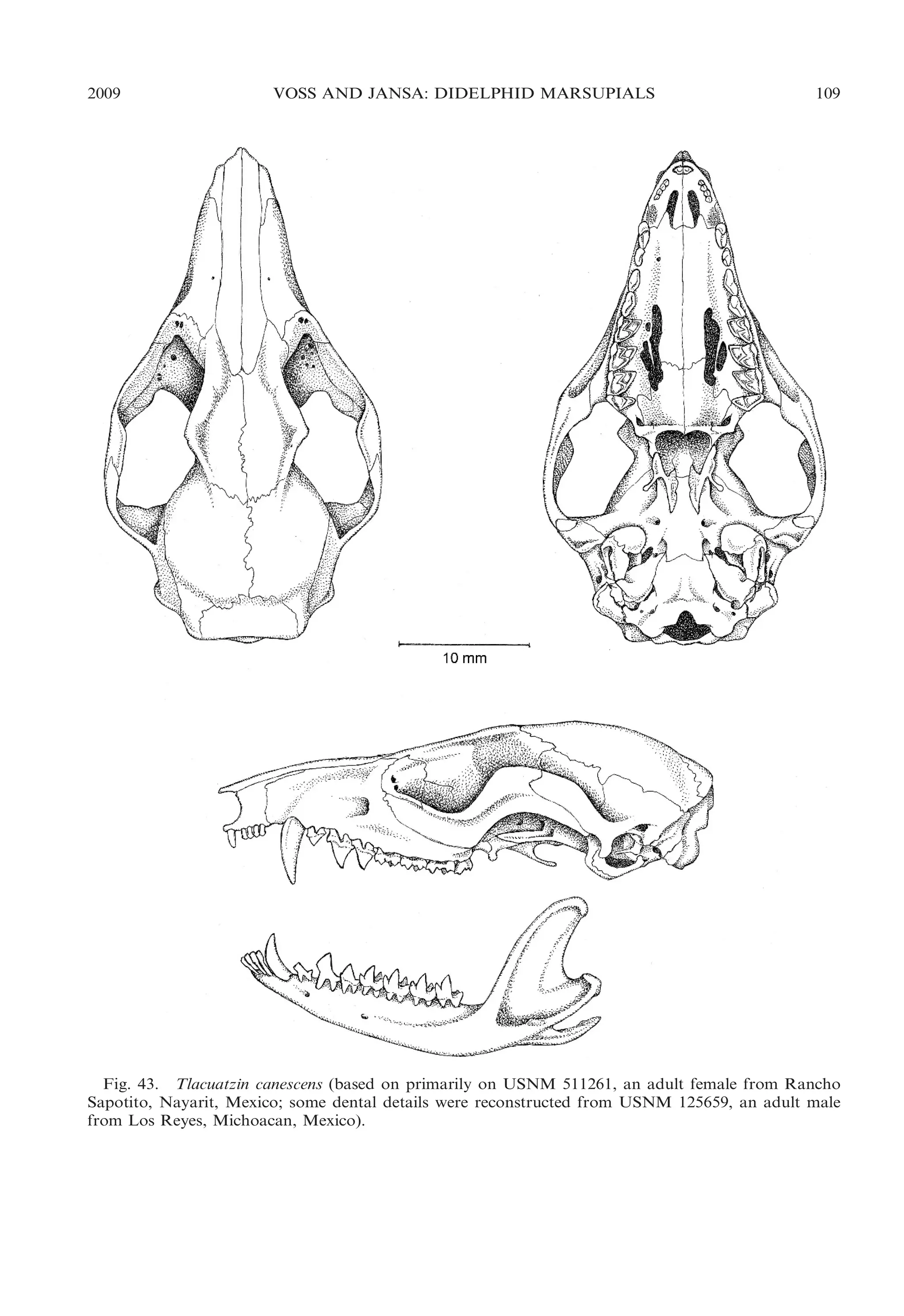
![2009
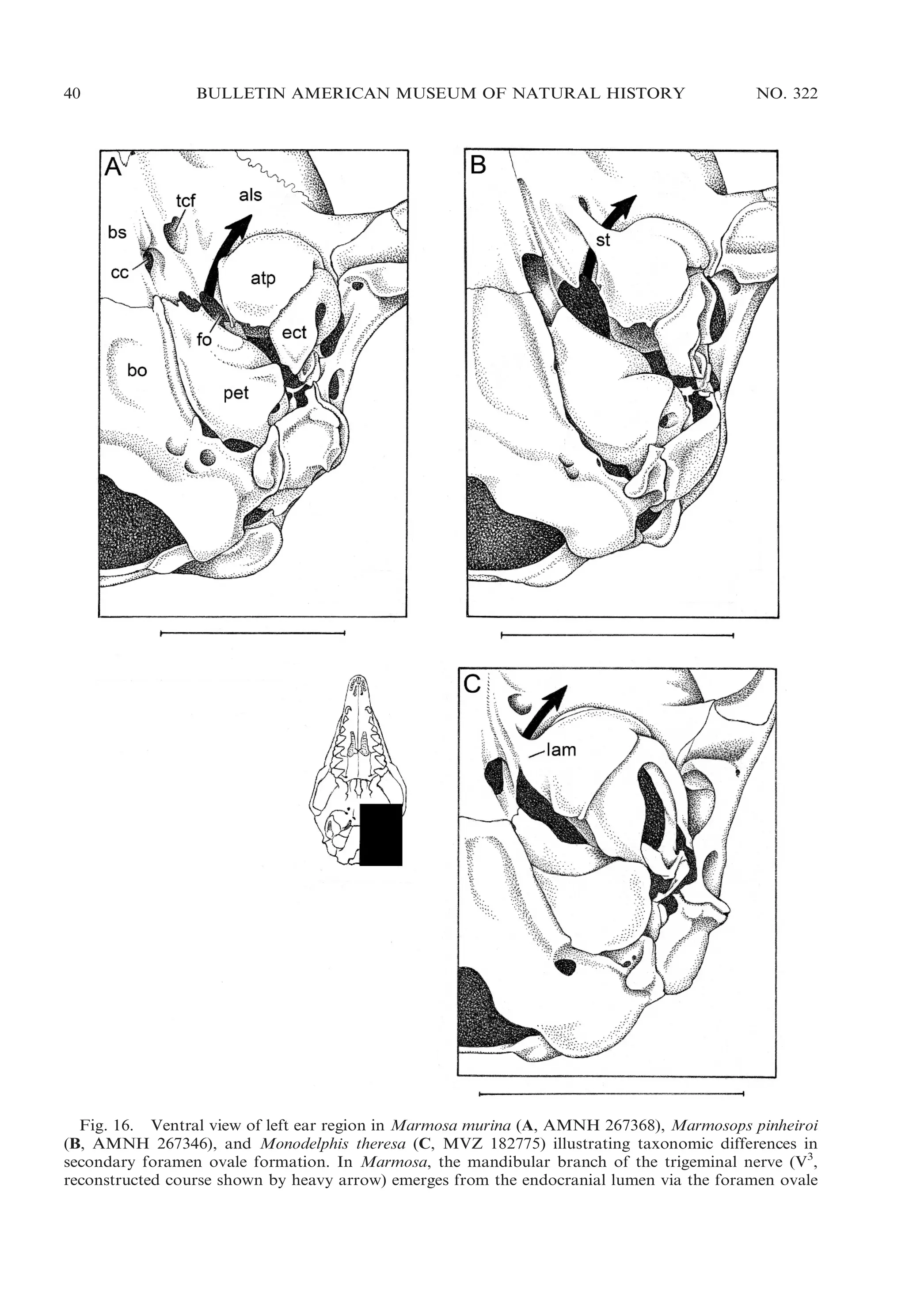
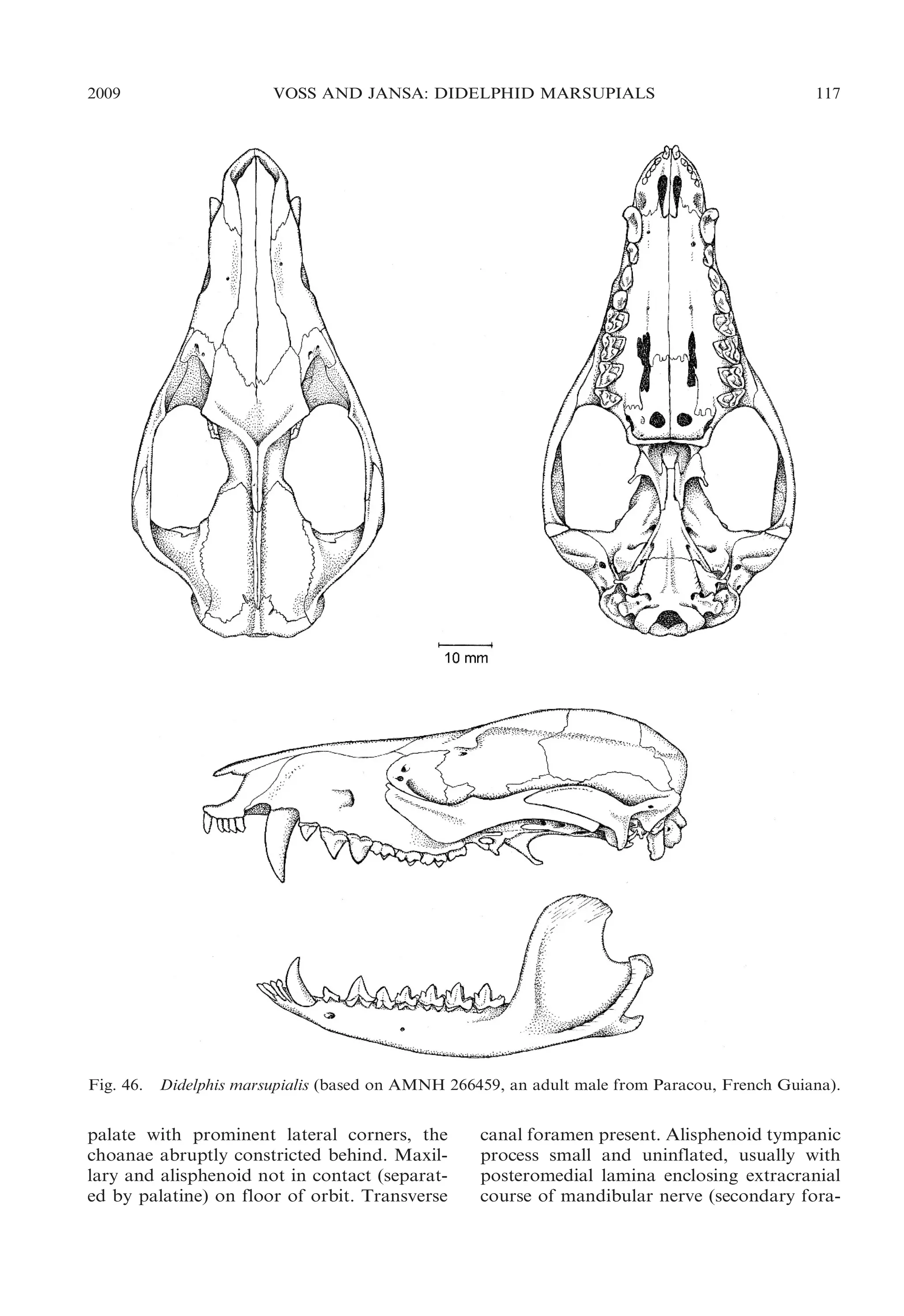
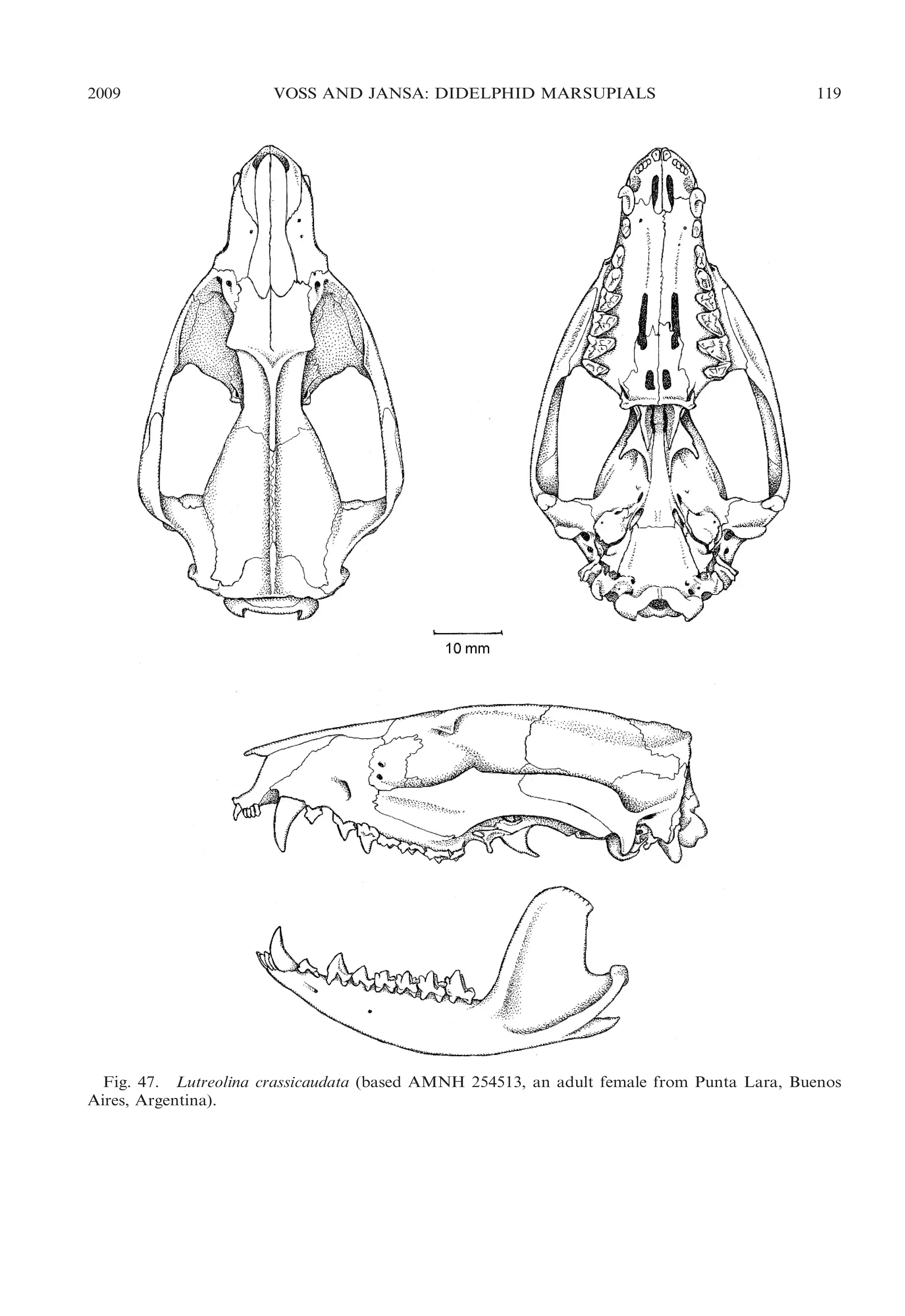
VOSS AND JANSA: DIDELPHID MARSUPIALS
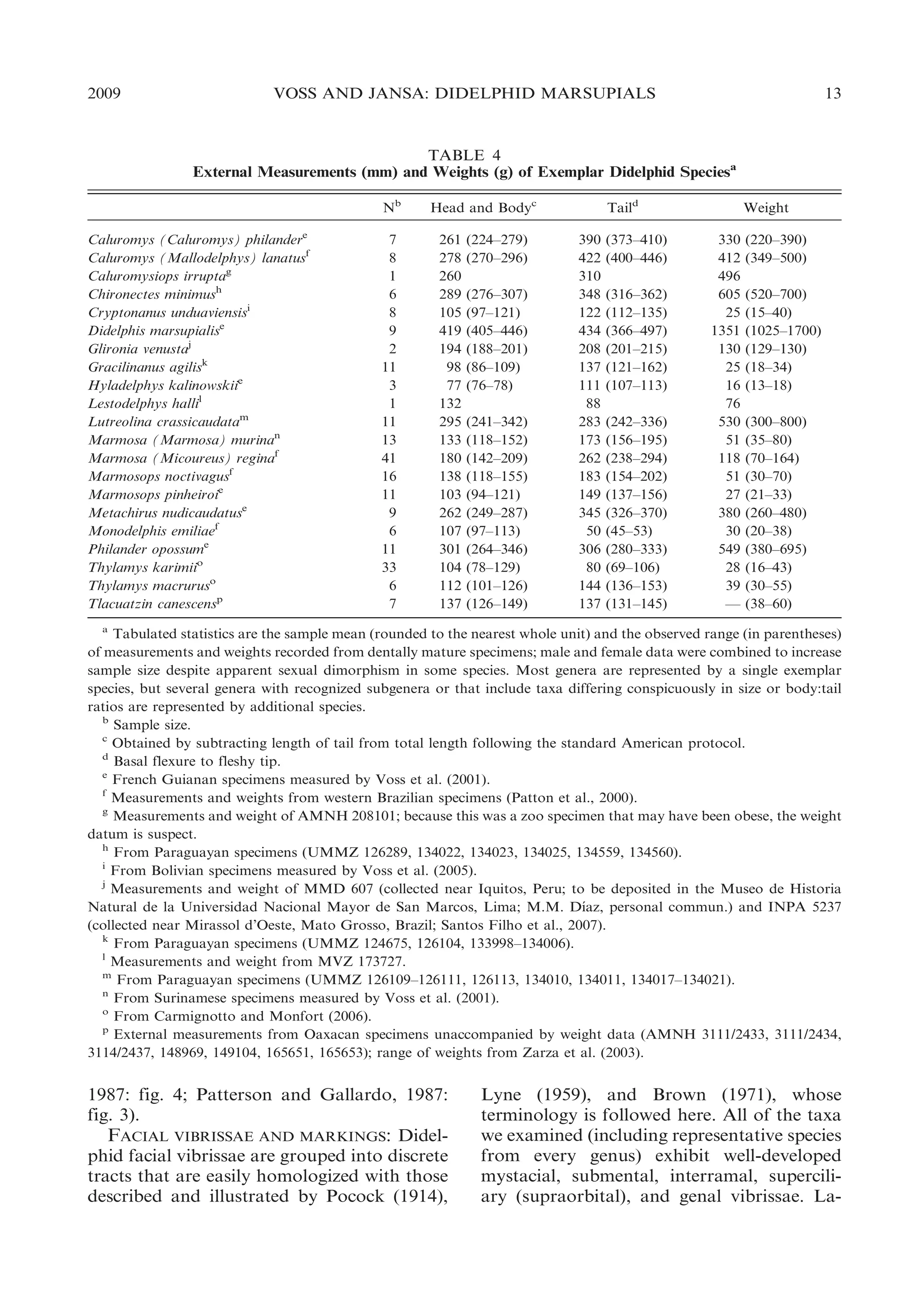
gular (throat) gland, the presence of which is
indicated on dried skins and fluid-preserved
specimens by a bare median patch of skin;
often, but not invariably, the surrounding fur
is discolored. According to Barnes (1977:
390), this secretory region contains ‘‘hypertrophied apocrine sudoriferous glands and
sebaceous glands, both confined to the
thickened dermis.’’ External signs of glandular activity tend to be maximally developed in
fully mature males (loc. cit.).
No unambiguously glandular throat patch
was observed in any examined specimens of
Caluromys, Caluromysiops, Chironectes, Hyladelphys, Lutreolina, Philander, or Tlacuatzin. By contrast, fully adult male specimens
of Cryptonanus, Gracilinanus, Lestodelphys,
and Thylamys usually exhibit well-developed
gular glands. A gular gland is also present on
the young adult male holotype (and only
known skin) of Chacodelphys formosa. Other
didelphid genera (Marmosa, Marmosops, and
Monodelphis) include some species that consistently develop adult male gular glands and
others that just as consistently show no trace
of such organs (Voss and Jansa, 2003).
Although adult male Didelphis often have
discolored gular fur, no glandular skin is
macroscopically distinguishable. Because we
were not able to examine any fully adult male
specimens of Glironia, the occurrence of gular
glands in this taxon is unknown.
Gular (or sternal) glands that are macroscopically and histologically similar to those
of didelphids are present in most dasyurids
(Cooper et al., 2005), but other plesiomorphic outgroup taxa (e.g., caenolestids,
peramelids, Dromiciops) seem to lack all
external traces of glandular activity on the
throat or chest.
BODY PELAGE: All didelphids have one or
more tracts of postcranial vibrissae (Brown
and Yalden, 1973), including ulnar-carpal
vibrissae (at the wrist), medial antebrachial
vibrissae (at or near the middle of the
forearm), anconeal vibrissae (at the elbow),
and/or calcaneal vibrissae (on the ankle).
Lyne (1959) reported the occurrence of
postcranial vibrissae in several didelphid
species based on his examination of pouch
young, whose sensory hair follicles are easily
seen because they are not obscured by coat
hairs. Unfortunately, postcranial vibrissae
15
are much harder to observe on fully furred
adult specimens, the only material commonly
available for most species. We found ulnarcarpal and medial antebrachial vibrissae on
most examined didelphids, whereas anconeal
and calcaneal vibrissae were often inapparent.
All didelphids have soft (nonspinous) fur
consisting of two or more distinct types of
hairs whose density and morphology determine the appearance and texture of the coat.
Some taxa (e.g., Caluromys) have somewhat
woolly fur that does not lie flat or exhibit the
glossy highlights typically seen in the pelts of
many other taxa, but textural differences are
hard to define by objective criteria that can
be used for character-state definitions or
taxonomic diagnoses. The only structural
(nonpigmental) feature of didelphid body
pelage that seems useful in this context is
the presence of uniquely long, coarse guard
hairs that project conspicuously from the
underfur in species of Didelphis.
The dorsal body pelage of most didelphids
is uniformly colored and unpatterned, usually some shade of brownish or grayish, but
some taxa are distinctively marked (see
illustrations in Eisenberg, 1989; Redford
and Eisenberg, 1992; Perez-Hernandez et
´
´
al., 1994; Reid, 1997; Eisenberg and Redford,
1999). Blackish transverse bars connected
middorsally on a pale-grayish background,
for example, characterize Chironectes; dark
scapular stripes are unique to Caluromysiops;
three longitudinal stripes are present in
several species of Monodelphis (e.g., M.
theresa); a grayish middorsum contrasting
with reddish flanks is exhibited by other
species in that genus (e.g., M. brevicaudata);
and a grayish midbody contrasting with
reddish head and rump is seen in others
(e.g., M. emiliae). The subtle but consistently
diagnostic ‘‘tricolor’’ shading of Thylamys
and Lestodelphys was described by Tate
(1933: 209):
Instead of the usual bicolor system composed of
a dorsal color, paling a little on the sides, which
is replaced at a generally well-marked transition
line by a distinct ventral color, the elegans group
[5 Thylamys] displays three distinct shades,
separated from each other along each side by
two lines of transition. The additional lines are
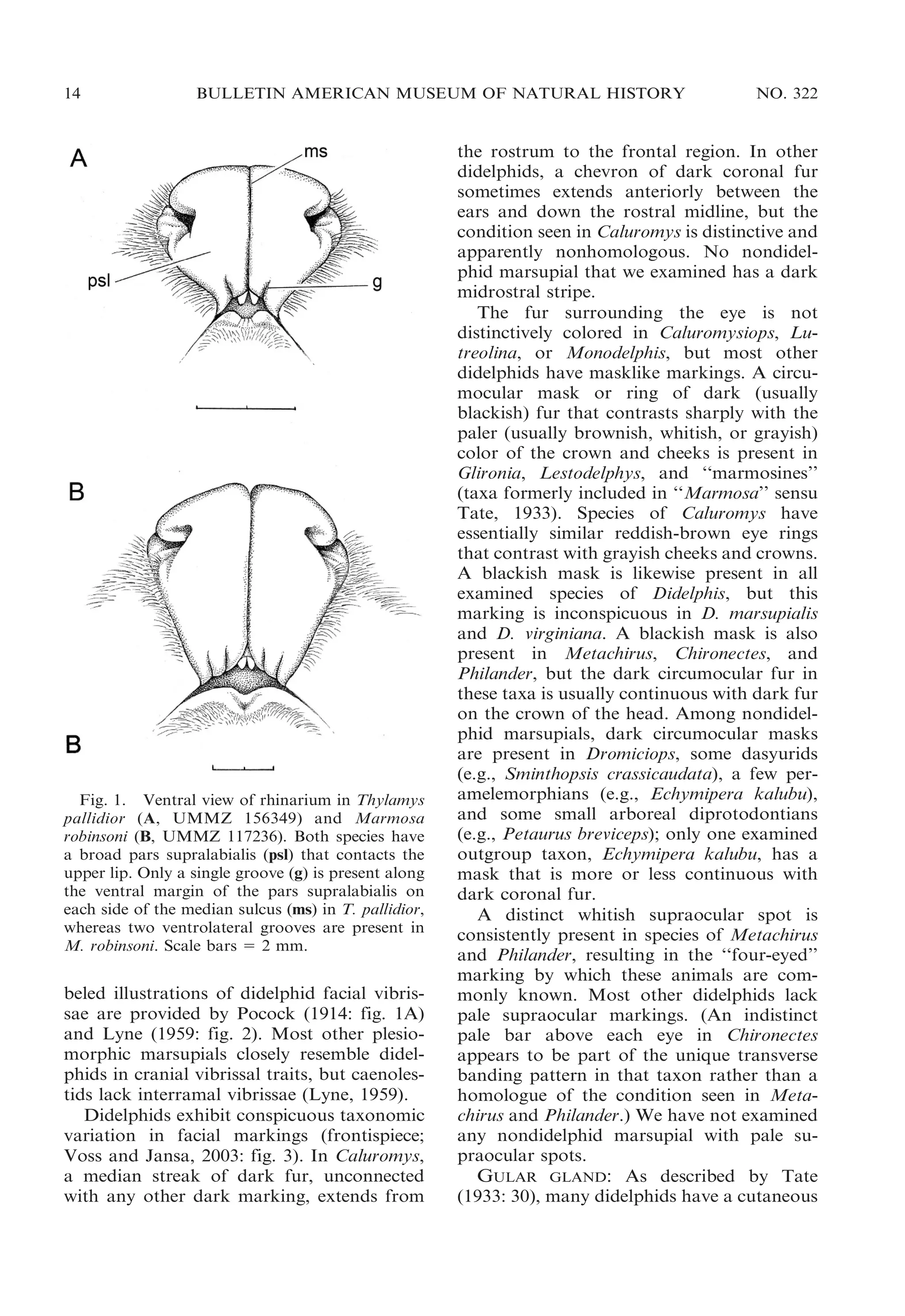
subdorsal, running from a point at the center of](https://image.slidesharecdn.com/vossjansa2009-140219090612-phpapp02/75/Voss-jansa-2009-15-2048.jpg)
![16
BULLETIN AMERICAN MUSEUM OF NATURAL HISTORY
the frons [forehead] past the inner edge of each
ear (not including it), and straight backward
through scapulae and hips, where they again
approach the median line of the body and
merge with the tail. This pair of lines encloses
the major part of the dorsal area of head and
body, the color of the area being very dark
brownish-gray or grayish fuscous. The fuscous
area is pointed at front, projecting forward
between the ears, and narrows again to a point
as it merges with the dark color of the upper
surface of the tail. The second [lateral] area,
light gray in color, frequently tinged with buffy
or yellowish, extends [on each side] between the
dark dorsal region and the edge of the belly
color at the normal transition line. Ventral color
either buffy, grayish, or snowy white.
The hair bases of the dorsal fur are heavily
pigmented, usually dark gray (or grayish), in
most didelphids, but species of Didelphis
uniquely exhibit white dorsal underfur.
The ventral pelage also exhibits noteworthy taxonomic variation among didelphids.
In some species the ventral fur is ‘‘graybased,’’ a descriptor that applies when the
individual hairs are grayish basally and
abruptly paler (usually whitish, yellowish,
or brownish) distally; the overall color of the
ventral fur then depends on the degree to
which the dark basal pigmentation shows
through the paler superficial hue. In other
species, the ventral fur is partially or entirely
‘‘self-colored,’’ a descriptor that applies when
the individual hairs have the same pigmentation (usually whitish) from root to tip.
Because marked differences in the patterning
of gray-based versus self-colored ventral pelage can occur among closely related (congeneric) species, such variation is often described
and illustrated in the revisionary taxonomic
literature (e.g., Patton et al., 2000: fig. 41).
However, the existence of numerous intermediate conditions spanning the entire range of
taxonomic variation in ventral color patterns
(e.g., from entirely gray-based to completely
self-white ventral fur) precludes meaningful
phylogenetic scoring of this character.
Whereas most of the pelage colors (and
color patterns) described above are preserved
on museum skins that have been protected in
dark cabinets from the bleaching effects of
light, other colors that can be striking in life
fade quickly after death. The ventral pelage
NO. 322
of live Monodelphis emiliae, for example, has
been described as ‘‘bright glowing violaceous
pink’’ (Emmons, 1997: 34), a lurid hue that is
not retained in any examined museum
specimens. What little is known about such
fugitive pigments (some of which fluoresce
under ultraviolet light) was summarized by
Pine et al. (1985).
Most other marsupials have unremarkable
body pelage that essentially resembles the
common didelphid condition as described
above, but postcranial vibrissae are reduced
or absent in a few clades (Lyne, 1959) and
peramelemorphians have stiff, dorsally grooved
guard hairs (illustrated in cross section by
Lyne and McMahon, 1951: figs. 51, 60) that
impart a distinctively harsh, spinous texture
to their fur. Most other marsupials also
resemble didelphids in having unpatterned
dorsal fur, although there are noteworthy
examples of apparently convergent markings
(e.g., those of Dromiciops somewhat resemble
Chironectes’; Marshall, 1978b) and some
strikingly divergent ones (e.g., the transverse
sacral barring seen in Perameles gunnii; Lyne,
1951: pl. 1B). All examined outgroup taxa
have dark dorsal underfur.
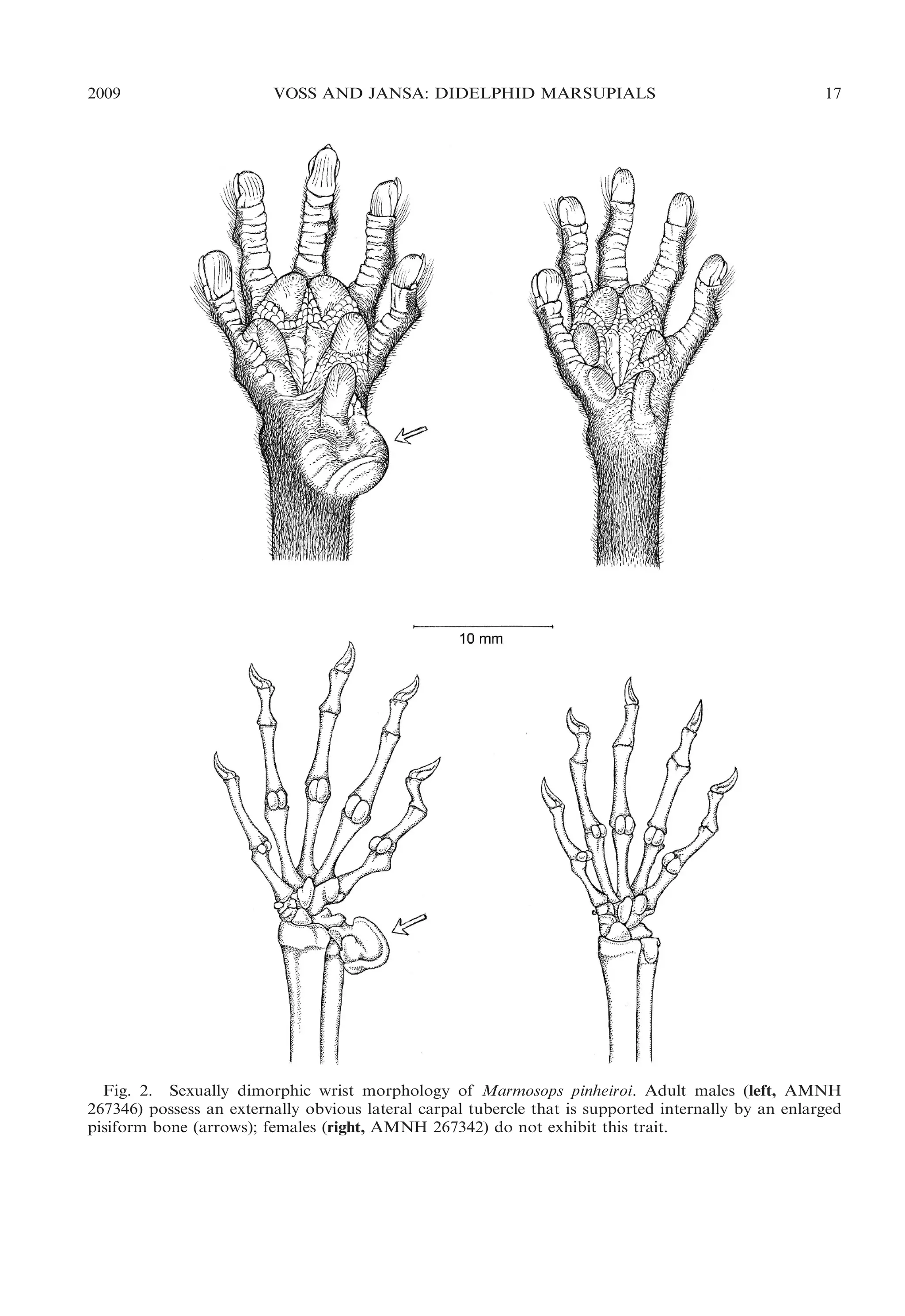
WRIST: The wrists of males and females
are morphologically similar and externally
featureless in most didelphids, but striking
sexual dimorphism is present in certain small
arboreal and scansorial forms (Lunde and
Schutt, 1999). Grossly enlarged glabrous
tubercles supported internally by carpal
ossifications are exhibited by large adult
males of Cryptonanus, Gracilinanus, Marmosops, Tlacuatzin, and some species of Marmosa. Two distinct kinds of tubercles can be
distinguished, consisting of lateral (‘‘ulnar’’)
tubercles supported internally by the pisiform, and medial (‘‘radial’’) tubercles supported by the prepollex (op. cit.). Although
some intraspecific variation in the development of carpal tubercles has been documented, most of it can be attributed to ontogeny:
tubercles are consistently present in the
largest adult male specimens of species in
which such structures occur, whereas they
may be lacking in some smaller (presumably
younger) conspecific males. Lunde and
Schutt (1999) plausibly suggest that these
structures function as clasping devices during
copulation.](https://image.slidesharecdn.com/vossjansa2009-140219090612-phpapp02/75/Voss-jansa-2009-16-2048.jpg)
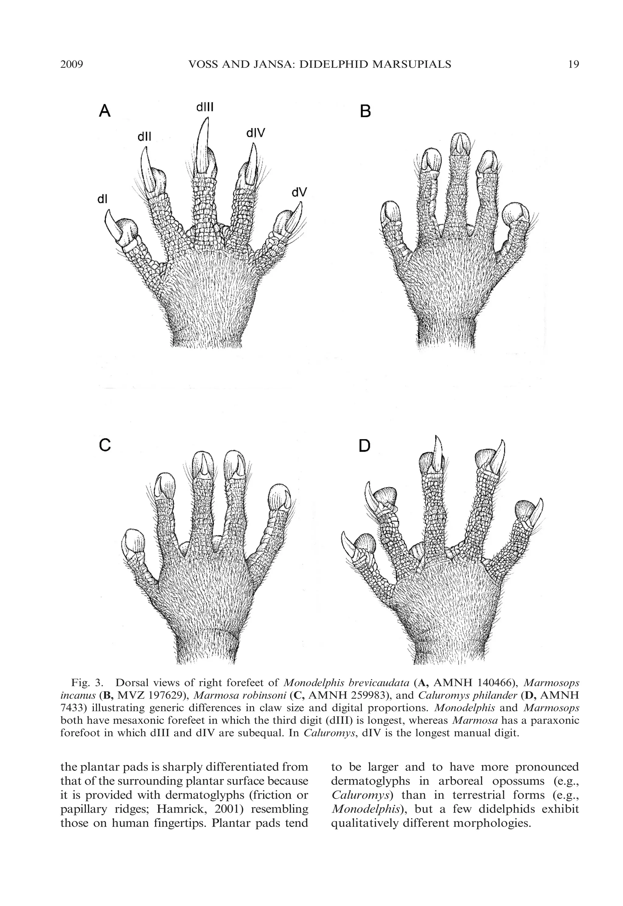
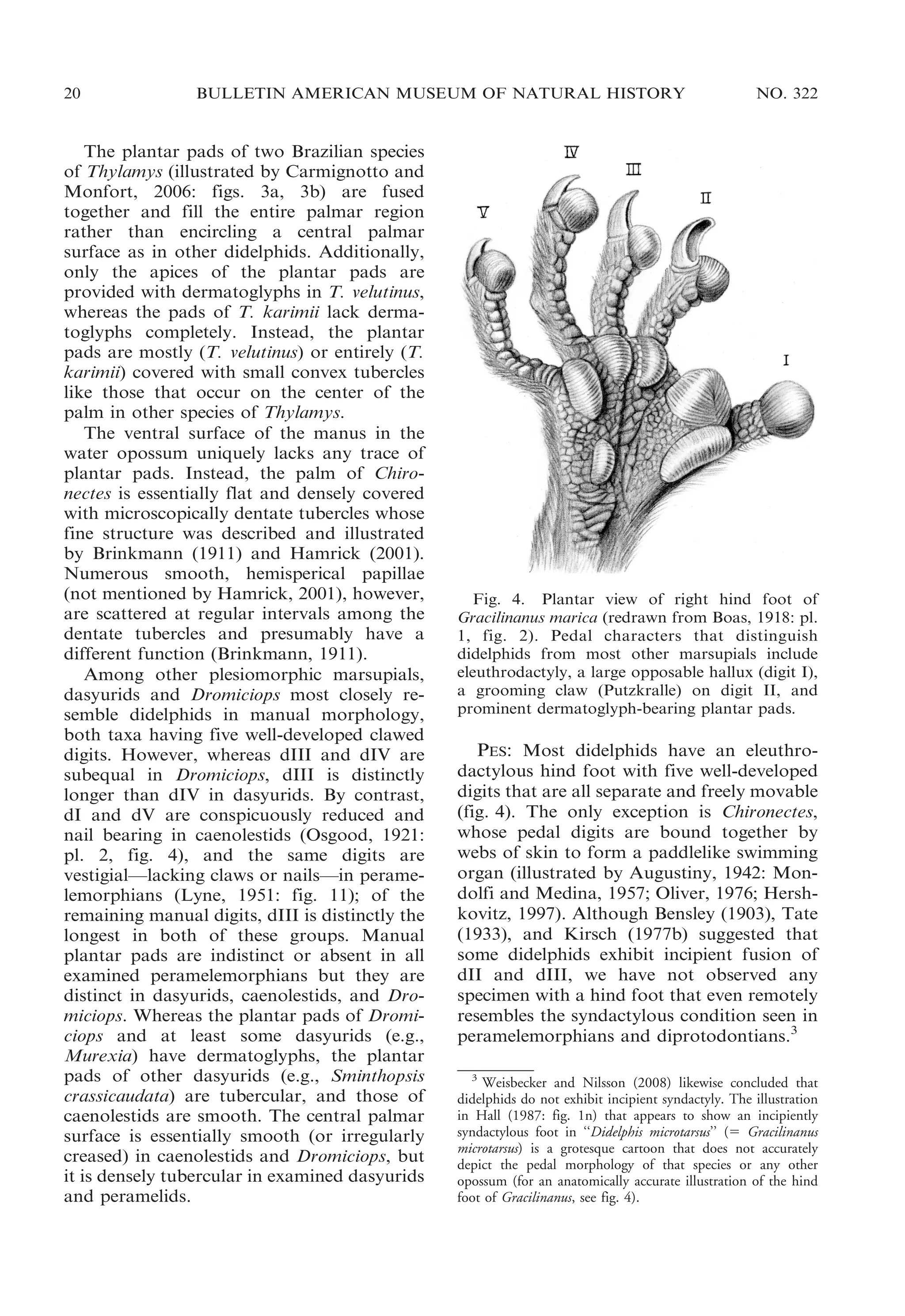
![22
BULLETIN AMERICAN MUSEUM OF NATURAL HISTORY
ular morphology, Chironectes is unique
among didelphids in lacking any trace of
pedal plantar pads.
The eleuthrodactylous hind foot of Dromiciops resembles the common didelphid
morphotype in having a large opposable
hallux, a grooming claw on dII, dIV . dIII,
and dermatoglyph-bearing plantar pads
(Hershkovitz, 1999: fig. 21). By contrast, the
hind foot of peramelids is syndactylous (with
fused dII and dIII; Hall, 1987; Weisbecker
and Nilsson, 2008); the hallux is small and
effectively nonopposable in caenolestids (Osgood, 1921: pl. 2, fig. 3), dasyurids (Thomas,
1888: pl. 23, fig. 8), and peramelids (Lyne,
1951: fig. 12); dIII and dIV are subequal in
examined caenolestids and dasyurids, both of
which groups also lack a grooming claw on
dII; peramelids do not have distinct plantar
pads; the plantar pads of caenolestids are
smooth; and the plantar pads of some
dasyurids are tubercular. The plantar epithelium of the heel is macroscopically naked in
most of these outgroup taxa, but the underside of the heel is coarsely furred in some
dasyurids (e.g., Sminthopsis crassicaudata).
POUCH AND MAMMAE: Based on our firsthand examination of parous adult female
specimens, pouchlike enclosures for nursing
young are unequivocally present or absent
among didelphids. Although our sample sizes
were always small, we observed no intraspecific variation in this aspect of female
reproductive morphology, nor did we observe any intermediate condition between
absence and presence of a pouch. Despite
the fact that several distinctly different pouch
configurations can be recognized among
opossums, we provisionally recognize all
pouches as homologous in the absence of a
priori evidence to the contrary.
We found no trace of a pouch in suitable
material (fluid-preserved specimens and carefully prepared skins) of parous adult female
Cryptonanus, Glironia, Gracilinanus, Hyladelphys, Lestodelphys, Marmosa, Marmosops,
Metachirus, Monodelphis, Thylamys, and
Tlacuatzin. By contrast, well-developed
pouches were consistently found to be
present in suitably prepared parous adult
females of Caluromys, Chironectes, Didelphis,
and Philander. Although Lutreolina was
described as pouchless by Thomas (1888),
NO. 322
Cabrera (1919), and Marshall (1978a), two
fluid-preserved parous females that we examined (UMMZ 166634, USNM 536827)
had pouches exactly resembling the morphology illustrated and described by Krieg (1924:
fig. 11). Caluromysiops is said to have a
pouch (Izor and Pine, 1987; Reig et al., 1987),
but no explicit description or illustration of
the female reproductive anatomy of this
genus is available, nor were we able to
examine suitably preserved parous female
specimens. The presence or absence of a
pouch remains undocumented for many
opossums, notably Chacodelphys. Contradictory literature accounts of a pouch as present
or absent in Metachirus were discussed by
Voss and Jansa (2003).
The marsupium of Caluromys philander
uniquely consists of deep lateral skin folds
that enclose the nursing young and open in
the midline (resembling the morphology that
Tyndale-Biscoe and Renfree [1987: fig. 2.8]
incorrectly attributed to didelphids in general). In Caluromys lanatus, Didelphis, and
Philander, however, the lateral pockets are
joined posteriorly, forming a more extensive
enclosure that opens anteriorly (Enders,
1937: fig. 19). Yet another condition is
exhibited by Chironectes and Lutreolina, in
which the lateral pockets are connected
anteriorly, forming a marsupium that opens
posteriorly (Krieg, 1924: fig. 11A; Oliver,
1976: fig. 1B).
In all marsupials that possess a pouch the
mammae are contained within it, but the
mammae of pouchless taxa are variously
distributed (Tate, 1933: fig. 3). In most
pouchless didelphids (e.g., Glironia, Marmosa, Metachirus) the mammae are confined
to a more or less circular inguinal/abdominal
array that occupies the same anatomical
position as the pouch in taxa that possess a
marsupium. However, other pouchless opossums (e.g., Cryptonanus guahybae, Marmosops incanus) have bilaterally paired mammae
that extend anteriorly well beyond the pouch
region. Although most of these anterior teats
are not actually located on the upper chest,
they are usually referred to as ‘‘pectoral’’ or
‘‘thoracic’’ mammae (e.g., by Tate, 1933;
Reig et al., 1987).
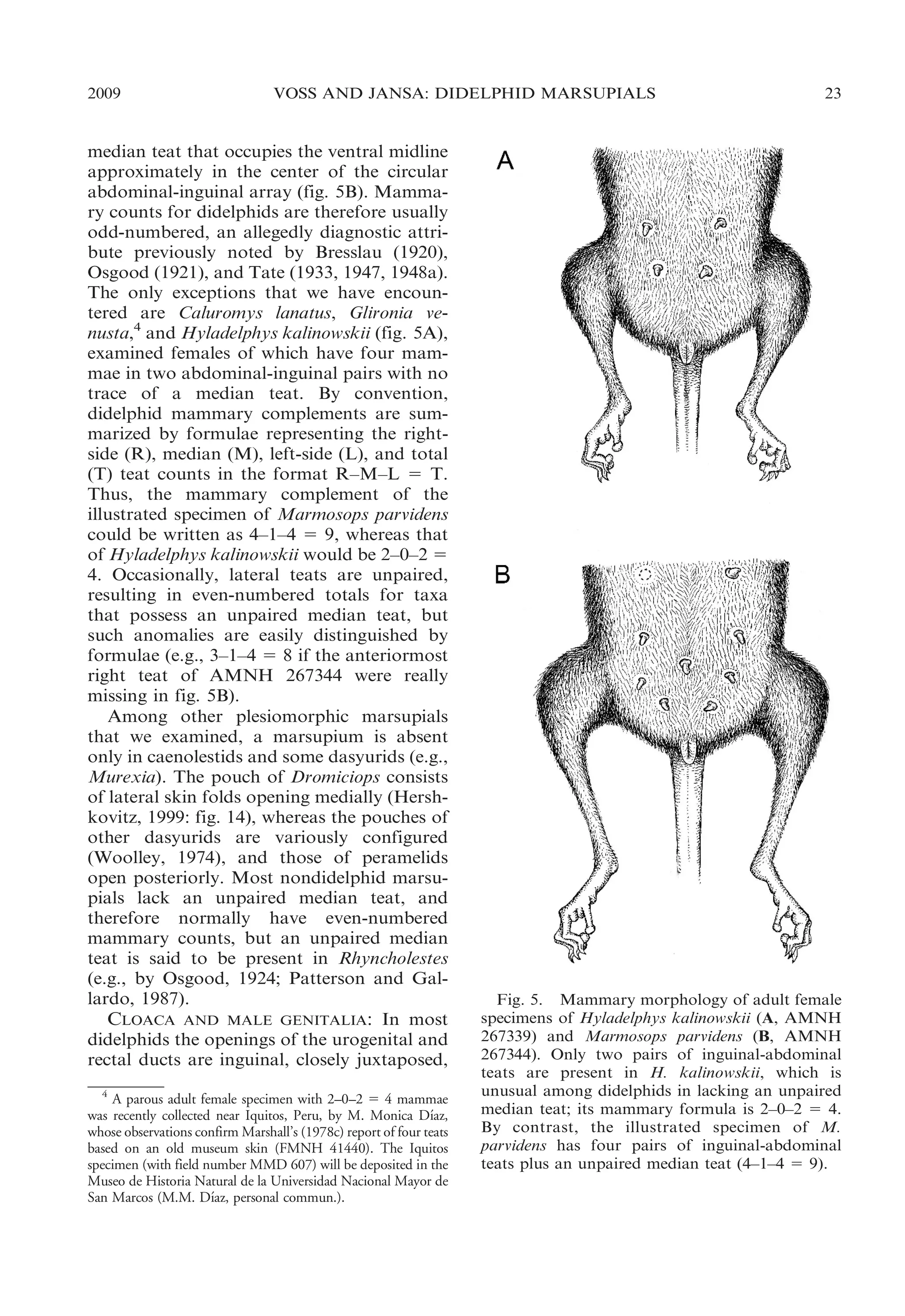
Most didelphids have, in addition to
bilaterally paired mammae, an unpaired](https://image.slidesharecdn.com/vossjansa2009-140219090612-phpapp02/75/Voss-jansa-2009-22-2048.jpg)
![30
BULLETIN AMERICAN MUSEUM OF NATURAL HISTORY
pials. However, some peramelemorphians
(e.g., Echymipera, Perameles) appear to have
simple (unbranched) maxilloturbinals, whereas the maxilloturbinals of examined dasyurids (e.g., Murexia, Sminthopsis) and Dromiciops closely resemble the elaborately dendritic condition seen in most didelphids. A
monographic study of marsupial endonasal
features based on high-resolution X-ray
computed tomography (Macrini and Voss,
in preparation) will doubtless provide additional points of comparison among didelphids and other marsupial clades.
ZYGOMATIC ARCH: The bones comprising
the zygomatic arch exhibit few variable
features among didelphids. The maxillaryjugal suture is always more or less straight or
irregularly crescentic, a frontal process of the
jugal is invariably present, and the jugalsquamosal suture is always deeply inflected
(typically ,- or ,- shaped). Likewise, a
faceted preglenoid process of the jugal and a
well-developed postglenoid process of the
squamosal are always present, and a distinct
glenoid (entoglenoid) process of the alisphenoid consistently forms part of the posterior
zygomatic root. Although the zygomatic arch
tends to be more gracile, to have a more
pronounced suborbital deflection, and to
have a more distinct frontal process in small
opossums (with relatively large eyes and
weakly developed masticatory muscles; e.g.,
Hyladelphys; fig. 40) than in large opossums
(with relatively smaller eyes but massive
masticatory muscles; e.g., Lutreolina; fig.
47), most didelphids exhibit intermediate
zygomatic morphologies.
Contrasting features of the zygomatic
region among other marsupial groups include: (1) the deeply inflected maxillary-jugal
suture of peramelemorphians, which divides
the jugal into distinct anterodorsal and
anteroventral processes flanking a well-developed nasolabial fossa (Filan, 1990); (2) the
absence of a frontal process of the jugal in
caenolestids and some peramelemorphians
(Osgood, 1921); (3) the absence of a faceted
preglenoid process of the jugal in several Old
World clades (e.g., Thylacinus and macropodoids); (4) the absence of a distinct
postglenoid process of the squamosal in
Hypsiprymnodon, Tarsipes, and vombatids;
and (5) the absence of a glenoid process of
NO. 322
the alisphenoid in Myrmecobius and many
diprotodontians.
ORBITAL MOSAIC: The anteriormost part
of the didelphid orbit is formed by the
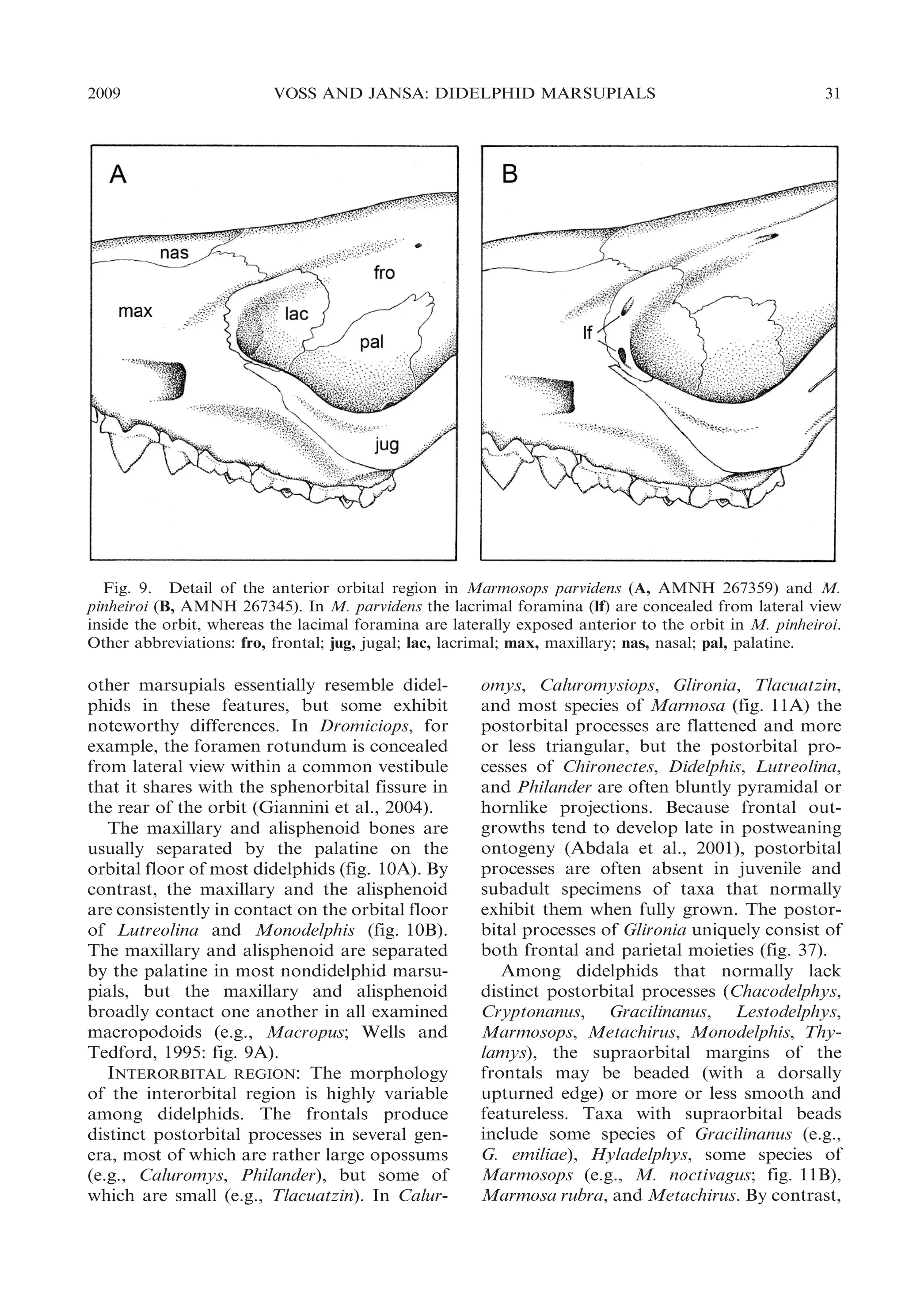
lacrimal, which is always prominently exposed in lateral view. The lacrimal is
perforated by one or more lacrimal foramina
that are sometimes concealed within the orbit
(fig. 9A) but usually open laterally on or near
the orbital margin (fig. 9B). Most didelphids
normally have two lacrimal foramina on each
side, but Chironectes, Hyladelphys, and some
populations of Didelphis virginiana usually
have just one lacrimal foramen, and many
other didelphids that normally have two
lacrimal foramina occasionally have a single
foramen on one or both sides of the skull.
The orbital margin formed by the lacrimal is
smoothly rounded in all didelphids, none of
which exhibits lacrimal tubercles (e.g., like
those seen in macropodids; Wells and Tedford, 1995: fig. 9) or distinct crests (as in
Myrmecobius and some peramelemorphians).
Unlike the condition seen in some Old World
marsupials with maxillary-frontal contact on
the medial wall of the orbit (Flannery et al.,
1987: fig. 3), the didelphid lacrimal is always
in posteroventral contact with the palatine.6
The medial wall of the didelphid orbit is
perforated by several openings, including the
sphenopalatine foramen (always in the palatine bone), the ethmoid foramen (in the suture
between the orbitosphenoid and frontal), the
sphenorbital fissure (between the palatine,
orbitosphenoid, alisphenoid, presphenoid,
and sometimes the pterygoid), and the foramen rotundum (in the alisphenoid). The
configuration of these orbital perforations in
all examined taxa is essentially similar to that
illustrated for Monodelphis by Wible (2003:
fig. 4). In particular, the foramen rotundum is
always exposed to lateral view behind the
sphenorbital fissure, from which it is invariably separated by a bony partition.7 Many
6
Note that elements of the didelphid orbital mosaic are
incorrectly labeled in some published illustrations (e.g.,
Hershkovitz, 1992b [fig. 19], 1997 [fig. 12]), where the orbital
process of the palatine that contacts the lacrimal is misidentified
as the ‘‘sphenoid.’’
7
According to Novacek (1986, 1993), the foramen rotundum is confluent with the sphenorbital fissure in didelphids, but
these foramina are unambiguously separate openings in all of
the material examined by us and by Wible (2003).](https://image.slidesharecdn.com/vossjansa2009-140219090612-phpapp02/75/Voss-jansa-2009-30-2048.jpg)

![34
BULLETIN AMERICAN MUSEUM OF NATURAL HISTORY
NO. 322
Fig. 12. Lateral view of posterior braincase in Marmosa murina (A, AMNH 272816) and Marmosops
impavidus (B, AMNH 272709), illustrating the presence of a fenestra (fen) that exposes the petrosal (pet)
between the parietal (par) and squamosal (sq) in Marmosops. The petrosal is not laterally exposed by a
fenestra in the parietal-squamosal suture of Marmosa. Other abbreviations: fc, fenestra cochleae; ssf,
subsquamosal foramen.
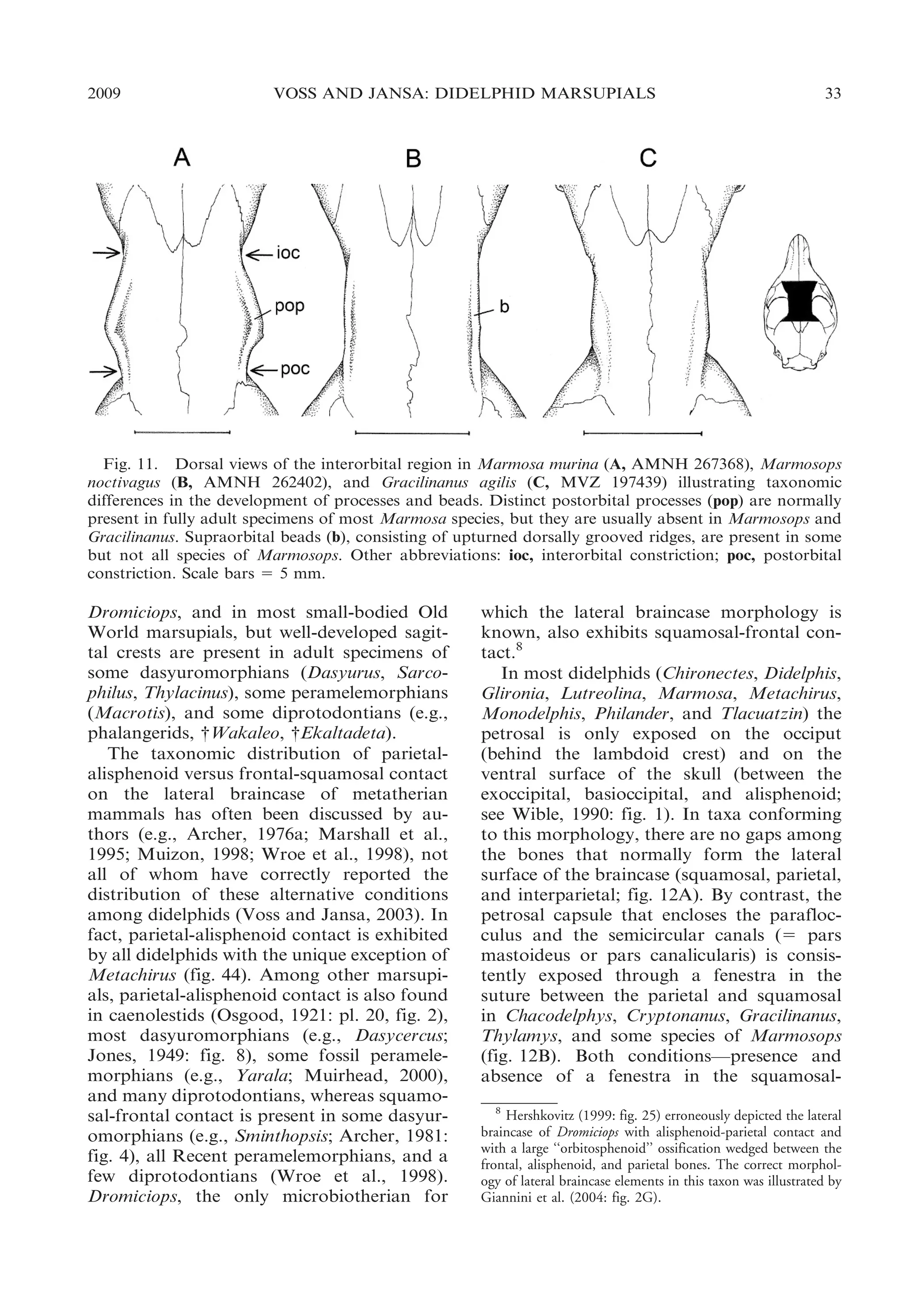
parietal suture—occur as balanced polymorphisms (neither state clearly predominating)
in Lestodelphys, Marmosops incanus, and M.
noctivagus. No nondidelphid marsupial that
we examined has a fenestrated squamosalparietal suture.
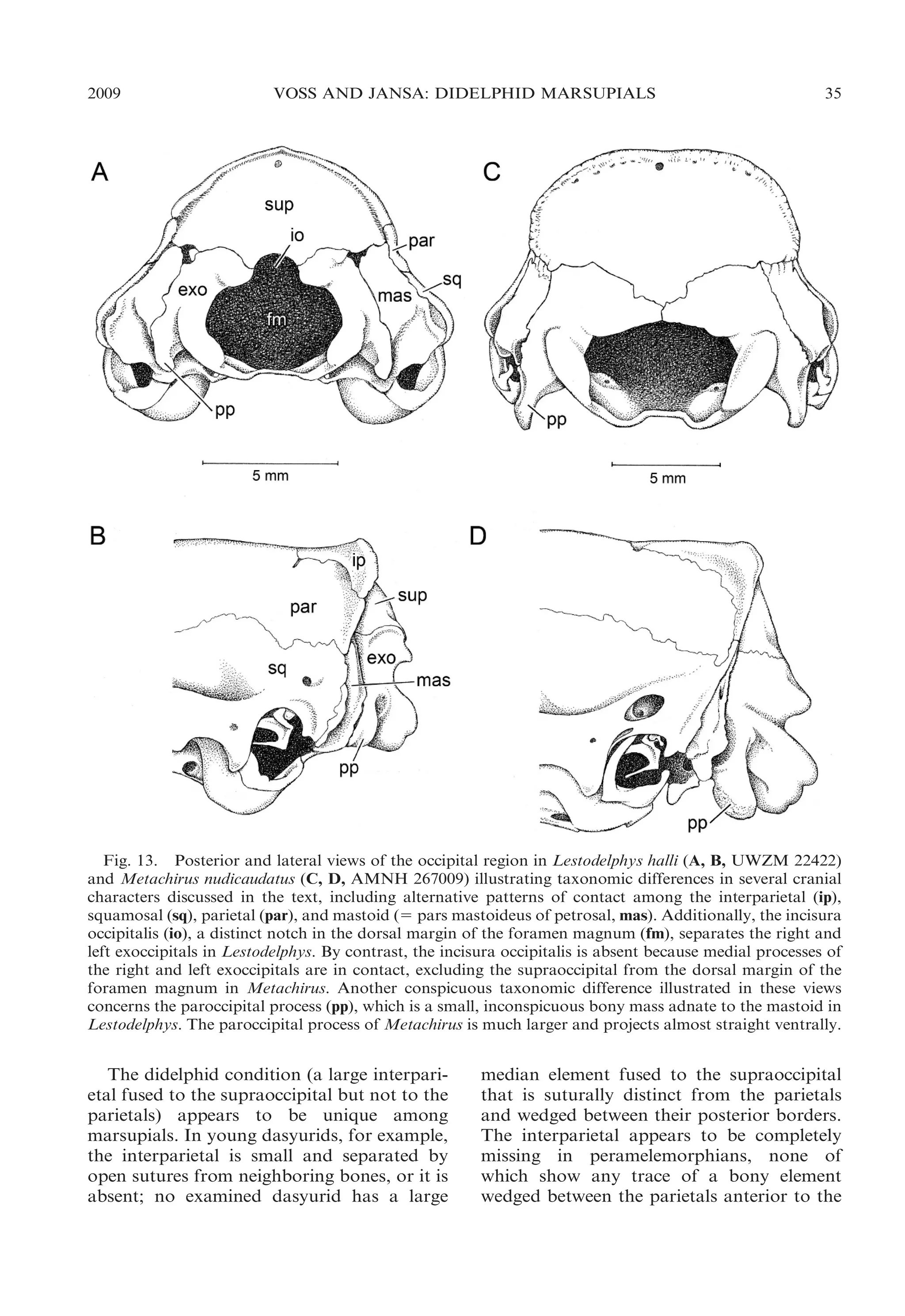
INTERPARIETAL: Although some authors
(e.g., Novacek, 1993) have stated that marsupials lack an interparietal bone, a large
interparietal is unequivocally present in
Dromiciops (see Giannini et al., 2004: fig.
3), many diprotodontians (e.g., Tarsipes;
Parker, 1890: fig. 2), and some dasyuromorphians (e.g., Myrmecobius). In these taxa, the
sutures that separate the interparietal from
adjacent bones (supraoccipital and parietals)
are clearly visible and ontogenetically persistent. In all of the taxa we examined with such
distinctly sutured interparietals, the interparietal-supraoccipital boundary coincides
closely with the transverse (lambdoid) crest
that marks the dorsalmost insertion of the
neck extensor musculature.
An interparietal is also unambiguously
present in didelphids, all of which exhibit a
large, unpaired, wedge-shaped or oblong
element between the left and right parietals
in the same position (anterior to the lamb-
doid crest) as the sutured interparietals of
other marsupials. The didelphid interparietal,
however, is fused with the supraoccipital in
all of the skulls we examined (including
postweaning juveniles assignable to Gardner’s [1973] age class 1), few of which show
any trace of a suture.9 Fortunately, developmental studies of didelphid pouch young are
available to prove the existence of a distinct
interparietal center of ossification. In Didelphis the interparietal first appears around day
8 postpartum (p) and fuses with the supraorbital by day 28p (Nesslinger, 1956), whereas in Monodelphis these events occur on days
3p and 8p, respectively (Clark and Smith,
1993). By contrast, the didelphid interparietal
never appears to fuse with the parietals, the
sutures between them persisting even in the
largest adult specimens we examined.
9
Vestiges of the interparietal-supraoccipital suture in juvenile
specimens of Monodelphis brevicaudata were described by Wible
(2003). What appears to be a well-defined suture between the
interparietal and supraoccipital of Didelphis albiventris in an
illustration published by Abdala et al. (2001: fig. 3) was
intended to represent the hypothetical boundary between bones
that were indistinguishably fused in all of the specimens
examined by the authors of that report (D.A. Flores, personal
commun.).](https://image.slidesharecdn.com/vossjansa2009-140219090612-phpapp02/75/Voss-jansa-2009-34-2048.jpg)
![2009
VOSS AND JANSA: DIDELPHID MARSUPIALS
43
Fig. 17. Oblique ventrolateral view of left ear region in Marmosops impavidus (A, MUSM 13284) and
Philander mcilhennyi (B, MUSM 13299) illustrating taxonomic differences in ectotympanic suspension.
Whereas the ectotympanic (ect) is suspended from the skull by attachments both to the petrosal (pet) and
to the malleus (mal) in Marmosops, the ectotympanic of Philander is suspended only from the malleus
(there is no attachment to the petrosal). Other abbreviations: atp, alisphenoid tympanic process; pro,
promontorium (of petrosal); rtp, rostral tympanic process (of petrosal); sq, squamosal.
squamosal. This structure is a cup-shaped
cavity that, in all skulls retaining vestiges of
auditory soft tissues, appears to be covered
by the membrana Shrapnelli (pars flaccida of
the tympanic membrane). Archer (1976a:
304) remarked that, ‘‘[i]n some didelphids
(e.g., Metachirus) there is a depression in the
squamosal which is clearly the homologue of
this sinus,’’ but nothing resembling the
squamosal epitympanic sinus of dasyurids
occurs in any living opossum.
ECTOTYMPANIC AND OSSICLES: Although
didelphids were described by van der Klaauw
(1931: 26) as having a completely free
ectotympanic, two distinct patterns of ectotympanic attachment can be recognized in
the family. In most didelphids the anterior
limb (or crus) of the ectotympanic annulus is
directly connected to the skull near the point
where the squamosal, alisphenoid, and petrosal are juxtaposed behind the postglenoid
process. Where the connection can be seen
clearly (dried remnants of soft tissues frequently obscure this feature), the actual
attachment seems to be to the petrosal (fig.
17A). In six genera (Caluromys, Caluromysiops, Chironectes, Didelphis, Lutreolina, and
Philander), however, the anterior limb of the
ectotympanic is not directly attached to the
skull, and the suspension is indirect, via the
anterior (tympanic) process of the malleus
(fig. 17B). In all didelphids with an indirect
dorsal connection between the ectotympanic
and the skull, the tympanic annulus is more
or less ringlike because the posterior (ventral)
limb is not expanded to form part of the floor
of the middle ear cavity. By contrast, in some
taxa with direct ectotympanic suspension, the](https://image.slidesharecdn.com/vossjansa2009-140219090612-phpapp02/75/Voss-jansa-2009-43-2048.jpg)
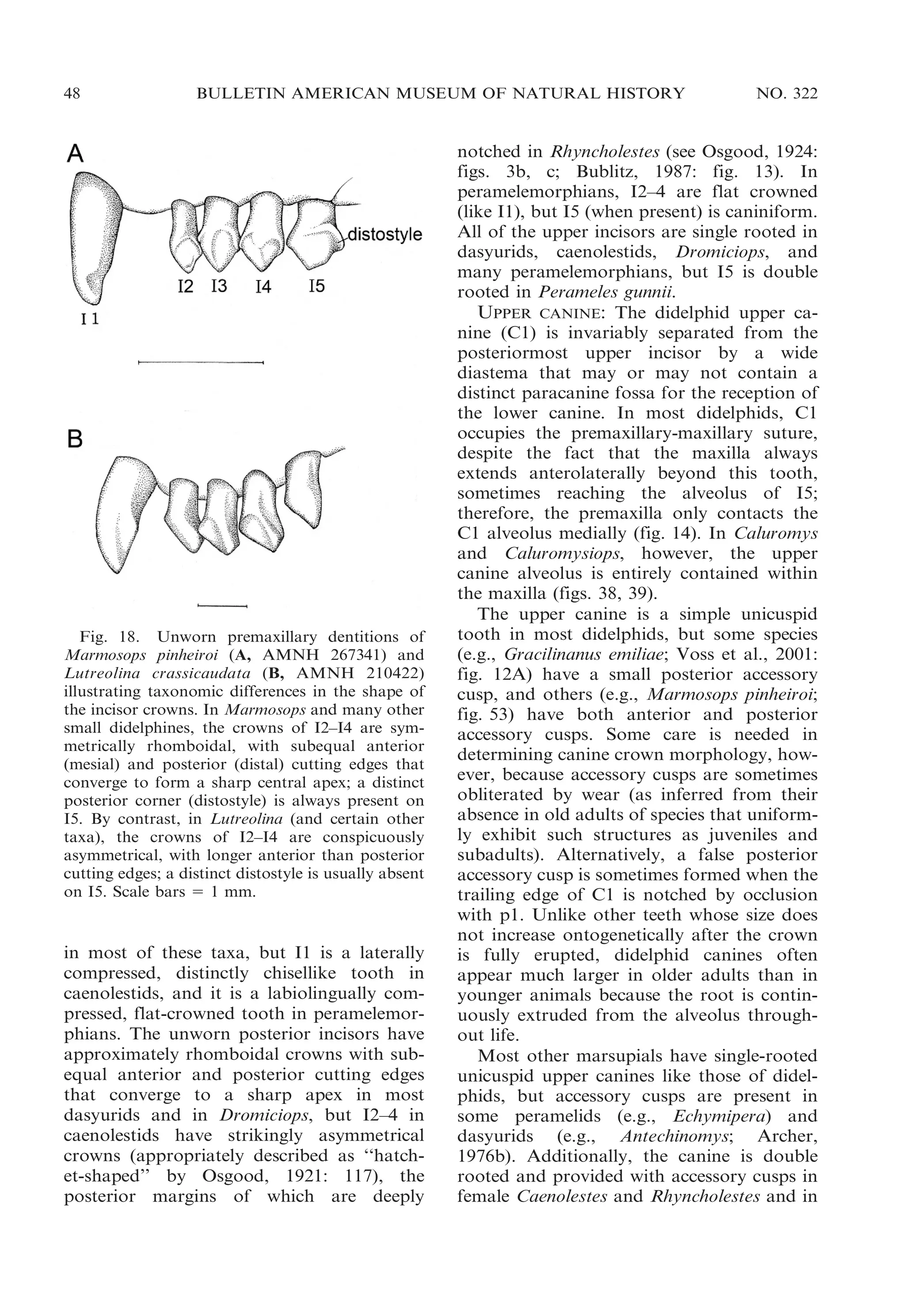
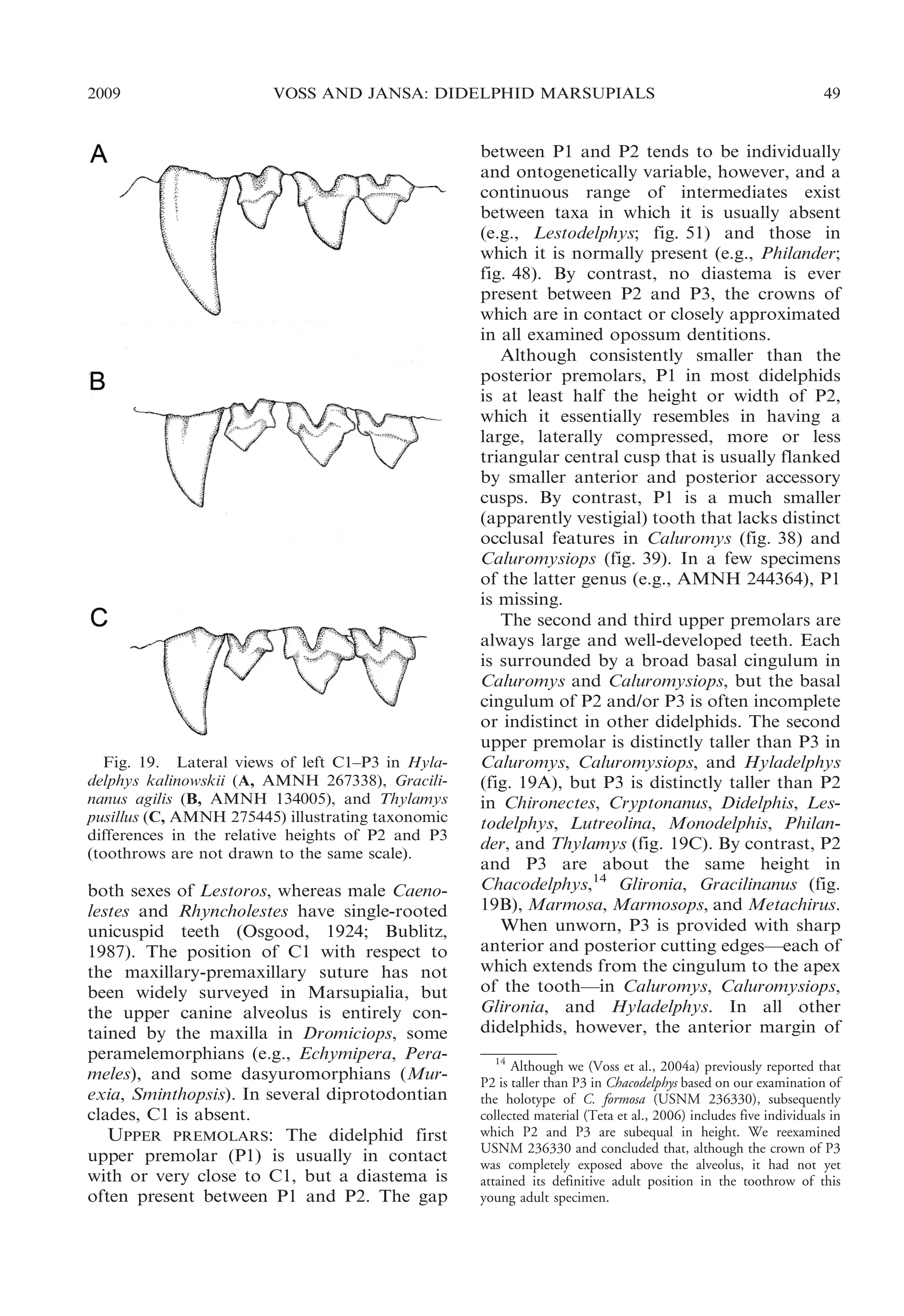
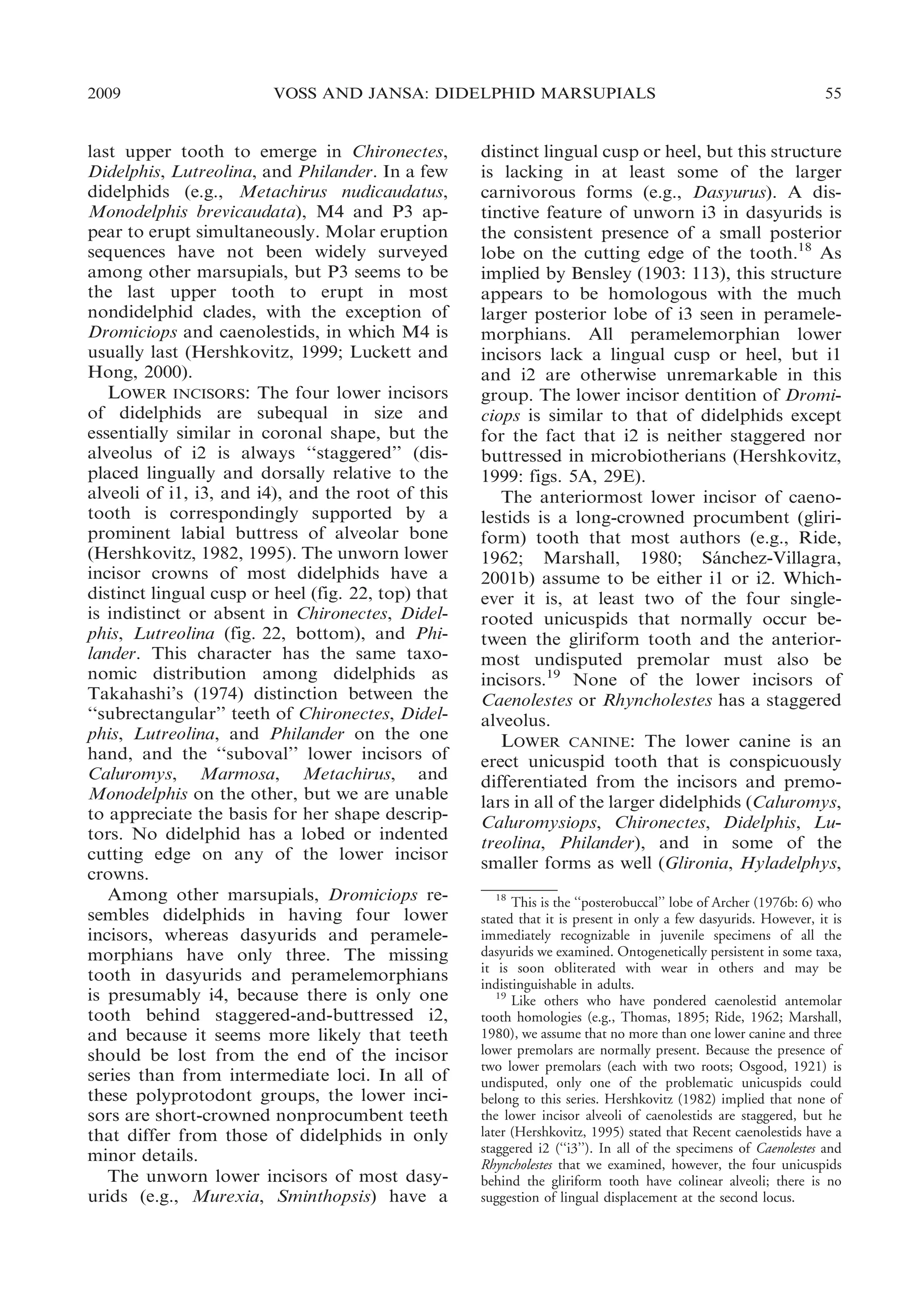
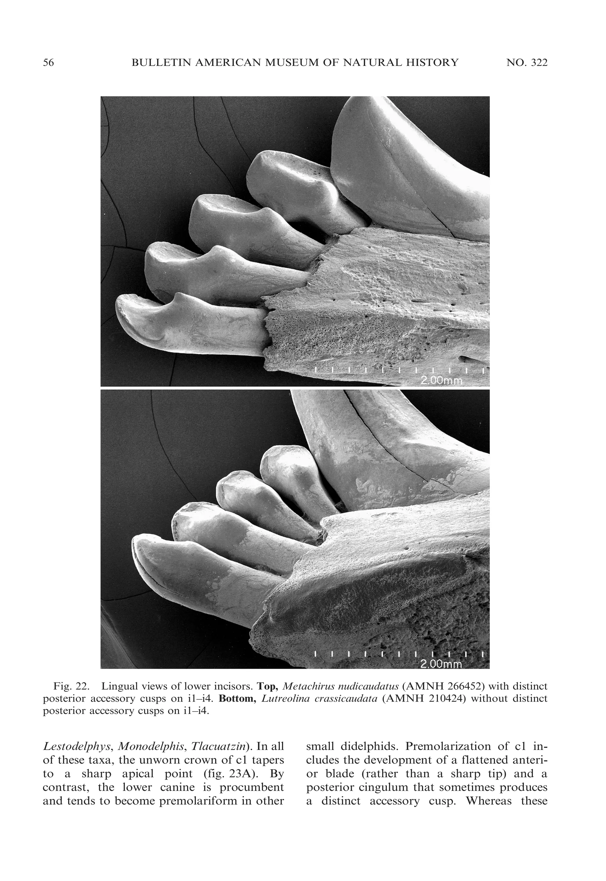
![50
BULLETIN AMERICAN MUSEUM OF NATURAL HISTORY
the tooth is rounded, and only the posterior
cutting edge is well developed. Small anterior
blades are variably present near the base of
P3 in Chironectes and Philander, but the apex
of the tooth is always rounded anteriorly as
in the other taxa with single-bladed P3s.
Most other plesiomorphic marsupials have
three upper premolars, but some dasyurids
(e.g., Dasyurus) have only two. In addition to
the usual diastema between P1 and P2, some
adult dasyurids (e.g., Murexia) and all adult
peramelemorphians have a diastema between
P2 and P3. The third upper premolar (P3) is
taller than P2 in caenolestids, Dromiciops,
and some dasyurids (e.g., Murexia, Sminthopsis), but P2 is taller than P3 in other dasyurids
(e.g., Myoictis). An anterior cutting edge is
apparently absent from P3 in all nondidelphid
marsupials.
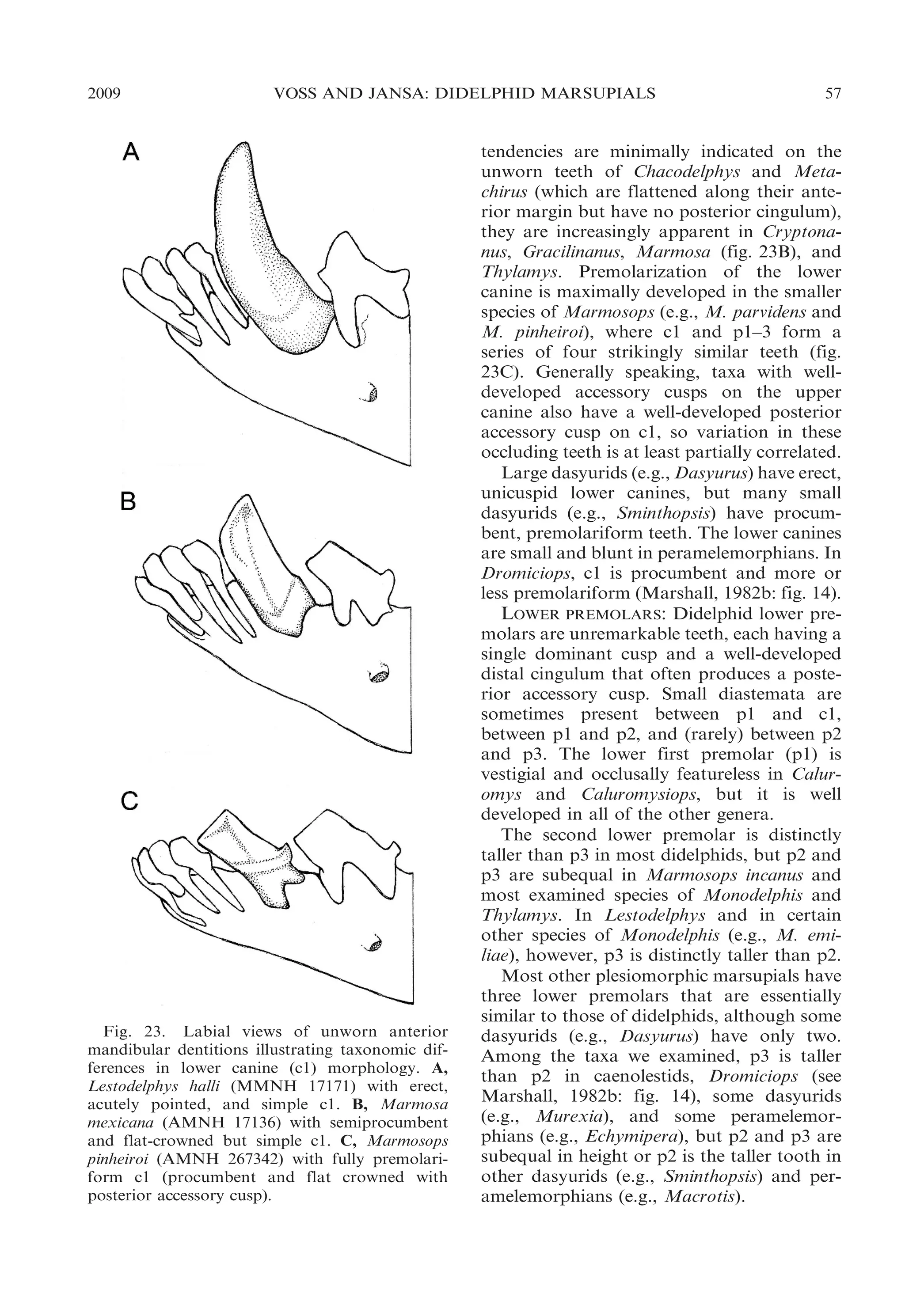
UPPER MILK PREMOLAR: Although the
deciduous upper premolar (dP3) of didelphids has consistently been described as large
and molariform (Flower, 1867; Thomas,
1888; Bensley, 1903; Tate, 1948b; Archer,
1976b), there is noteworthy taxonomic variation in the morphology of this tooth. In
most opossums dP3 is, indeed, a sizable
tooth: its crown area ranges from about 42%
to 96% of the crown area of M1, the tooth
immediately behind it (Voss et al., 2001: table
5). An obviously functional element of the
upper toothrow, dP3 occludes with both the
deciduous lower premolar (dp3) and with m1.
Hyladelphys, however, has a very small upper
milk premolar (,10% of the crown area of
M1; op. cit.) that appears to be functionally
vestigial because it does not occlude with any
lower tooth.
Although molariform, the didelphid upper
milk premolar does not always match the
teeth behind it in all occlusal details. In most
specimens, the paracone of dP3 is located on
the labial margin of the crown, and the stylar
shelf is correspondingly incomplete, whereas
the paracone is lingual to a continuous stylar
shelf on all didelphid molars (see below).
Among those didelphids whose milk premolars we examined, only Caluromys has a dP3
in which the paracone is usually lingual to a
continuous stylar shelf.
The morphology of milk premolars remains to be widely surveyed in Marsupialia,
but of those taxa that we examined, only
NO. 322
Dromiciops has a large, molariform dP3
resembling the common didelphid condition
(Marshall, 1982b: fig. 17; Hershkovitz, 1999:
fig. 32). By contrast, dP3 is much smaller,
more or less vestigial, and structurally
simplified in caenolestids, dasyurids, and
peramelemorphians (Tate, 1948a; Archer,
1976b; Luckett and Hong, 2000).
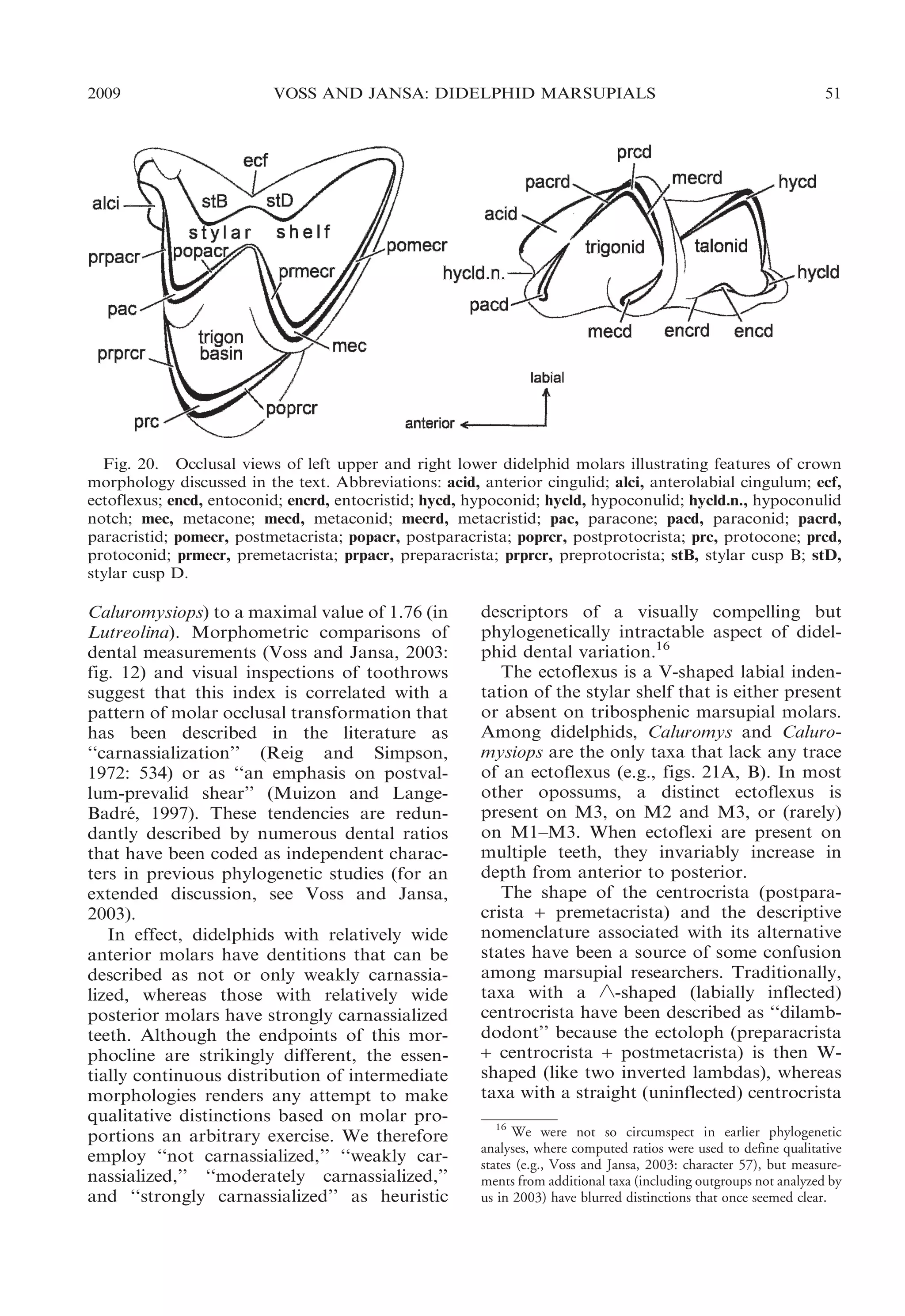
UPPER MOLARS: Didelphid upper molars
conform to the basic tribosphenic bauplan
(Simpson, 1936) in having three principal
cusps—paracone, protocone, and metacone—connected by the usual crests in a
more or less triangular array (fig. 20). A
broad stylar shelf and an anterolabial cingulum are invariably present; the centrocrista
(postparacrista + premetacrista) and the
ectoloph (preparacrista + centrocrista +
postmetacrista) are uninterupted by gaps;
the para- and metaconules are indistinct or
absent;15 and there is no posterolingual talon.
In addition, most didelphids have several
(usually five or six) small cusps on the stylar
shelf, for which most authors employ alphabetical labels (after Bensley, 1906; Simpson,
1929; Clemens, 1966). Of these, stylar cusp B
(labial to the paracone) is more consistently
recognizable than the others, but a stylar
cusp in the D position (labial to the
metacone) is often subequal to it in size.
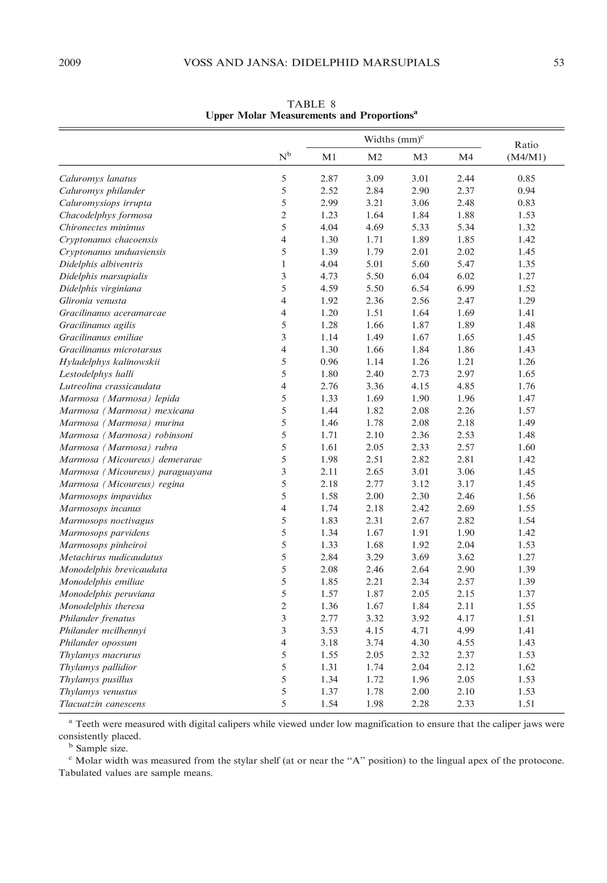
Didelphids differ conspicuously in the
relative width (transverse or labial-lingual
dimension) of successive molars within toothrows. In some taxa, the anterior molars tend
to be wide in proportion to more posterior
teeth, but in others the posterior molars are
relatively wider (fig. 21). The ratio obtained
by dividing the width of M4 by the width of
M1 (M4/M1; table 8) conveniently indexes
this size-independent shape variation and
ranges from a minimal value of 0.83 (in
15
The literature is inconsistent on this point, with some
authors claiming to have observed significant taxonomic
variation among Recent didelphids in the occurrence of conules
(see Voss and Jansa, 2003: appendix 4). Indeed, careful
examination of unworn teeth usually reveals a tiny enameled
chevron on the postprotocrista that is presumably homologous
with the metaconule of stem metatherians; corresponding
structures on the preprotocrista, presumably vestigial paraconules, are much less frequently observed. The fact that conules
have been scored as present and absent in the same taxon (e.g.,
Didelphis) by different authors (e.g., Reig et al. [1987] versus
Wroe et al. [2000]) sufficiently illustrates the ambiguous
interpretation of such indistinct features.](https://image.slidesharecdn.com/vossjansa2009-140219090612-phpapp02/75/Voss-jansa-2009-50-2048.jpg)
![52
BULLETIN AMERICAN MUSEUM OF NATURAL HISTORY
Fig. 21. Occlusal views of left upper molar
rows illustrating taxonomic differences in relative
widths of teeth (not drawn to the same scale). A,
Caluromysiops irrupta (FMNH 84426); B, Caluromys philander (AMNH 267334); C, Glironia
venusta (AMNH 71395); D, Marmosa regina
(MVZ 190333); E, Marmosops impavidus (MUSM
13284); F, Lestodelphys halli (MMNH 15708).
have usually been called ‘‘predilambdodont’’
(e.g., by Reig et al., 1987). Unfortunately,
neither descriptor applies unambiguously to
some didelphids (Goin, 1997), and certain
taxa have been coded with contradictory
NO. 322
character states in different phylogenetic
datasets (e.g., Didelphis; see Reig et al.,
1987 [character 1]; Wroe et al., 2000 [character 10]; Wible et al., 2001 [character 31]). Our
observations agree with Johanson’s (1996),
that the shape (linearity versus labial inflection) of this crest is correlated with its
occlusal relief (apical height above the trigon
basin), and we recognize an intermediate
condition for taxa that do not conform with
either traditionally recognized morphotype.
Among didelphids, only Caluromysiops
has a truly linear centrocrista on M1–M3.
In this taxon, the apex of the centrocrista is
essentially level with the floor of the trigon
basin, clearly conforming to the predilambdodont condition defined by Johanson
(1996). By contrast, the centrocrista is
strongly inflected labially (buccally)—and
therefore distinctly ‘-shaped—in Chacodelphys, Cryptonanus, Gracilinanus, Lestodelphys, Marmosa, Marmosops, Metachirus,
Monodelphis, Thylamys, and Tlacuatzin. The
apex of the crest is elevated well above the
trigon floor in these taxa, which are unambiguously dilambdodont sensu Johanson
(1996). The intermediate condition occurs in
the seven remaining genera (Caluromys,
Chironectes, Didelphis, Glironia, Hyladelphys,
Lutreolina, Philander) in which the centrocrista has a weak labial inflection with a
slightly elevated apex; these taxa can appropriately be described as weakly dilambdodont.
In many didelphids—Caluromys, Caluromysiops, Glironia, Gracilinanus, Hyladelphys,
Marmosa, Tlacuatzin, and some species of
Cryptonanus and Marmosops—the preprotocrista passes labially around the base of the
paracone to join with the anterolabial cingulum. This results in the formation of a
continuous shelf along the anterior margin
of the tooth (fig. 21A–D), and taxa possessing this feature are sometimes said to have a
‘‘complete’’ anterior cingulum (Archer,
1976b: 3) or to exhibit ‘‘double rank prevallum-postvallid shearing’’ (Cifelli, 1993:
213). In the alternative morphology—exhibited by Chacodelphys, Chironectes, Didelphis,
Lestodelphys, Lutreolina, Metachirus, Monodelphis, Philander, Thylamys, and other
species of Cryptonanus and Marmosops—the
preprotocrista extends only to a point at or](https://image.slidesharecdn.com/vossjansa2009-140219090612-phpapp02/75/Voss-jansa-2009-52-2048.jpg)
![54
BULLETIN AMERICAN MUSEUM OF NATURAL HISTORY
near the base of the paracone; the anterolabial cingulum does not converge toward the
preprotocrista in these taxa, but passes
obliquely dorsally such that the two crests
are discontinuous on the anterior surface of
the tooth crown (fig. 21E, F). Voss and Jansa
(2003: 39) discussed several inconsistencies in
the published literature concerning the distribution of these traits among didelphids.
The postprotocrista also exhibits noteworthy variation among didelphids. In most
genera, this crest decreases in height and
width as it passes posterolabially around the
base of the metacone before merging with the
posterior surface of the tooth crown, leaving
a distinct groove or gap in the posterior wall
of the trigon basin. By contrast, the postprotocrista connects directly with the base of
the metacone, such that the posterior wall of
the trigon basin has no gap or groove, in
Chironectes, Didelphis, Lutreolina, and Philander; instead, the unworn postprotocrista
of these taxa exhibits a distinct carnassial
notch near the base of the metacone.
Dasyurids and Dromiciops have tribosphenic upper molars that differ from those of
didelphids in only minor details. Indeed,
some dasyurids and didelphids are indistinguishable in published matrices of upper
molar characters (e.g., Archer, 1976b: table
1), although there is a tendency for stylar
cusp D to be larger than stylar cusp B when
both structures are present in the former
group (Wroe, 1997), and a few dasyurids
(e.g., Sarcophilus) are conspicuously divergent in other upper molar traits. The upper
molars of Dromiciops differ from those of
most didelphids by having a linear centrocrista, indistinct stylar cusps, and a stylar
shelf that is conspicuously reduced in width
labial to the paracone on M1.17
17
These distinctive features are not apparent in some
published illustrations of the dentition of Dromiciops, notably
Marshall’s (1982b) figure 16b, where an occlusal view of the
upper teeth of FMNH 22671 shows ‘-shaped centrocristae and
an unreduced stylar shelf on M1. However, in another
illustration (based on FMNH 22673; op. cit.: fig. 17b), M1
is correctly shown with a reduced stylar shelf labial to the
paracone, and with a linear centrocrista. We borrowed FMNH
22671 to determine whether this specimen is unusual in any
way and found that it is not: the paracone of M1 is almost on
the labial margin of the tooth, and the centrocristae of M1–M3
are linear; therefore, Marshall’s figure 16b is inaccurate.
NO. 322
The upper molars of Recent peramelemorphians differ from those of didelphids in
several respects, most notably by having a
discontinuous centrocrista: instead of forming a ‘-shaped (labially inflected) crest with
an apex that is lingual to the stylar shelf, the
postparacrista and the premetacrista of
modern peramelemorphians terminate on
the labial margin of the tooth, where they
are separated by a small gap (Muirhead and
Filan [1995] referred to this trait as the result
of the centrocrista ‘‘breaching’’ the ectoloph).
Additionally, all modern peramelemophians
have a large posterolingual cusp on M1–M3;
in most genera (e.g., Echymipera, Perameles)
this is a hypertrophied metaconule, but in
Macrotis it is the metacone (Archer, 1976b).
Another peramelemorphian trait is that the
preparacrista of M1 is a tall crest that passes
posterolabially to a large stylar cusp at or
near the C position (Archer, 1976b). The
anterolabial cingulum is present in some
peramelemorphians (e.g., Echymipera) but
not in others (e.g., Perameles).
On caenolestid molars the principal labial
cusps are hypertrophied stylar elements in the
B and D positions (Osgood, 1921; Marshall,
1987; Goin and Candela, 2004; Goin et al.,
2007). In both Caenolestes and Rhyncholestes, the paracone is absent (or indistinguishably fused with stylar cusp B). The
metacone is also absent in Rhyncholestes but,
although fused to the lingual aspect of stylar
cusp D, the metacone is still clearly recognizable in Caenolestes. Given this interpretation of caenolestid cusp homologies, it
follows that a low crest along the labial base
of the tooth crowns in both genera is a
neomorphic cingulum rather than a vestigial
stylar shelf. The first two caenolestid upper
molars also have a well-developed posterolingual cusp that has been interpreted either
as a hypertrophied metaconule (Marshall,
1987; Goin and Candela, 2004; Goin et al.,
2007) or as a neomorphic outgrowth of the
postprotocingulum (Hunter and Jernvall,
1995). Another distinctive trait of the upper
molars of Recent caenolestids is the complete
absence of an anterolabial cingulum.
UPPER DENTAL ERUPTION SEQUENCES: As
described by Tribe (1990), P3 is the last upper
tooth to erupt in most didelphids. By
contrast, P3 erupts before M4, which is the](https://image.slidesharecdn.com/vossjansa2009-140219090612-phpapp02/75/Voss-jansa-2009-54-2048.jpg)
![2009
VOSS AND JANSA: DIDELPHID MARSUPIALS
83
TABLE 16
Classifications of Recent Opossums, 1888–1958a
Thomas (1888)
Didelphidae
Didelphis
Didelphisb
Metachirusb
Philanderb
Micoureusb
Peramysb
Chironectes
Matschie (1916)
Didelphidae
Didelphis
Didelphisb
Metachiropsb
Metachirusb
Peramysb
Micoureusb
Caluromysb
Marmosab
Grymaeomysb
Marmosopsb
Thylamysb
Dromiciopsb
Glironiab
Monodelphisb
Monodelphiopsb
Microdelphysb
[Chironectes]
Cabrera (1919)
Simpson (1945)
Cabrera (1958)
Didelphidae
Dromiciops
Glironia
Philander
Marmosa
Marmosab
Thylamysb
Peramys
Minuania
Lutreolina
Metachirus
Holothylax
Didelphis
Chironectes
Didelphidae
Philander
Monodelphis
Dromiciops
Glironia
Notodelphisc
Marmosa
Metachirops
Metachirus
Lutreolina
Didelphis
Chironectes
Didelphidae
Caluromys
Caluromysiops
Glironia
Dromiciops
Monodelphis
Monodelphisb
Minuaniab
Lestodelphys
Marmosa
Marmosab
Thylamysb
Philander
Metachirus
Lutreolina
Didelphis
Chironectes
a
Incorrect spellings (e.g., ‘‘Didelphys’’ for Didelphis; Thomas, 1888) have been changed to conform with current usage
throughout, but the sequence of names in each classification has been preserved as originally published. Note that some
names (e.g., Philander) were used by early taxonomists for species that are now placed in other genera (see text).
b
Ranked as subgenera.
c
Not an available name. Notodelphys Thomas, 1921, originally proposed for the taxon now known as Lestodelphys, is
preoccupied by Notodelphys Allman, 1847, a copepod (Tate, 1934).
mark catalog of the marsupials in the British
Museum of Natural History.
By comparison with Old World marsupials, didelphids appeared to Thomas (1888:
315) to be ‘‘an exceedingly homogeneous
[family], its members presenting a very small
range of differentiation.’’ Accordingly, he
recognized only Didelphis and Chironectes as
full genera, but several other taxa were ranked
as subgenera of Didelphis (table 16). These
included Metachirus (containing species now
referred to Metachirus, Philander, and Lutreolina), Philander (for Caluromys), Micoureus (containing species now referred to
Marmosa, Marmosops, Thylamys, and Gracilinanus), and Peramys (for Monodelphis).
Although knowledge of didelphid diversity
increased rapidly in the years following
Thomas’s classification, Matschie (1916) persisted in referring all nonaquatic opossums to
the genus Didelphis. However, Matschie
recognized more subgenera of Didelphis than
Thomas did, resurrecting old names or
describing new ones to suit his needs.
Dromiciops (described as a didelphid by
Thomas, 1894) was also included. Matschie’s
taxonomy is noteworthy for his early recognition that the so-called ‘‘murine’’ opossums
(pouchless mouse- and rat-sized species with
circumocular masks and long tails) that
Thomas (1888) had lumped together in
Micoureus actually represent several distinct
groups, and for treating those groups as
coordinate taxa.
Cabrera’s (1919) classification was not the
first to reject Linnaeus’s inclusive concept of
Didelphis, but it was influential in establishing modern binomial usage. His taxonomy
included 11 genera, only one of which
contained subgenera. Like Thomas (1888)
and Matschie (1916), Cabrera made no use of
subfamilies, tribes, or other suprageneric
categories to indicate relationships among
living opossums. Unlike Matschie, Cabrera
grouped all of the ‘‘murine’’ opossums
together, using Marmosa as the name for
the taxon that Thomas called Micoureus.
Simpson (1945) recognized several subfamilies of didelphids, but he placed all of the
living opossums (plus Dromiciops) in the](https://image.slidesharecdn.com/vossjansa2009-140219090612-phpapp02/75/Voss-jansa-2009-83-2048.jpg)
![2009
VOSS AND JANSA: DIDELPHID MARSUPIALS
the best of our knowledge, the first familygroup name based on Marmosa is technically
available from Hershkovitz (1992b). An
alternative name for this clade is Monodelphini, which McKenna and Bell (1997)
attributed to Talice et al. (1961). However,
Talice et al. did not mention any characters
purported to differentiate Monodelphini
from other didelphids, so their name is
unavailable (ICZN, 1999: Article 13). To
the best of our knowledge, the first familygroup name based on Monodelphis is also
available from Hershkovitz (1992b). As first
revisors in the sense of the Code (ICZN,
1999: Article 24), we select Marmosini
Hershkovitz, 1992, to have precedence over
Monodelphini Hershkovitz, 1992.
Our reluctance to recognize any subtribal
distinction between Marmosa and Monodelphis is based on the uncertain position of
Tlacuatzin, which is not convincingly resolved despite the large amount of data at
hand (.2000 parsimony-informative characters; table 12). In the event that a sister-group
relationship between Marmosa and Tlacuatzin (weakly indicated by supermatrix analyses; figs. 33–36) were strongly supported by
some future dataset, it would then make
sense to recognize one subtribe (e.g., Marmosina) for those taxa and another (Monodelphina) for Monodelphis and {Thylatheridium. Although it seems indisputable that
Monodelphis and {Thylatheridium are closely
related (Goin and Rey, 1997), we note that
this hypothesis has yet to be tested analytically, nor is it certainly known whether
or not these taxa are reciprocally monophyletic.25
Marmosa Gray, 1821
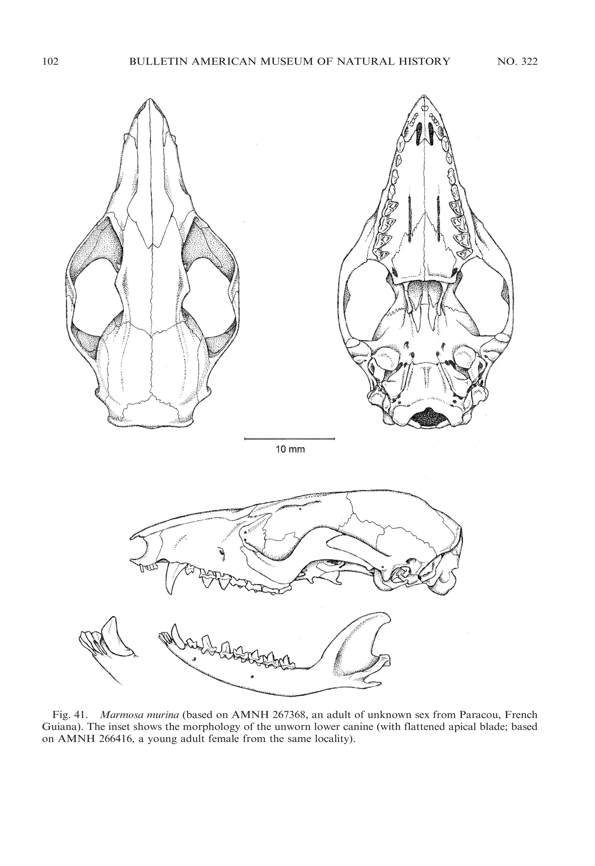
Figure 41
CONTENTS: We recognize two subgenera,
Marmosa Gray, 1821, and Micoureus Lesson,
1842 (see Remarks, below).
The subgenus Marmosa contains andersoni
Pine, 1972; lepida Thomas, 1888 (including
25
In fact, it has been suggested that they are not: ‘‘Considero
´
que Thylatheridium es un genero derivado, en el Plioceno
inferior, de una de las especies de Monodelphis que, ante
´
´
posibilidades ecologicas favorables, acelero su ritmo evolutivo,
´
´
´
´
apartandose rapidamente de sus congeneres de evolucion lenta
…’’ (Reig, 1958: 90).
101
grandis Tate, 1931); mexicana Merriam, 1897
(including mayensis Osgood, 1913; savannarum Goldman, 1917; and zeledoni Goldman, 1917); murina Linnaeus, 1758 (including
bombascarae Anthony, 1922; chloe Thomas,
1907; dorsigera Linnaeus, 1758; duidae Tate,
1931; guianensis Kerr, 1792; klagesi J.A.
Allen, 1900; macrotarsus Wagner, 1842;
madeirensis Cabrera,. 1913; maranii Thomas,
1924; meridionalis Miranda-Ribeiro, 1936;
moreirae Miranda-Ribeiro, 1936; muscula
Cabanis, 1848; parata Thomas, 1911; roraimae Tate, 1931; tobagi Thomas, 1911; and
waterhousei Tomes, 1860); quichua Thomas,
1899 (including musicola Osgood, 1913);
robinsoni Bangs, 1898 (including casta Thomas, 1911; chapmani J.A. Allen, 1900; fulviventer Bangs, 1901; grenadae Thomas, 1911;
isthmica Goldman, 1912; luridavolta Goodwin, 1961; mimetra Thomas, 1921; mitis
Bangs, 1898; nesaea Thomas, 1911; pallidiventris Osgood, 1912; ruatanica Goldman,
1911; and simonsi Thomas, 1899); rubra Tate,
1931; tyleriana Tate, 1931 (including phelpsi
Tate, 1939); and xerophila Handley and
Gordon, 1979.
The subgenus Micoureus contains alstoni
J.A. Allen, 1900 (including nicaraguae Thomas, 1905); constantiae Thomas, 1904 (including budini Thomas, 1920); demerarae Thomas, 1905 (including areniticola Tate, 1931;
domina Thomas, 1920; esmeraldae Tate,
1931; limae Thomas, 1920; and meridae Tate,
1931); paraguayana Tate, 1931 (including
cinerea Temminck, 1824 [preoccupied]);
phaea Thomas, 1899 (including perplexa
Anthony, 1922); and regina Thomas, 1898
(including germana Thomas, 1904; mapiriensis Tate, 1931; parda Tate, 1931; rapposa
Thomas, 1899; and rutteri Thomas, 1924).
MORPHOLOGICAL DESCRIPTION: Combined
length of adult head and body ca. 100–
210 mm; adult weight ca. 20–170 g. Rhinarium with two ventrolateral grooves on each
side of median sulcus; dark circumocular
mask present; pale supraocular spot absent;
dark midrostral stripe absent; throat gland
absent in some species (e.g., M. murina) but
present in adult males of other species (e.g.,
M. mexicana). Dorsal pelage unpatterned,
superficially brownish, reddish, or grayish,
but dorsal hair bases always dark gray;
dorsal guard hairs short and inconspicuous;](https://image.slidesharecdn.com/vossjansa2009-140219090612-phpapp02/75/Voss-jansa-2009-101-2048.jpg)

![2009
VOSS AND JANSA: DIDELPHID MARSUPIALS
of marsupial mammal interrelationships. Molecular Phylogenetics and Evolution 33: 240–250.
Astua, D. 2007 (‘‘2006’’). Range extension and
´
first Brazilian record of the rare Hyladelphys
kalinowskii (Hershkovitz, 1992) (Didelphimorphia, Didelphidae). Mammalia 2006: 174–176.
Astua de Moraes, D., E. Hingst-Zaher, L.F.
´
Marcus, and R. Cerqueira. 2000. A geometricmorphometric analysis of cranial and mandibular shape variation of didelphid marsupials.
Hystrix 10: 115–130.
Augustiny, G. 1942. Die Schwimmanpassung von
Chironectes. Zeitschrift fur Morphologie und
¨
¨
Okologie der Tiere 39: 276–319.
Baker, M.L., J.P. Wares, G.A. Harrison, and R.D.
Miller. 2004. Relationships among the families
and orders of marsupials and the major
mammalian lineages based on recombination
activating gene-1. Journal of Mammalian Evolution 11: 1–16.
Barker, F.K., G.F. Barrowclough, and J.G. Groth.
2002. A phylogenetic hypothesis for passerine
birds: taxonomic and biogeographic implications of an analysis of nuclear DNA sequence
data. Proceedings of the Royal Society of
London B 269: 295–308.
Barkley, L.J. 2008. Genus Glironia O. Thomas,
1912. In A.L. Gardner (editor), Mammals of
South America. Vol. 1. Marsupials, xenarthrans, shrews, and bats, 12–14. Chicago:
University of Chicago Press.
Barnes, R.D. 1977. The special anatomy of
Marmosa robinsoni. In D. Hunsaker (editor),
The biology of marsupials, 387–413. New York:
Academic Press.
Beck, R.M.D. 2008. A dated phylogeny of
marsupials using a molecular supermatrix and
multiple fossil constraints. Journal of Mammalogy 89: 175–189.
Bensley, B.A. 1903. On the evolution of the
Australian Marsupialia; with remarks on the
relationships of marsupials in general. Transactions of the Linnaean Society of London (2nd.
Series, Zoology) 9: 83–217 + pls. 5–7.
Bensley, B.A. 1906. The homologies of the stylar
cusps in the upper molars of the Didelphyidae.
University of Toronto Studies (Biological Series) 5: 149–159.
Biggers, J.D., H.I. Fritz, W.C.D. Hare, and R.A.
McFeeley. 1965. Chromosomes of American
marsupials. Science 148: 1602–1603.
Birney, E.C., J.A. Monjeau, C.J. Phillips, R.S.
Sikes, and I. Kim. 1996. Lestodelphys halli: new
information on a poorly known Argentine
´
marsupial. Mastozoologıa Neotropical 3: 171–
181.
Boas, J.E.V. 1918. Zur Kenntnis des Hinterfusses
der Marsupialier. Kongelige Danske Videnska-
143
bernes Selskaber Biologiske Meddelelser 1(8):
1–23 + 2 pls.
Bown, T.M., and J.G. Fleagle. 1993. Systematics,
biostratigraphy, and dental evolution of the
Palaeothentidae, later Oligocene to early-middle
Miocene (Deseadan–Santacrucian) caenolestoid
marsupials of South America. Paleontological
Society Memoir 29: i–v + 1–76.
Braun, J.K., R.A. Van Den Bussche, P.K. Morton,
and M.A. Mares. 2005. Phylogenetic and
biogeographic relationships of mouse opossums
Thylamys (Didelphimorphia, Didelphidae) in
southern South America. Journal of Mammalogy 86: 147–159.
Bresslau, E. 1920. The mammary apparatus of the
Mammalia in the light of ontogenesis and
phylogenesis. London: Methuen.
Brinkmann, A. 1911. Om Hudens Bygning paa
Haand og Fod hos Chironectes variegatus.
Videnskabelige Meddelelser den naturhistoriske
Forening i København 1910: 1–17, 1 pl.
Brisson, M.J. 1762. Regnum Animale in classes IX
distributum, sive synopsis methodica … (2nd
ed.). Lugduni Batavorum [Leiden]: Theodorum
Haak.
Brown, B.E. 2004. Atlas of New World marsupials. Fieldiana Zoology (New Series) 102: i–vii,
1–308.
Brown, J.C. 1971. The description of mammals. 1.
The external characters of the head. Mammal
Review 1: 151–168.
Brown, J.C., and D.W. Yalden. 1973. The
description of mammals. 2. Limbs and locomotion of terrestrial mammals. Mammal Review 3:
107–134.
Bublitz, J. 1987. Untersuchungen zur Systematik
der rezenten Caenolestidae Trouessart, 1898:
unter Verwendung craniometerischer Methoden. Bonner zoologische Monographien 23:
1–96.
Bucher, J.E., and R.S. Hoffmann. 1980. Caluromys derbianus. Mammalian Species 140: 1–4.
Butler, W.T., and H. Ritchie. 1995. The nature and
functional significance of dentin extracellular
matrix proteins. International Journal of Developmental Biology 39: 169–179.
Cabrera, A. 1919. Genera mammalium: Monotremata, Marsupialia. Madrid: Museo Nacional
de Ciencias Naturales.
Cabrera, A. 1958 (‘‘1957’’). Catalogo de los
´
´
mamıferos de America del Sur [part 1]. Revista
´
del Museo Argentino de Ciencias Naturales
‘‘Bernardino Rivadavia’’ (Ciencias Zoologicas)
´
4(1): i–iv, 1–307.
Carmignotto, A.P., and T. Monfort. 2006. Taxonomy and distribution of the Brazilian species
of Thylamys (Didelphimorphia: Didelphidae).
Mammalia 2006: 126–144.](https://image.slidesharecdn.com/vossjansa2009-140219090612-phpapp02/75/Voss-jansa-2009-143-2048.jpg)
![2009
VOSS AND JANSA: DIDELPHID MARSUPIALS
Douady, C.J., P.I. Chatelier, and O. Madsen., et
al. 2002. Molecular phylogenetic evidence confirming the Eulipotyphla concept and in support
of hedgehogs as the sister group to shrews.
Molecular Phylogenetics and Evolution 25:
200–209.
Eden, R. 1555. The Decades of the newe worlde or
west India, conteynyng the navigations and
conquests of the Spanyardes, with the particular
description of the moste ryche and large landes
and Ilandes lately founde in the west Ocean
perteynyng to the inheritaunce of the kinges of
Spayne [etc.]. Londini: In aedibus Guilhelmi
Powell. [Reprinted and edited by Edward Arber
(1885) in ‘‘The first three English books on
America’’ (Edinburgh: Turnbull and Spears).]
Efron, B., E. Halloran, and S. Holmes. 1996.
Bootstrap confidence levels for phylogenetic
trees. Proceedings of the National Academy of
Sciences of the United States of America 93:
7085–7090.
Eisenberg, J.F. 1989. Mammals of the Neotropics.
Vol. 1. Panama, Colombia, Venezuela, Guyana,
Suriname, French Guiana. Chicago: University
of Chicago Press.
Eisenberg, J.F., and K.H. Redford. 1999. Mammals
of the Neotropics. Vol. 3. Ecuador, Peru, Bolivia,
Brazil. Chicago: Chicago University Press.
Emmons, L.H. 1997. Neotropical rainforest mammals: a field guide. 2nd ed. Chicago: Chicago
University Press.
Emmons, L.H. 1998. Mammal fauna of Parque
Nacional Noel Kempff Mercado. In T.J. Killeen
and T.S. Schulenberg (editors), A biological
assessment of Parque Nacional Noel Kempff
Mercado, Bolivia. (RAP Working Papers 10),
129–135. Washington, DC: Conservation International.
Emmons, L.H. 2008. Genus Caluromysiops Sanborn, 1951. In A.L. Gardner (editor), Mammals
of South America. Vol. 1. Marsupials, xenarthrans, shrews, and bats, 11–12. Chicago:
Chicago University Press.
Enders, R.K. 1937. Panniculus carnosus and
formation of the pouch in didelphids. Journal
of Morphology 61: 1–26.
Engstrom, M.D., and A.L. Gardner. 1988. Karyotype of Marmosa canescens (Marsupialia:
Didelphidae): a mouse opossum with 22 chromosomes. Southwestern Naturalist 33: 231–233.
Erixon, P., B. Svennblad, T. Britton, and B.
Oxelman. 2003. Reliability of Bayesian posterior probabilities and bootstrap frequencies in
phylogenetics. Systematic Biology 52: 665–673.
Fantin, C., and M.N.F. da Silva. In press.
Karyotype of the bushy-tailed opossum Glironia
venusta (Didelphidae, Didelphimorphia) from
145
the western Brazilian Amazon. Journal of
Mammalogy.
Felsenstein, J. 1985. Confidence limits on phylogenies: an approach using the bootstrap. Evolution 39: 783–791.
Feng, J.Q., H. Huang, and Y. Lu., et al. 2003. The
dentin matrix Protein 1 (DMP1) is specifically
expressed in minerallized, but not soft, tissues
during development. Journal of Dental Research 82: 776–780.
Filan, S.L. 1990. Myology of the head and neck of
the bandicoot (Marsupialia: Peramelemorphia).
Australian Journal of Zoology 38: 617–634.
Flannery, T., M. Archer, and G. Maynes. 1987.
The phylogenetic relationships of living phalangerids (Phalangeroidea: Marsupialia) with a
suggested new taxonomy. In M. Archer (editor),
Possums and opossums: studies in evolution.
Vol. 2, 477–506. Sydney: Surrey Beatty.
Flores, D.A. 2009. Phylogenetic analyses of
postcranial skeletal morphology in didelphid
marsupials. Bulletin of the American Museum
of Natural History 320: 1–81.
Flores, D.A., and F. Abdala. 2001. Diferencias
morfologicas de craneo y denticion en Didelphis
´
´
´
albiventris y D. marsupialis (Didelphimorphia:
Didelphidae) de Argentina y Bolivia. Comunicacoes do Museu de Ciencias e Tecnologia da
¸˜
ˆ
PUCRS (Serie Zoologia) 14: 101–110.
´
´
Flores, D.A., R.M. Barquez, and M.M. Dıaz.
2008. A new species of Philander Brisson, 1762
(Didelphimorphia, Didelphidae). Mammalian
Biology 73: 14–24.
´
Flores, D.A., M.M. Dıaz, and R.M. Barquez.
2007. Systematics and distribution of marsupials
in Argentina: a review. University of California
Publications in Zoology 134: 579–669.
Flores, D.A., N.P. Giannini, and F. Abdala. 2003.
Cranial ontogeny of Lutreolina crassicaudata
(Didelphidae): a comparison with Didelphis
albiventris. Acta Theriologica 48: 1–9.
Flores, D.A., N.P. Giannini, and F. Abdala. 2006.
Comparative postnatal ontogeny of the skull in
the Australidelphian metatherian Dasyurus albopunctatus (Marsupialia: Dasyuromorphia:
Dasyuridae). Journal of Morphology 267:
426–440.
Flower, W.H. 1867. On the development and
succession of the teeth in the Marsupialia.
Philosophical Transactions of the Royal Society
of London 157: 631–641 + pls. 29, 30.
Fong, S.-L., W.-B. Fong, T.A. Morris, K.M.
Kedzie, and C.D.B. Bridges. 1990. Characterization and comparative structural features of
the gene for human interstitial retinol-binding
protein. Journal of Biological Chemistry 265:
3648–3653.](https://image.slidesharecdn.com/vossjansa2009-140219090612-phpapp02/75/Voss-jansa-2009-145-2048.jpg)
![146
BULLETIN AMERICAN MUSEUM OF NATURAL HISTORY
Freedman, L. 1967. Skull and tooth variation in
the genus Perameles. Part 1. Anatomical features. Records of the Australian Museum 27:
147–65, pls. 16–23.
Gabbert, S.L. 1998. Basicranial anatomy of
Herpetotherium (Marsupialia: Didelphimorphia) from the Eocene of Wyoming. American
Museum Novitates 3235: 1–13.
Gardner, A.L. 1973. The systematics of the genus
Didelphis (Marsupialia: Didelphidae) in North
and Middle America. Special Publications of the
Museum Texas Tech University 4: 1–81.
Gardner, A.L. 1981. Mammals of Surinam [review]. Journal of Mammalogy 62: 445–448.
Gardner, A.L. 2005. Order Didelphimorphia. In
D.E. Wilson and D.M. Reeder (editors), Mammal species of the world: a taxonomic and
geographic reference. 3rd ed.: 3–18. Baltimore:
Johns Hopkins University Press.
Gardner, A.L. (editor). 2008 (‘‘2007’’). Mammals
of South America. Vol. 1. Marsupials, xenarthrans, shrews, and bats. Chicago: Chicago
University Press.
Gardner, A.L., and G.K. Creighton. 1989. A new
generic name for Tate’s microtarsus group of
South American mouse opossums (Marsupialia:
Didelphidae). Proceedings of the Biological
Society of Washington 102: 3–7.
Gardner, A.L., and G.K. Creighton. 2008a
(‘‘2007’’). Genus Marmosops Matschie, 1916.
In A.L. Gardner (editor), Mammals of South
America. Vol. 1. Marsupials, xenarthrans, shrews,
and bats, 61–74. Chicago: Chicago University
Press.
Gardner, A.L., and G.K. Creighton. 2008b (‘‘2007’’).
Genus Micoureus Lesson, 1842. In A.L. Gardner
(editor), Mammals of South America. Vol. 1.
Marsupials, xenarthrans, shrews, and bats, 74–82.
Chicago: Chicago University Press.
Gardner, A.L., and M. Dagosto. 2008 (‘‘2007’’).
Tribe Metachirini Reig, Kirsch, and Marshall,
1985. In A.L. Gardner (editor), Mammals of
South America. Vol. 1. Marsupials, xenarthrans, shrews, and bats, 35–39. Chicago:
Chicago University Press.
Gaudin, T.J., J.R. Wible, J.A. Hopson, and W.D.
Turnbull. 1996. Reexamination of the evidence
for the Cohort Epitheria (Mammalia, Eutheria).
Journal of Mammalian Evolution 3: 31–79.
Gellert, M. 2002. V(D)J recombination: RAG
proteins, repair factors, and regulation. Annual
Review of Biochemistry 71: 101–132.
George, A., B. Sabsay, P.A.L. Simonian, and A.
Veis. 1993. Characterization of a novel dentin
matrix acidic phosphoprotein. Journal of Biological Chemistry 268: 12624–12630.
George, T.K., S.A. Marques, M. de Vivo, L.C.
Branch, N. Gomes, and R. Rodrigues. 1988.
NO. 322
´
Levantamento de mamıferos do Parna–Tapajos.
´
Brasil Florestal 63: 33–41.
Giannini, N.P., F. Abdala, and D.A. Flores. 2004.
Comparative postnatal ontogeny of the skull in
Dromiciops gliroides (Marsupialia: Microbiotheria). American Museum Novitates 3460:
1–17.
Ginsburg, D., and E.J.W. Bowie. 1992. Molecular
genetics of von Willebrand disease. Blood 79:
2507–2519.
Goin, F.J. 1997. New clues for understanding
Neogene marsupial radiations. In R.F. Kay,
R.H. Maddeen, R.L. Cifelli, and J.J. Flynn
(editors), Vertebrate paleontology in the Neotropics: the Miocene fauna of La Venta,
Colombia, 187–206. Washington, DC: Smithsonian Institution Press.
Goin, F.J., and A.M. Candela. 2004. New
Paleogene marsupials from the Amazon basin
of eastern Peru. Natural History Museum of
Los Angeles County Science Series 40: 15–60.
Goin, F.J., and U.F.J. Pardinas. 1996. Revision de
˜
´
las especies del genero Hyperdidelphys Ame´
ghino, 1904 (Mammalia, Marsupialia, Didelphidae). Su significacion, filogenetica, estratogra´
´
´
fica, y adaptativa en el Neogeno del Cono Sur
sudamericano. Estudios Geolologicos 52: 327–
´
359.
Goin, F.J., and P. Rey. 1997. Sobre las afinidades
de Monodelphis Burnett, 1830 (Mammalia:
Marsupialia: Didelphidae: Marmosinae). Neotropica 43: 93–98.
Goin, F.J., M.R. Sanchez-Villagra, A. Abello, and
´
R.F. Kay. 2007. A new generalized paucituberculatan marsupial from the Oligocene of Bolivia
and the origin of ‘shrew-like’ opossums. Paleontology 50: 1267–1276.
Gonzalez, E.M., and G. Fregueiro. 1998. Primer
´
registro de Chironectes minimus para Uruguay
(Mammalia, Didelphidae). Comunicaciones Zoologicas del Museo de Historia Natural de
´
Montevideo 12(192): 1–8.
Gonzalez, E.M., A.M. Saralegui, and G. Fre´
gueiro. 2000. The genus Thylamys Gray, 1843,
in Uruguay (Didelphimorphia, Didelphidae).
´
Boletın de la Sociedad Zoologica del Uruguay
´
12: 44–45.
Graipel, M., P.R.M. Miller, and A. Ximenez.
´
1996. Contribucao a identificao e distribuicao
¸˜ `
¸˜
¸˜
das subespecies de Lutreolina crassicaudata
´
(Desmarest) (Marsupialia, Mammalia). Revista
Brasileira de Biologia 13: 781–790.
Groth, J.G., and G.F. Barrowclough. 1999. Basal
divergences in birds and the phylogenetic utility
of the nuclear RAG-1 gene. Molecular Phylogenetics and Evolution 12: 115–123.
Gruber, K.F., R.S. Voss, and S.A. Jansa. 2007.
Base-compositional heterogeneity in the RAG1](https://image.slidesharecdn.com/vossjansa2009-140219090612-phpapp02/75/Voss-jansa-2009-146-2048.jpg)
![152
BULLETIN AMERICAN MUSEUM OF NATURAL HISTORY
Pine, R.H. 1973. Anatomical and nomenclatural
notes on opossums. Proceedings of the Biological Society of Washington 86: 391–402.
Pine, R.H., and C.O. Handley, Jr. 2008 (‘‘2007’’).
Genus Monodelphis Burnett, 1830. In A.L.
Gardner (editor), Mammals of South America.
Vol. 1. Marsupials, xenarthrans, shrews, and
bats, 82–107. Chicago: Chicago University
Press.
Pine, R.H., J.E. Rice, J.E. Bucher, D.H. Tank, Jr.,
and A.M. Greenhall. 1985. Labile pigments and
fluorescent pelage in didelphid marsupials.
Mammalia 49: 249–256.
Pocock, R.I. 1914. On the facial vibrissae of
Mammalia. Proceedings of the Zoological
Society of London 1914: 889–912.
Pocock, R.I. 1926. The external characters of
Thylacinus, Sarcophilus, and some related marsupials. Proceedings of the Zoological Society of
London 1926: 1037–1084.
Porter, C.A., M. Goodman, and M.J. Stanhope.
1996. Evidence on mammalian phylogeny from
sequences of exon 28 of the von Willebrand
factor gene. Molecular Phylogenetics and Evolution 5: 89–101.
Posada, D., and T. Buckley. 2004. Model selection
and model averaging in phylogenetics: advantages of Akaike Information Criterion and
Bayesian approaches over likelihood ratio tests.
Systematic Biology 53: 793–808.
Posada, D., and K.A. Crandall. 1998. ModelTest:
testing the model of DNA substitution. Bioinformatics 14: 817–818.
Prochel, J., and M. Sanchez-Villagra. 2003. Carpal
´
ontogeny in Monodelphis domestica and Caluromys philander (Marsupialia). Zoology 106:
73–84.
Raterman, D., R.W. Meridith, L.A. Ruedas, and
M.S. Springer. 2006. Phylogenetic relationships
of the cuscuses and brushtail possums (Marsupialia: Phalangeridae) using the nuclear gene
BRCA1. Australian Journal of Zoology 54:
353–361.
Redford, K.H., and J.F. Eisenberg. 1992. Mammals of the Neotropics. Vol. 2. Chile, Argentina, Uruguay, Paraguay. Chicago: Chicago
University Press.
Reeder, S.A., and R.D. Bradley. 2004. Molecular
systematics of neotomine-peromyscine rodents
based on the dentin matrix protein 1 gene.
Journal of Mammalogy 85: 1194–1200.
Reid, F.A. 1997. A field guide to the mammals of
Central America and southeast Mexico. New
York: Oxford University Press.
Reig, O.A. 1955. Noticia preliminar sobre la
presencia de microbiotherinos vivientes en la
fauna sudamericana. Investigaciones Zoologicas
´
Chilenas 2: 121–130.
NO. 322
Reig, O.A. 1958. Comunicacion preliminar sobre
´
nuevas especies del genero Thylatheridium Reig
´
(Mammalia, Didelphidae). Neotropica 4: 89–95.
Reig, O.A., A.L. Gardner, N.O. Bianchi, and J.L.
Patton. 1977. The chromosomes of the Didelphidae (Marsupialia) and their evolutionary
significance. Biological Journal of the Linnaean
Society of London 9: 91–216.
Reig, O.A., J.A.W. Kirsch, and L.G. Marshall.
1985. New conclusions on the relationships of
the opossum-like marsupials, with an annotated
classification of the Didelphimorphia. Ameghiniana 21: 335–343.
Reig, O.A., J.A.W. Kirsch, and L.G. Marshall.
1987. Systematic relationships of the living and
Neocenozoic American ‘‘opossum-like’’ marsupials (suborder Didelphimorphia), with comments on the classification of these and of the
Cretaceous and Paleogene New World and
European metatherians. In M. Archer (editor),
Possums and opossums: studies in evolution.
Vol. 1, 1–89. Sydney: Surrey Beatty.
Reig, O.A., and G.G. Simpson. 1972. Sparassocynus (Marsupialia, Didelphidae), a peculiar
mammal from the late Cenozoic of Argentina.
Journal of Zoology (London) 167: 511–539.
Ribeiro, M.G., and J.C. Nogueira. 1990. The penis
morphology of the four-eyed opossum Philander opossum. Anatomischer Anzeiger 171:
65–72.
Ride, W.D.L. 1962. On the evolution of Australian
marsupials. In: G.E. Leeper (editor), The
evolution of living organisms, 281–306. Melbourne: Melbourne University Press.
´
Rodrıguez, J.A., and B.R. Henderson. 2000.
Identification of a functional nuclear export
sequence in BRCA1. Journal of Biological
Chemistry 275: 38589–38596.
Rofe, R., and D. Hayman. 1985. G-banding
evidence for a conserved complement in the
Marsupialia. Cytogenetics and Cell Genetics 39:
40–50.
Ronquist, F., and J.P. Huelsenbeck. 2003.
MrBayes 3: Bayesian phylogenetic inference
under mixed models. Bioinformatics 19: 1572–
1574.
Rossi, R.V. 2005. Revisao taxonomica de Mar˜
ˆ
mosa Gray, 1821 (Didelphimorphia, Didelphidae). Unpublished Ph.D. dissertation, Universidade de Sao Paulo. 2 vols.
˜
Rougier, G.W., J.R. Wible, and M.J. Novacek.
1998. Implications of Deltatheridium specimens
for early marsupial history. Nature 396:
459–463. [Data matrix and character descriptions were published electronically as supplementary information at www.nature.com. The
first paper publication of this information
appeared as appendix 1 of Wible et al. (2001).]](https://image.slidesharecdn.com/vossjansa2009-140219090612-phpapp02/75/Voss-jansa-2009-152-2048.jpg)
![156
BULLETIN AMERICAN MUSEUM OF NATURAL HISTORY
genero Marmosa: M. pusilla bruchi, M. agilis
´
chacoensis, y M. microtarsus (Marsupialia:
Didelphidae). Physis 38: 33–38.
Weisbecker, V., and M. Nilsson. 2008. Integration,
heterochrony, and adaptation in pedal digits of
syndactylous marsupials. BMC Evolutionary
Biology 8: 14 pp. [no pagination; published
online only], doi:10.1186/1471-2148-8-160.
Weksler, M. 2003. Phylogeny of Neotropical
oryzomyine rodents (Muridae: Sigmodontinae)
based on the nuclear IRBP exon. Molecular
Phylogenetics and Evolution 29: 331–349.
Wells, R.T., and R.H. Tedford. 1995. Sthenurus
(Macropodidae: Marsupialia) from the Pleistocene of Lake Callabonna, South Australia.
Bulletin of the American Museum of Natural
History 225: 1–111.
Whipple, I.L. 1904. The ventral surface of the
mammalian chiridium, with special reference to
the conditions found in man. Zeitschrift fur
¨
Morphologie und Anthropologie 7: 261–368.
Wible, J.R. 1990. Petrosals of Late Cretaceous
marsupials from North America and a cladistic
analysis of the petrosal in therian mammals.
Journal of Vertebrate Paleontology 10: 183–205.
Wible, J.R. 2003. On the cranial osteology of the
short-tailed opossum Monodelphis brevicaudata
(Didelphidae, Marsupialia). Annals of the Carnegie Museum 72: 137–202.
Wible, J.R., G.W. Rougier, M.J. Novacek, and
M.C. McKenna. 2001. Earliest eutherian ear
region: a petrosal referred to Prokennalestes
from the Early Cretaceous of Mongolia. American Museum Novitates 3322: 1–44.
Wiens, J.J. 2000. Coding morphological variation
within species and higher taxa for phylogenetic
analysis. In J.J. Wiens (editor), Phylogenetic
analysis of morphological data, 115–145. Washington, DC: Smithsonian Institution Press.
´
Wilson, D.E. 1991. Mammals of the Tres Marıas
Islands. Bulletin of the American Museum of
Natural History 206: 214–250.
Wilson, D.R. and D.M. Reeder (editor). 2005.
Mammal species of the world. 3rd ed. Baltimore: Johns Hopkins University Press, 2 vols.
NO. 322
Winge, H. 1893. Jordfundne og nulevende Pungdyr (Marsupialia) fra Lagoa Santa, Minas
Geraes, Brasilien. E Museo Lundii 2(2): 1–132,
pls. 1–4.
Woolley, P. 1974. The pouch of Planigale subtilissima and other dasyurid marsupials. Journal
of the Royal Society of Western Australia 57:
11–15.
Wroe, S. 1997. A reexamination of proposed
morphology-based synapomorphies for the
families of Dasyuromorphia (Marsupialia). I.
Dasyuridae. Journal of Mammalian Evolution
4: 19–52.
Wroe, S., J. Brammall, and B.N. Cooke. 1998. The
skull of Ekaltadeta ima (Marsupialia, Hypsoprymnodontidae?): an analysis of some marsupial cranial features and a re-investigation of
propleopine phylogeny, with notes on the
inference of carnivory in mammals. Journal of
Paleontology 72: 738–751.
Wroe, S., M. Crowther, J. Dortch, and J. Chong.
2004. The size of the largest marsupial and why
it matters. Proceedings of the Royal Society of
London B (suppl.) 271: S34–S36.
Wroe, S., M. Ebach, S. Ahyong, C. de Muizon,
and J. Muirhead. 2000. Cladistic analysis of
dasyuromorphian (Marsupialia) phylogeny using cranial and dental characters. Journal of
Mammalogy 81: 1008–1024.
Wroe, S., and A. Musser. 2001. The skull of
Nimbacinus dicksoni (Thylacinidae: Marsupialia). Australian Journal of Zoology 49: 487–514.
Ximenez, A. 1967. Contribucion al conocimiento
´
´
de Lutreolina crassicaudata (Desmarest, 1804) y
sus formas geograficas. Comunicaciones Zool´
ogicas del Museo de Historia Natural de
´
Montevideo 9(112): 1–7.
Zarza, H., G. Ceballos, and M.A. Steele. 2003.
Marmosa canescens. Mammalian Species 725:
1–4.
Zwickl, D.J. 2006. Genetic algorithm approaches
for the phylogenetic analysis of large biological
sequence datasets under the maximum likelihood criterion. Unpublished Ph.D. dissertation,
University of Texas at Austin.](https://image.slidesharecdn.com/vossjansa2009-140219090612-phpapp02/75/Voss-jansa-2009-156-2048.jpg)
![160
BULLETIN AMERICAN MUSEUM OF NATURAL HISTORY
APPENDIX 2
Specimens Sequenced for Nuclear Genes
Nuclear gene sequences were obtained
from the following specimens, listed alphabetically by family, genus, and species. The
institutional catalog number of each voucher
specimen is listed first (in parentheses),
followed by a tissue identifier (if any, in
square brackets). See appendix 1 for institutional abbreviations.
Caenolestidae
´
Caenolestes fuliginosus: Ecuador, Bolıvar, 4 km E
Cruz de Lizo (MSB 70587 [NK 27708]).
Rhyncholestes raphanurus: Chile, Osorno, La Picada
(Universidad Austral de Chile, unvouchered [LG
420]).
Dasyuridae
Murexia longicaudata: Papua New Guinea, Morobe,
Mt. Missim (ANWC M29669 [‘‘female 3’’]); Papua
New Guinea, Southern Highlands Province, Bobole (AMS M18453 [ABTC 45110]).
Sminthopsis crassicaudata: Australia, New South
Wales, Remington Station (ANWC M16816);
Australia, South Australia, Ngarkat Conservation
Park (SAM M20207 [ABTC 27560]).
Didelphidae
Caluromys lanatus: Ecuador, Napo, Parque Nacional
´
Yasunı (ROM 104570).
Caluromys philander: French Guiana, Les Nouragues
(V-823 [T-1754], V-960 [T-2020]).
Caluromysiops irrupta: New York Zoological Society
(AMNH 244364).
Chironectes minimus: Guyana, Barima-Waini, Waikerebi (ROM 98855 [FN 31677]).
Cryptonanus chacoensis: Paraguay, Caazapa, Estancia
´
Dos Marias (uncataloged specimen returned to
Paraguay [GD 521]).
Cryptonanus unduaviensis: Bolivia, Pando, Independencia (AMNH 262401 [NK 14234]); Bolivia,
Santa Cruz, Santiago de Chiquitos (AMNH
260032 [NK 12313]).
Didelphis albiventris: Paraguay, Canendiyu, 13.3 km
N Curuguaty (UMMZ 134041 [GKC 783]); Paraguay, Presidente Hayes, 24 km NW Villa Hayes
(UMMZ 134058 [GKC 816]).
´
Didelphis marsupialis: Peru, Loreto, Rıo Galvez
´
(AMNH 272836 [RSV 2357], MUSM 13282 [RSV
2273]).
Didelphis virginiana: Mexico, Yucatan, 1.5 km N
´
Labna (ROM 96483 [FN 30300]).
Glironia venusta: Brazil, Amazonas, alto Rio Urucu
(INPA 2570 [MNFS 75]).
Gracilinanus aceramarcae: Peru, Cusco, Cordillera
Vilcabamba (MUSM 13002 [LHE 1342]).
Gracilinanus agilis: Brazil, Minas Gerais, Mata do
Vasco, 12 km W Nova Ponte (MVZ 197438 [LPC
363], MVZ 197439 [LPC 364]).
´
Gracilinanus emiliae: Peru, Loreto, Rıo Ga lvez,
´
Nuevo San Juan (MUSM 15292 [DWF 413]).
NO. 322
Gracilinanus microtarsus: Brazil, Sao Paulo, Fazenda
˜
Intervales (MVZ 182055 [MAM 38], MVZ 182056
[MAM 49]).
Hyladelphys kalinowskii: French Guiana, Route de
Kaw, 30 km SE Cayenne (MNHN, uncataloged
[T4385]).
´
Lestodelphys halli: Argentina, Rıo Negro, 10 km by
road E Clementi Onelli (UWZ 22422 [Kirsch lab
extract 775]).
Lutreolina crassicaudata: Paraguay, Misiones, 2 km
NE Ayolas (UMMZ 134018 [GKC 848], UMMZ
134019 [GKC 849]).
Marmosa (Marmosa) lepida: Guyana, Potaro-Siparuni, Iwokrama Reserve (ROM 107034 [F 38809]);
´
Peru, Amazonas, Huampami on Rıo Cenepa
(MVZ 155245 [FC 4928, JLP7844]).
Marmosa (Marmosa) mexicana: Guatemala, El Peten, Biotopo Cerro Cahui, El Ramate (ROM 99608
´
´
[FN 32277]); Guatemala, El Progresso, Rıo Uyus,
´
5 km E San Cristobal, Acasaguascatlan (ROM
´
´
99776 [FN 34135]).
´
Marmosa (Marmosa) murina: Peru, Loreto, Rıo
Galvez (AMNH 272816 [RSV 2303], AMNH
´
272870 [RSV 2413]); Surinam, Para, Zanderij
(CM 68346 [TK 17359], 68353 [TK 17387]).
Marmosa (Marmosa) robinsoni: Panama, Darien,
´
Cana (TTU 39117 [TK 22552], 39118 [TK 22555]).
Marmosa (Marmosa) rubra: Peru, Amazonas, 0.5 mi
´
W Huampami on Rıo Cenepa (MVZ 153280 [JLP
6930]).
´
Marmosa (Micoureus) demerarae: Peru, Loreto, Rıo
Galvez (AMNH 272667 [RSV 2029], MUSM
´
13294 [RSV 2085]).
Marmosa (Micoureus) paraguayana: Brazil, Sa o
˜
Paulo, Fazenda Intervales, Capao Bonito (MVZ
˜
182064 [MAM 46], 182065 [MAM 47]).
Marmosa (Micoureus) regina: Brazil, Amazonas,
Igarape Nova Empresa, left bank Rio Jurua
´
´
(MVZ 190323 [JLP 15435]); Brazil, Acre, Igarape
´
Porongaba, right bank Rio Jurua (MVZ 190332
´
[MNFS 1232]).
´
Marmosops impavidus: Peru, Loreto, Rıo Galvez
´
(AMNH 272760 [RSV 2202], MUSM 13284 [RSV
2114]).
Marmosops incanus: Brazil, Minas Gerais, Estacao
¸˜
Biologica de Caratinga, Fazenda Montes Claros,
´
54 km E Caratinga (MVZ 182768 [MAM 201],
182769 [MAM 202]).
Marmosops noctivagus: Ecuador, Napo, Parque Na´
cional Yasunı (ROM 105316 [F 37644]); Peru,
´
Loreto, Rıo Galvez (AMNH 272775 [RSV 2225],
´
AMNH 272782 [RSV 2242], AMNH 272809 [RSV
2294], MUSM 13289 [RSV 2224], MUSM 13292
[RSV 2131]).
Marmosops parvidens: Guyana, Upper Takutu-Upper
Essequibo, Karanambo (ROM 97938 [FN 33439]).
Marmosops pinheiroi: Guyana, Potaro-Siparuni,
Iwokrama Reserve (ROM 108920 [F 43900]).
´
Metachirus nudicaudatus: Peru, Loreto, Rıo Galvez
´
(AMNH 272780 [RSV 2236], MUSM 13293 [RSV
2329]).
Monodelphis brevicaudata: Surinam, Saramacca, Raleigh Falls (CM 63511 [TK 10244]); Surinam,
Nickerie, Kayserberg Airstrip (CM 68359 [TK
17069]).](https://image.slidesharecdn.com/vossjansa2009-140219090612-phpapp02/75/Voss-jansa-2009-160-2048.jpg)
![2009
VOSS AND JANSA: DIDELPHID MARSUPIALS
´
Monodelphis emiliae: Peru, Loreto, Rıo Ga lvez
´
(MUSM 13298 [RSV 2083]).
Monodelphis peruvianus (5 M. ‘‘adusta’’ of previous
´
reports; see Solari, 2007): Peru, Loreto, Rıo Galvez
´
(AMNH 272695 [RSV 2086]).
Monodelphis theresa: Brazil, Sao Paulo, Ilha de
˜
Sebastia o, Parque Estadual Ilhabela (MVZ
˜
182776 [MAM 160]).
Philander frenatus: Brazil, Sa o Paulo, Fazenda
˜
Intervales, Capao Bonito (MVZ 182066 [MAM
˜
41], 182067 [MAM 64]).
´
Philander mcilhennyi: Peru, Loreto, Rıo Ga lvez
´
(AMNH 272818 [RSV 2310], MUSM 13299 [RSV
2153]).
Philander opossum: Surinam, Para, Zanderij (CM
68365 [TK 17015]); Surinam, Suriname, Plantation
Clevia, 8 km NE Paramaribo (CM 76743 [TK
17524]).
Thylamys macrurus: Paraguay, Concepcion, 7 km NE
´
Concepcion (MSB 70700 [NK 27536).
´
Thylamys pallidior: Bolivia, Tarija, roadside above
Cieneguillas (FMNH 162495 [NBH 76-97]).
´
Thylamys pusillus: Bolivia, Tarija, Estancia Bolıvar
(MSB 67016 [NK 25139]).
Thylamys venustus: Bolivia, Tarija, Chuquiaca
(FMNH 162505 [BDP 3345]); Bolivia, Tarija, ca.
10 km by road W Narvaez (FMNH 162507 [BDP
´
3309]).
Tlacuatzin canescens: Mexico, Jalisco, 6 km SE
Chamela (TTU 37700 [TK 11826]); Mexico,
Michoacan, 1 km E Playa Azul (UMSNH 2993
[TK 45085]).
Microbiotheriidae
Dromiciops gliroides: Argentina, Neuquen, near Estancia Paso Coihue (MVZ 184914 [JLP 16545]).
Peramelidae
Echymipera kalubu: Papua New Guinea, Western
Highlands, Trauna Valley (MVZ 138476 [FC 868]).
Perameles gunni: Australia, Tasmania, Ridgley (SAM,
unvouchered [ABTC 66105]).
APPENDIX 3
Nonmolecular Character Descriptions
EXTERNAL MORPHOLOGY
Character 1: Pars supralabialis of rhinarium broad (0);
or reduced to a narrow philtrum (1). See text for a
discussion of this character and its taxonomic
distribution.
Character 2: Rhinarium with two ventrolateral grooves
flanking the median sulcus (0); or with a single
ventrolateral groove on each side (1). Information
about the distribution of these traits among
didelphid terminal taxa was summarized by Voss
and Jansa (2003: character 1). See text for
comments about traits expressed by outgroup taxa.
This character was scored as inapplicable (‘‘-’’) for
taxa with a very narrow pars supralabialis (see
above).
Character 3: Reciprocating lateral fleshy outgrowths of
upper and lower oral margins absent (0); or present
161
(1). See text for a discussion of this character and
its taxonomic distribution.
Character 4: Interramal vibrissae present (0); or absent
(1). See text for a discussion of this character and
its taxonomic distribution.
Character 5: Dark midrostral stripe absent (0); or
present (1). Scoring criteria and information about
the distribution of these traits among didelphid
terminal taxa were provided by Voss and Jansa
(2003: character 2). See text for comments about
traits expressed by outgroup taxa.
Character 6: Fur surrounding eye not distinctively
colored (0); or eye surrounded by mask of dark fur
(1). Scoring criteria and information about the
distribution of these traits among didelphid terminal taxa were provided by Voss and Jansa (2003:
character 3). See text for comments about traits
expressed by outgroup taxa.
Character 7: Circumocular mask contrasts with coloration of cheeks and crown (0); or dark fur around
eye continuous with dark coronal fur (1). This
character was modified from Voss and Jansa’s
(2003) character 4 by eliminating an intermediate
condition for polymorphism, which was recoded as
0/1 for this analysis. See text for comments about
traits expressed by outgroup taxa.
Character 8: Conspicuous pale spot above each eye
absent (0); or present (1). Scoring criteria and
information about the distribution of these traits
among didelphid terminal taxa were provided by
Voss and Jansa (2003: character 5). See text for
comments about traits expressed by outgroup taxa.
Character 9: Gular gland absent (0); or present (1).
Scoring criteria and information about the distribution of these traits among didelphid terminal
taxa were provided by Voss and Jansa (2003:
character 6). See text for comments about traits
expressed by outgroup taxa.
Character 10: Dorsal body pelage more or less
uniformly colored, unpatterned (0); or marked by
dark transverse bars connected middorsally (1); or
marked by paired scapular stripes (2); or marked by
one median and two lateral stripes (3); or grayish
middorsum contrasting with reddish or yellowish
flanks (4); or grayish midbody (including flanks)
contrasting with reddish head and rump (5); or
‘‘tricolored’’ (6); or with transverse sacral bars (7).
Scoring criteria and information about the distribution of states 0–6 among didelphid terminal taxa
were provided by Voss and Jansa (2003: character
7). See text for comments about traits expressed by
outgroup taxa. In the absence of any compelling
evidence for intermediacy among these alternative
conditions, this character was treated as unordered
in all of our analyses.
Character 11: Dorsal underfur dark (0); or white (1).
Scoring criteria and information about the distribution of these traits among didelphid terminal
taxa were provided by Voss and Jansa (2003:
character 8). See text for comments about traits
expressed by outgroup taxa.
Character 12: Dorsal pelage hairs not grossly dissimilar
in length or coarseness (0); or dorsal pelage with
guard hairs conspicuously longer and coarser than
underfur (1); or dorsal pelage with stiff, dorsally](https://image.slidesharecdn.com/vossjansa2009-140219090612-phpapp02/75/Voss-jansa-2009-161-2048.jpg)
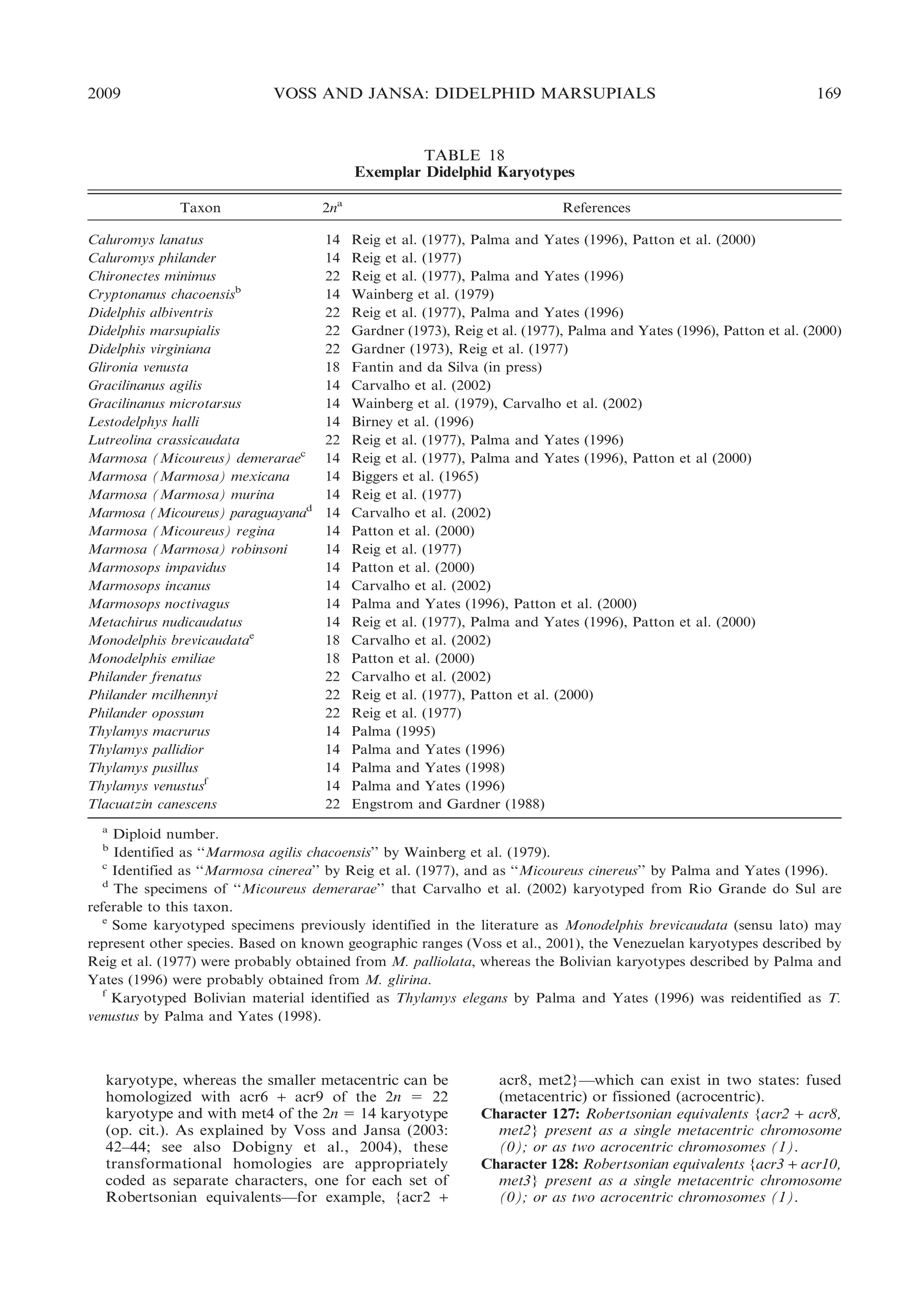
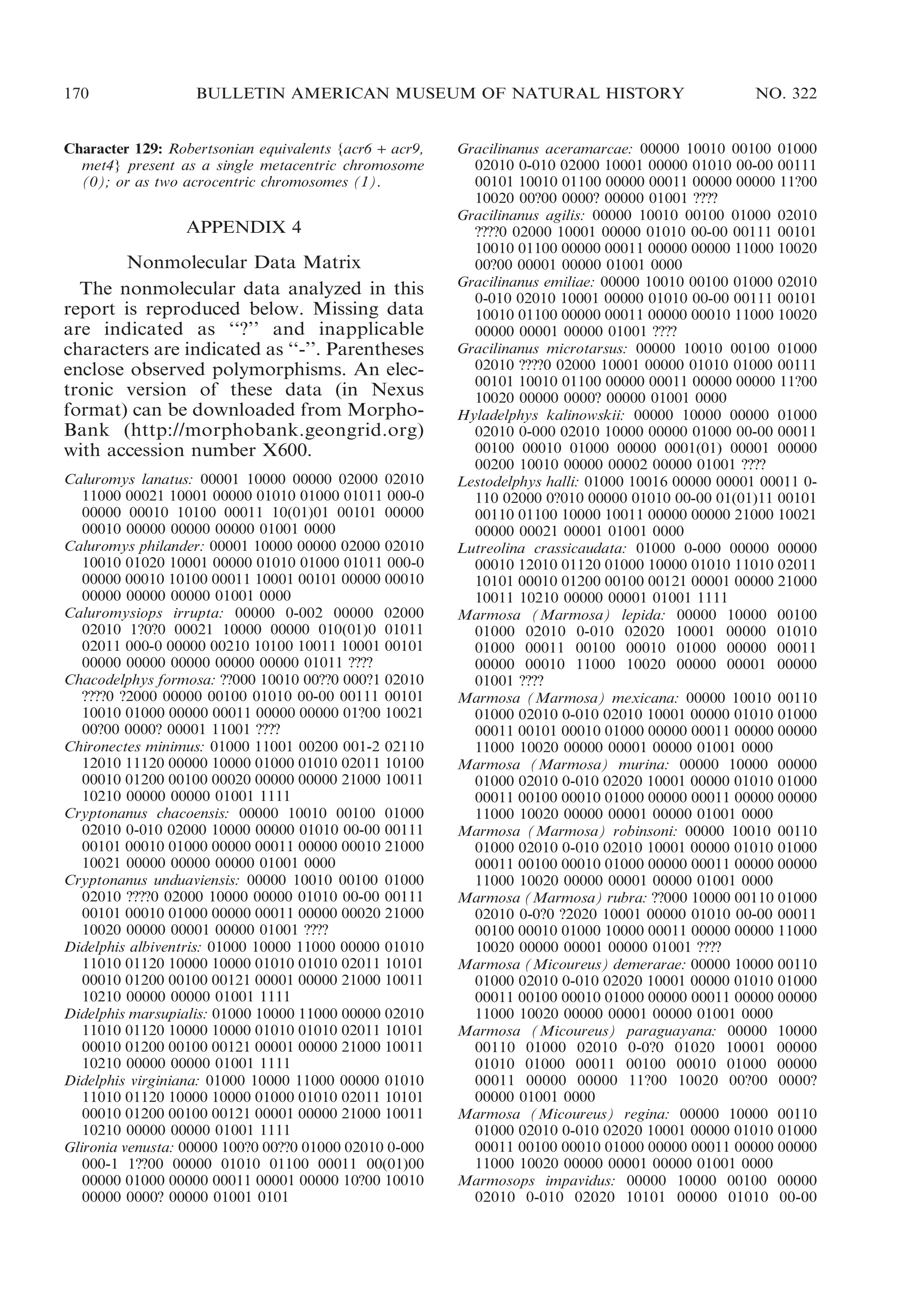
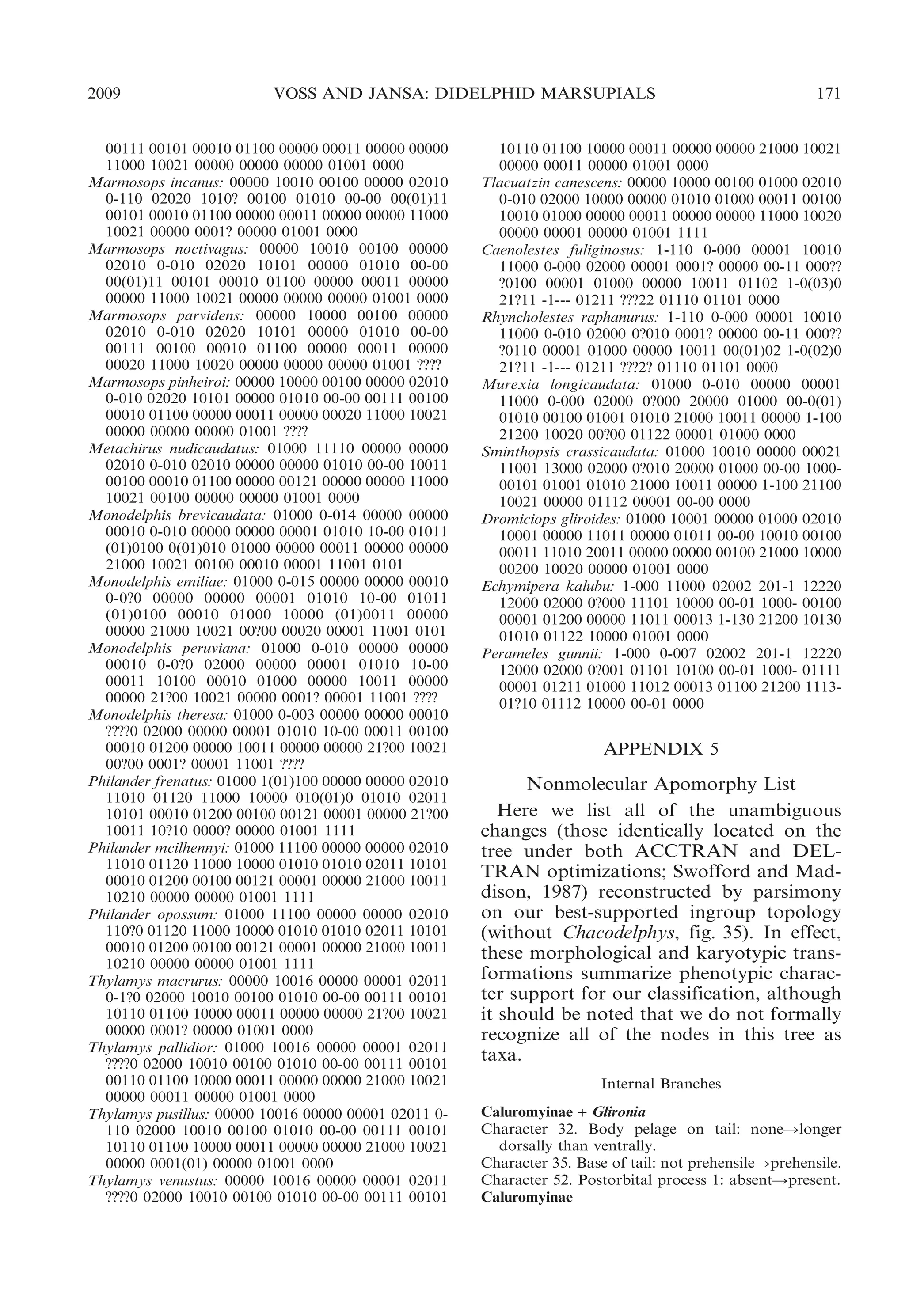
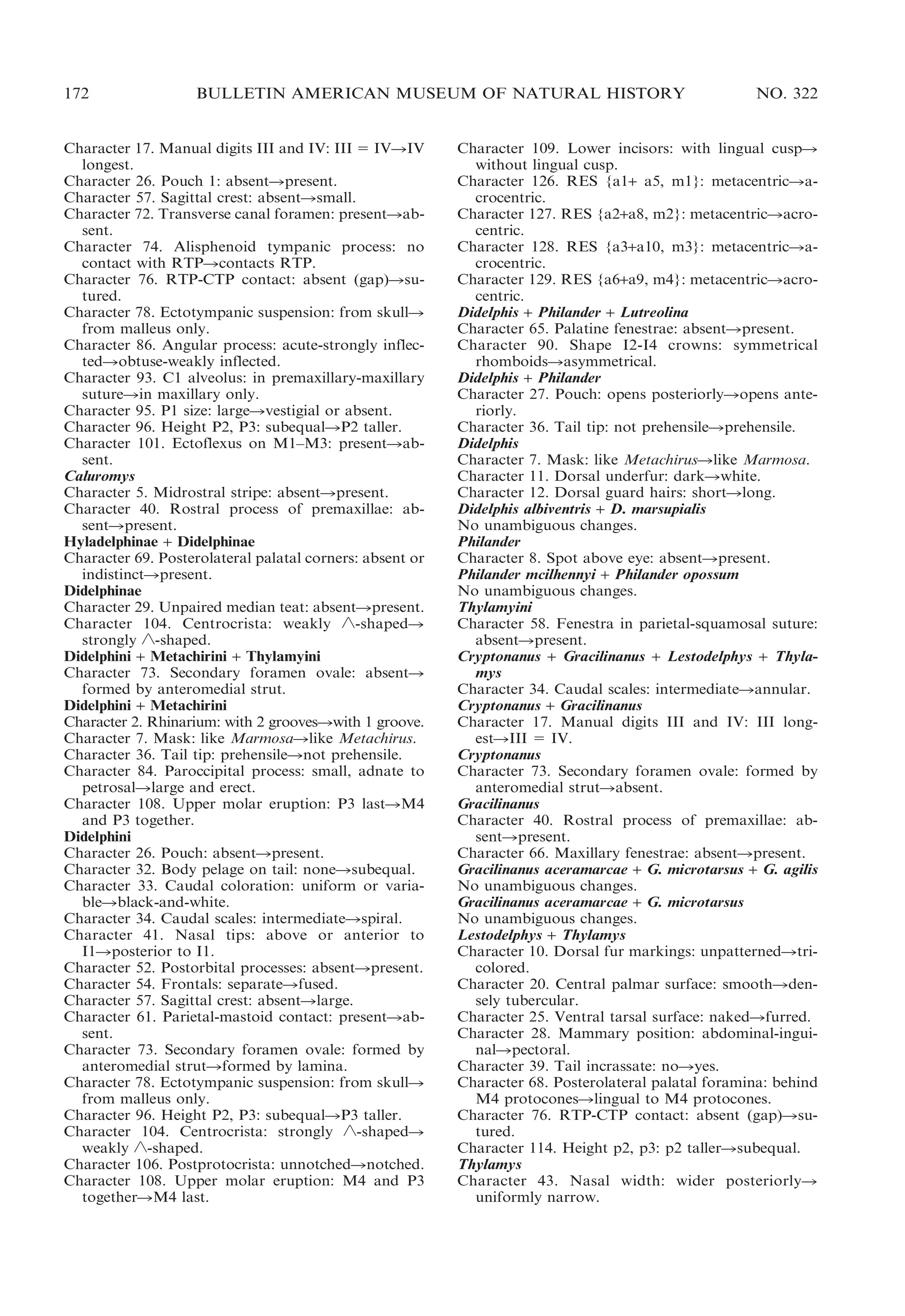
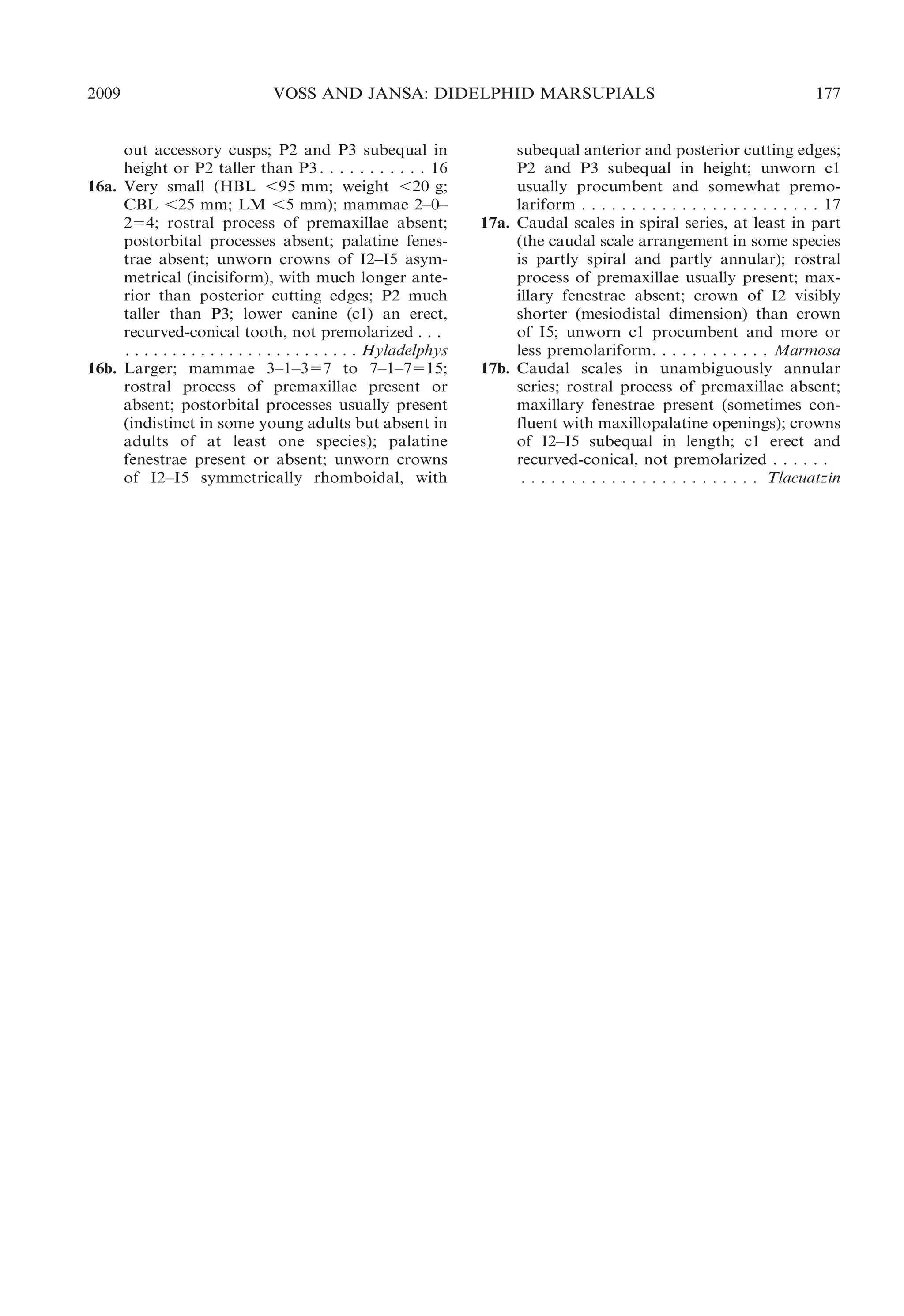

This document presents research on the phylogenetic relationships and classification of didelphid marsupials (opossums). The researchers analyzed morphological characteristics and sequences from 5 nuclear genes for over 40 opossum species. Separate analyses of the molecular and morphological data produced highly congruent estimates of opossum phylogeny. Analyses of combined data resolved most of the relationships with high statistical support. The researchers proposed a revised phylogenetic classification of opossums based on the results, with several new taxonomic subgroups defined.


