Vit d forms
•Download as PPTX, PDF•
2 likes•693 views

Vitamin D refers to a group of fat soluble secosteroids responsible for enhancing intestinal absorption of calcium, iron, magnesium, phosphate and zinc

Recommended
More Related Content
What's hot
What's hot (20)
Viewers also liked
Viewers also liked (7)
Similar to Vit d forms
Similar to Vit d forms (20)
Recently uploaded
Recently uploaded (20)
Vit d forms
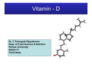
- 1. Vitamin - D Dr. T. Poongodi Vijayakumar Dept. of Food Science & Nutrition Periyar University Salem-11 Tamil Nadu
- 2. Introduction • Vitamin D refers to a group of fat- soluble secosteroids responsible for enhancing intestinal absorption of calcium, iron, magnesium, phosphate and zinc.
- 3. PRECURSOR D VITAMER Ergosterol D2 (Ergocalciferol) 7-Dehydrocholesterol D3 (cholecalciferol) 22,23-dehydroergosterol D4 7-Dehydrositosterol D5 7-Dehydrostigmasterol D6
- 4. CHEMISTRY • The chief structural prerequisite of compounds serving as D provitamins is the sterol structure which has been opened B ring that contains a D5,6 conjugated double bond. No vitamin activity is possessed by the compound until the B ring is opened. • This occurs as a result of exposure to ultraviolet light. In addition, vitamin activity is dependent on the presence of a hydroxyl group at carbon3 and upon the presence of conjugated double bonds at the 10-19, 5-6 & 7-8 positions. • If the location of these double bonds is shifted, vitamin activity is substantially reduced. A side chain of a length atleast equivalent to that of cholesterol is also a prequisite for vitamin activity. If the side chain is replaced by a hydroxyl group, for ex vitamin activity is lost. • The potency of the various D vitamins is determined by the side chain. • D5 for ex with its branched 10- carbon side chain, is much less active with respect to the calcification of bone cartilage than is D3 with its 9- membered sidechain. • Structurally to four - ring called compounds cyclopentanoperhydrophenanthrenes, from which they were derived by a photochemical reaction.
- 5. PHYSICAL CHARACTERISTICS • Under normal conditions, D3 is more stable than D2; however, both compounds undergo oxidation when exposed to air for period of 24 to 72 hrs. • In acid solutions, the D vitamins are unstable. • All the D vitamins are moderately soluble in fats, oils & ethanol, and very soluble in fat solvents such as chloroform, methanol and ether. • Other chemical alteration can result in decreased vitamin potency as well. Saturation of any of the double bonds or the substitution of a chloride, bromide, or mercaptan residue for the hydroxyl group attached to carbon 3 results in a loss of activity.
- 6. BIOPOTENCY • The comparative potency of the D vitamers depends on several factors : (1) the species consuming the vitamers ; and (2) the particular function assessed. • The activated forms of D3 (25- hydroxy and 1,25- dihydroxycholecalciferol) are far more potent than their parent vitamer D3. The synthetic analog of D3, 1α – hydroxycholecalciferol, likewise has 5 to 10 times the potency of cholecalciferol. • The analog 3- deoxy-1,25- dihydroxycholecalciferol is far more active as an agent to promote intestinal calcium uptake than as an agent to promote bone calcium mobilisation. This is also true for the analog, 25- hydroxy-5,6- cholecalciferol.
- 7. DIGESTION & ABSORPTION • Vitamin D3 (cholecalciferol) from the diet is absorbed from micelle, in association with fat and with the aid of bile salts, by passive diffusion into the intestinal cell. About 50% of dietary vitamin D3 is absorbed. Although the rate of absorption is most rapid in the duodenum, the largest amount of vitamin D is absorbed in the distal small intestine. • Absorption takes place primarily in the jejunum and ileum. The vitamin is absorbed in either the hydroxylated or the nonhydroxylated form. • Within the intestinal cell, vitamin D is incorporated primarily into chylomicrons (long chain fattyacids), which then enter the lymphatic system with subsequent entry into the blood. Chylomicrons transport about 40% of the cholecalciferol in the blood, although some vitamin D may be transferred from the chylomicron to DBP (Vitamin D Binding Protein) for delivery to extra hepatic tissues. Chylomicrons remnants deliver the vitamin to the liver. • Cholecalciferol reaching the liver either by way of chylomicrons remnants or by DBP typically is metabolized by a couple of different hydroxylases to generate the active form of the vitamin. • In the liver 25- hydroxylase functions in the mitochondria to hydroxylate cholecalciferol at carbon 25 to form 25-OH(vitamin)D3, also called calcidiol or 25-OH cholecalciferol.
- 8. • Following hydroxylation in the liver, 25-OH D3 bound to DBP is released into the blood and taken up by tissues, especially the kidney. Specifically, DBP- 25-OH D3 complex binds to megalin on the plasma membrane of the kidney and is transported into the renal cells. In the kidney, a second hydroxylation of 25-OH D3 occurs at position 1, resulting in 1,25- (OH)2 D3 (also called 1,25-dihydroxycholecalciferol or calcitriol) • Once synthesized, calcitriol is released from the kidney and bound to DBP for transport in the blood. DBP is one of the major proteins in the blood; the protein transports 1,25- (OH)2 D3 along with other metabolites to various target tissues. • Dietary phosphorus intake affects calcitriol production by 1-hydroxylase. A high intake of phosphorus causes a decrease in serum 1,25-(OH)2D3, whereas a low phosphorus intake stimulates its production.
- 9. EXCRETION • Calcitriol hydroxylation at carbon 24 generates the metabolite 1,24,25- (OH)2D3 which may be further oxidized to 1,25-(OH)2 24- oxo D3. Subsequent reactions including side chain clevage, yield calcitroic acid. • Other vitamin D metabolites are also formed after hydroxylation and oxidation. These other vitamin D metabolites may be conjugated and then excreted primarily in the bile. • Most vitamin D metabolites (more than 70%) are excreted in the feces, with lesser amount excreted in the urine.
- 10. PHYSIOLOGICAL FUNCTIONS • ABSORPTION OF CALCIUM FROM DIGESTED FOOD • REABSORPTION OF PHOSPHATE IN THE RENAL TUBULE • CALCIFICATION OF OSTEOBLAST CELLS OF GROWING SKELETAL STRUCTURES • CALCIUM HOMEOSTASIS • CALCITRIOL AND INTESTINE • CALCITRIOL AND KIDNEY • CALCITRIOL AND BONE • CELL DIFFERENTIATION , PROLIFERATION AND GROWTH