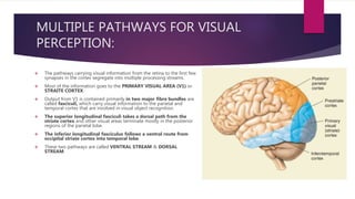
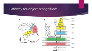
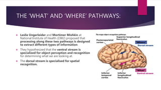
VISUAL COGNITION
ARIFA HUSSAIN
OBJECT RECOGNITION:
 The ability to accurately perceive and
recognise the objects is very crucial for visual
dependant organisms like humans.
 The visual system is able to extract a
reasonably constant representation of the
shapes despite their change in size or position.
 Object recognition is derived from a
perceptual ability to match an analysis of
shape and form of an object regardless of
colour, texture or motion cues.
Object recognition:
 Recognition may involve hierarchical representations in
which each successive stage adds complexity.
 Simple features such as lines can be combined into
edges, corners and intersections are grouped into parts
and parts are grouped into objects.
 One way to investigate how we encode shapes is to
identify areas of the brain that are active when we
compare contours that form a recognisable shape versus
contours that are just squiggles.
Visual processing can be sub divided into:
 Low level component
 Intermediate level component
 High level component
Low level component:
 In this stage brain extracts the information about the
physical properties of the stimulus such as edges,
brightness, colour and motion of the object present in
the scene.
 This stage is also known as bottom-up because it can
proceed from low(input)levels up through visual system
without any cognitive processes such as memory.
Intermediate level component:
 This stage involves combining information about
these physical properties in order to detect global
properties of an object, such as object shape and
orientation of the object in depth.
High level component:
 This stage include the stage that endow the
stimulus with meaning, leading to object
recognition and classification.
EXAMPLE:
Visual Agnosia:
 It is an impairment in recognition of visually
presented objects.
Earlier detection of Visual Agnosia:
 In 1881 Herman Munk noted that while lesions in occipital cortex caused
complete loss of vision, lesions in temporal cortex caused something completely
different.
 He noted in animals like monkeys and dogs lesion in temporal cortex caused them
to ignore food and water, even if they are hungry and thirsty. They were
completely fine with their vision acuity as they would avoid obstacles in the room
placed in front of them and navigate the rooms.
 Munk referred this as psychic blindness.
 Visual agnosia has since been demonstrated in humans who have suffered injury
in occipital and temporal lobes.
CASE OF G.S.
• The case of a patient G.S. is significantly important and famous in the history of
neurosciences.
• In his early 30s G.S. suffered from a stroke and nearly died from it, as he recovered, he
retrieved most of his cognitive abilities, but was not able to recognise objects.
• After performing several tests, it was observed that his language functioning was intact, it
was observed that there was no loss of visual acuity, recognition of colours or judgment
between two lines weren’t compromised either.
• For example: he was given a salad bowl and a candle for recognition, he wasn’t able to
recognise them, but was able to recall the shapes of them (he recalled candle as long and
thin and salad bowl as curved and brown).
• When asked to name a round, wooden object with lettuce in it, he recognised and
remembered the salad bowl. He was able to recognise the objects through touch, smell
and other senses, which confirmed that his memory and vision were intact.
Diagnosis:
 Associative agnosia can be diagnosed by following criteria:
 First, the patient must show difficulty recognising visually presented
objects, as measured by both naming and non verbal tests of
recognition, such as sorting objects by category. Example: grouping
kitchen utensils together, separate them from the sport equipment.
 Second, the patient must show knowledge about the object through
sensory modalities other than vision & and be able to recognise
objects by sound or touch or through verbal questioning.
Types of visual agnosia:
 APPERCEPTIVE AGNOSIA
 ASSOCIATIVE AGNOSIA
APPERCEPTIVE AGNOSIA:
 It affects the person at an early stage of life.
 Patients with such deficits are relatively rare. Usually they
have suffered with diffused brain damage through carbon
monoxide poisoning.
 Patients suffering from apperceptive agnosia have difficulty
with simple discrimination tasks, copying images, naming
and categorizing objects.
ASSOCIATIVE AGNOSIA:
 Patients suffering from associative agnosia can identify and
copy shapes easily, however their ability to associate any
meaning with the images is impaired.
 Associative agnosia often involves damage to the inferior
portion of the posterior cortex, ventral occipital cortex
or fusiform gyri in the posterior temporal lobe.
 Its diagnosed according to the following criteria:
 First the patient must show difficulty recognising visually
presented objects, as measured by both naming and non
verbal tests of recognition.
VISUAL PATHWAYS FOR
OBJECT RECOGNITION
PROCESSING IN
NON-HUMAN PRIMATES
MULTIPLE PATHWAYS FOR VISUAL
PERCEPTION:
 The pathways carrying visual information from the retina to the first few
synapses in the cortex segregate into multiple processing streams.
 Most of the information goes to the PRIMARY VISUAL AREA (V1) or
STRAITE CORTEX.
 Output from V1 is contained primarily in two major fibre bundles are
called fasciculi, which carry visual information to the parietal and
temporal cortex that are involved in visual object recognition.
 The superior longitudinal fasciculi takes a dorsal path from the
striate cortex and other visual areas terminate mostly in the posterior
regions of the parietal lobe.
 The inferior longitudinal fasciculus follows a ventral route from
occipital striate cortex into temporal lobe.
 These two pathways are called VENTRAL STREAM & DORSAL
STREAM.
Pathway for object recognition:
THE ‘WHAT’ AND ‘WHERE’ PATHWAYS:
 Leslie Ungerleider and Mortimer Mishkin at
National Institute of Health (1982) proposed that
processing along these two pathways is designed
to extract different types of information.
 They hypothesized that the ventral stream is
specialized for object perception and recognition
for determining what we are looking at.
 The dorsal stream is specialised for spatial
recognition.
CONTINUED…
 Initially the what and where pathways of ventral and dorsal streams came from
lesions in monkeys.
 Animals with bilateral lesions in temporal lobe that disrupted the ventral
system had great difficulty discriminating between different shapes.
 For example: they made errors while discriminating from one object to another.
However these same animals had no problem discriminating where an object was
in relation to other objects.
 Opposite was true for the animals with lesions in parietal lobe.
OBJECT RECOGNITION PATHWAY:
 The object recognition in both monkeys
and humans consist of interconnected
set of cortical areas.
 The pathways lie adjacent to the primary
visual cortex (V1) in the occipital region.
 The cortical analysis of object begins at
V1, where information like contour,
orientation, colour composition and
brightness is processed.
 The information then projects forward to
the interdigitating thin, thick and
interstriped region in V2.
 After the V2 region the neural signals
proceeds from V2 to V4 to the lateral
and ventromedial hemispheres.
 From V4 the signals transfer to posterior
temporal region just in front of V4
called TEO.
 From both V4 and TEO signals related to
object form, colour and texture transfers
to TE region.
 Together TEO and TE forms IT cortex.
CONTINUED…
 Recent evidences have shown that the separation of
what and where pathways are not limited to the visual
system.
 Studies with various species including humans have shown
that auditory processing is similarly divided into dorsal
and ventral streams.
 The anterior part of auditory cortex are specialised for
auditory pattern processing (what is the sound?) belongs
to the ventral pathway and posterior region is specialised
for identifying the spatial location of sounds belongs to
the dorsal pathway.
REPRESENTATIONAL DIFFERENCES BETWEEN
DORSAL AND VENTRAL STREAMS:
 Neurons in both parietal and dorsal streams have large receptive fields (the receptive field of
an individual sensory neuron is the particular region of the sensory space (e.g., the body surface,
or the visual field) in which a stimulus will trigger the firing of that neuron), but the physiological
properties of the neurons within each lobe are quite different.
 Although 40% of these neurons have receptive fields near the central regions of vision
(fovea) the remaining cells have receptive fields that exclude the foveal regions.
 These cells are ideally suited for detecting the presence and location of the stimulus,
especially one that has entered the receptive field of view.
 The response of neurons in the ventral stream of the temporal lobe is quite different.
 The receptive field of these neurons always encompasses the fovea, and most of these
neurons can be activated by the stimulus that falls within either the left or the right visual field.
Thus we make use of the greater acuity of the foveal vision by looking directly at things we want
to identify.
NEURONAL PROPERTIES IN OBJECT
RECOGNITION PATHWAY
 Different cortical areas in occipitotemporal pathway share a number of physiological characteristics.
 All areas in the occipitotemporal pathway contains neurons sensitive to shape, colour, texture etc.
 however at higher order properties are usually attributed to neurons in higher tier. For example many V1 neurons
act as a spatial filter, and respond to the contours of the object based on light and dark contrast, but the neurons
in higher tier respond to illusory contours.
 Another of these properties is the increasingly strong silent suppressive zones surrounding the classical
receptive field of the neurons. Silent zones are basically neurons that do not get stimulated by visual input if it
doesnot cause any change from the baseline neuronal activity.
 For example: many V4 neurons respond to maximal stimulus only if it stands out from its background.
 When we view an object such as a dog, whether it’s a real dog, a drawing of a dog, a statue of a dog, or an outline
of a dog made of flashing lights, we recognize it as a dog. This insensitivity to the specific visual cues that define
an object is known as cue invariance.
 .
 At the higher level, it became difficult to know the optimal stimulus of a given single cell, by stimulating a group
of them by a visual stimulus. Later in 1960s it was believed that every object would be coded by maximal firing of
a single neuron in IT cortex, it was named as grandmother neuron hypothesis. as it was believed that each
grandmother was supposed to caused maximal firing, and if that neuron was destroyed we would be unable to
recognise our grandmother.
 Later this theory was dismissed because there weren’t as much neurons present as there are object in the
world. Instead the idea of population code was introduces.
 A POPULATION CODE is in which each object corresponds to a unique set of neuron firing that share
connections and overlapping functional selectivity
 Tanaka (1996) has suggested that IT neurons that respond to common visual features are grouped together
in cortical columns that run perpendicular to the cortical surface.
 New evidences from fMRI has proved the same.
 Biederman (1987) postulates that objects can be defined as 30 or so primitive shapes called Geon.
GEONS:
 Geons are 3D building blocks that when combined can represent any arbitrary
object.
INVARIANT REPRESENTATION IN THE
OCCIPITOTEMPORAL PATHWAY:
 The function of occipitotemporal pathway is to determine the invariant features of the
object that are important for object recognition.
 Recent experiment of Li and DiCarlo (2008,2010) has shown how stimulus invariance
might be achieved through an associative learning process based on repeated
exposures to the same stimulus under viewing conditions that change in a
predictable way.
 Under normal conditions viewing, movement of object will result in a rapid shift in the
location of the object on the retina and yet to identity of the object has not changed.
 Through repeated exposures, the brain can theoretically learn to associate these
different positions, thus generating a position invariant representation.
Continued…
 They predicted that if such an associative learning process were responsible for
generating invariant representations, this manipulation should result in these two
stimuli being confused as the same object.
 Initially, the neurons responded to the first object only and not the second.
 However, overtime it responded to both, suggesting that the neuron perceived
the two stimuli as the same.
 The ability of downstream neurons to discriminate between the two stimuli has
been abolished.
 The experiments showed the same effect for object size, suggesting that a similar
mechanism might be responsible for generating size invariance.
FACE SELECTIVE NEURONS IN IT CORTEX:
 One of the characteristics monkey temporal cortex is the presence of neurons with
responses selective for face stimuli, discovered by Gross, Bender and Rocha
Miranda in 1969.
 These neurons have received considerable attention, not only for their role in
social communication but also for their theories concerning object recognition.
 Early studies reported that face selective neurons could be found throughout the
temporal cortex. But latest studies that included the fMRI, have shown that in non
human primates that face selective neurons are concentrated in small patches
located along the lower bank of the superior temporal sulcus and extending
onto the adjacent IT gyrus.
CONTINUED…
 It is currently hypothesised that these patches
maybe homologous to the face selective neurons
in human cortex.
 Some face selective neurons respond well to
both real faces and face pictures, but give little
or no response to any other stimuli tested,
including other complex objects, texture patterns
and images in which the features making up the
face are rearranged and scrambled.
CONTINUED…
 Other neurons respond to specific face components, such as presence of eyes per se,
distance between the eyes or extent of the forehead, direction of gaze of the
eyes, which is important social signal for both monkeys and humans.
 Other neurons are sensitive to different face expression (open mouth threat or
fearful faces).
 These observations have led to the proposal that primary temporal lobe has evolved
specialized mechanisms for the encoding and recognition of biologically
significant stimuli, especially face.
 Some examples suggests that face selectivity maybe innate, but experience likely
improve such selectivity as face recognition improves throughout development.
RESPONSE PROPERTIES OF NEURONS IN
THE OBJECT RECOGNITION PATHWAY
ARE AFFECTED BY EXPERIENCE:
 Experiments by Miller and Desimone (1994) have shown how short-term changes
in response properties of neurons in temporal cortex.
 Repeated exposure to stimuli results in weaker brain responses. Such reduced
responses gives the redirection of attention towards novel objects. This is called
repetition suppression.
FUNCTIONAL NEUROIMAGING OF
OBJECT RECOGNITION:
 Various visual impairments produced by focal lesions in clinical cases suggest that
the human visual cortex like that of a monkeys, has parietal and temporal streams.
 The specific clinical syndromes produced by occipitotemporal lesions can
include visual object agnosia, prosopagnosia (inability to recognise familiar
faces) and achromatopsia or cortical colour blindness.
 Syndromes produced by occipitoparietal lesions can include optic ataxia (mis
reaching), visuospatial neglect, constructional apraxia, gaze apraxia,
akinetopsia (inability to perceive movement) and disorders of spatial
cognition.
CONTINUED…
 Development of fMRI and PET has made it possible to map the organisation of
the human visual cortex with far greater precision.
 fMRI has revealed a number of distinct visual areas within the ventral and dorsal
streams in humans. Many of these areas appear to be equivalent (perhaps
homologous) to specific monkey visual areas, including V1, V2, V3, V3A, V4 and the
middle temporal region.
 Receptive fields size increases and stimulus requirements become more stringent as
one progresses from V1 through various stages of ventral stream.
 Regions responding more to objects than to scrambled stimuli were interpreted
as being involved in object related processing, collectively these regions were
named as LOC (lateral occipital complex) because of their anatomical locations.
PERCEPTION AND RECOGNITION OF
SPECIFIC CLASSES OF OBJECTS:
 PROSOPAGNOSIA:
 It is a selective deficit in recognising familiar faces, this has been known since
the end of nineteenth century.
 Patients with this syndrome are aware that faces are faces but they fail to achieve
a sense of familiarity.
 These patients have trouble forming memories of new faces even if the other
new objects can be learned.
 Because the voices of the visually unrecognised person usually enables them to
identify and feel familiar with that person, prosopagnosia appears to be
specifically visual impairment.
CONTINUED…
 The damage common to a number of cases lay within the lingual and fusiform gyri, ventrally and medially
within the cortex at the occipitotemporal junction.
 Developmental prosopagnosia is characterized by severely impaired face recognition with no detectable brain
damage. Prosopagnosia is usually observed in patients who have lesions in the ventral pathway, especially
occipital regions associated with face perception and the fusiform face area. In many cases, the lesions are
bilateral, resulting from the unfortunate occurrence of two strokes affecting the territory of the posterior cerebral
artery. Or the bilateral damage might be from encephalitis or carbon monoxide poisoning. Prosopagnosia can
also occur after unilateral lesions; in these cases, the damage is usually in the right hemisphere.
 Early PET and fMRI studies revealed a region on the fusiform gyrus of the right hemisphere that is more
active when subjects view faces, compared to when they view common nonface objects, this area was called
‘fusiform face area’ by Kanwisher, McDermott and Chun (1997).
 To give a sense of how bizarre prosopagnosia can be, one widely cited case involved a patient with bilateral
occipital lesions who not only was prosopagnosic for friends and family, but also failed to recognize an even more
familiar person—himself. As he reported, “At the club I saw someone strange staring at me, and asked the steward
who it was. You’ll laugh at me. I’d bee n looking at myself in the mirror”.
CONTINUED…
 FFA is more active during face matching then location matching and during the
viewing of faces than during viewing of scrambled faces, letters and textures.
 Other face selective regions have since been identified in ventral occipital cortex
(the occipital face area OFA), the superior temporal sulcus and the anterior portion
of the temporal lobe.
 Damage to one or more of these areas can lead to deficits in face processing
including prosopagnosia.
ELECTROPHYSIOLOGICAL APPROACHES:
 Evidence from electrophysiological recordings in patients with implanted
electrodes.
 A large evoked potential called N200 (because it occurs roughly 200ms after
stimulus onset) is generated in small regions in the ventral occipitotemporal
cortex.
 The N200 amplitude is largest in response to full faces and progressively
decreases to eyes, face contours, lips and noses viewed in isolation.
 A region just lateral to face selective N200 sites is more responsive to internal face
parts than to faces and some sites in ventral occipitotemporal cortex appear to be
face part specific.
CONTINUED…
 Other electrophysiological approaches have been applied to the study of face
processing in humans.
 Liu, Harris and Kanwisher (2002) used MEG to associate two separate two separate
evoked potentials (M100, M170) in the occipitotemporal cortex to different stages
of face processing: face detection and face identification.
VENTRAL PATHWAYS CONTAINS
REGIONS SPECIALIZED FOR OTHER
VISUAL CATAGORIES:
 Subsequent neuroimaging studies have identified regions selective for other visual
categories:
 Medial to the fusiform gyrus is an area within parahippocampal cortex that
is significantly more active when subjects view complex scenes (such as
rooms, landscapes, landmarks and city streets) compared when they view objects,
faces or other kinds of visual stimuli. This region is named as parahippocampal
place areas (PPA).
 And damage to lingual/parahippocampal region may results in landmark
agnosia.
CONTINUED…
 Along the lateral occipitotemporal cortex (near the middle occipital gyrus) and
area selectively activated by images of human body parts (such as isolated foot or
arm) relative to viewing objects has been identified.
 This region is called extrastraite body area (EBA).
 An additional body part selective region is identified in the fusiform gyrus
(Fusiform body area)
 Alexia (unable to read despite intact visual capabilities) is especially a visual
agnosia for verbal materials.
BINOCULAR RIVALRY:
 Binocular rivalry refers to the presentation of mutually incongruent images to the
left and the right eye, which typically results in the perceptual alternation of the two
stimuli instead of their perceptual fusion.
 People with normal binocular vision usually experience a single stable visual world
even though objects throughout most of the visual field produce two retinal images,
one in each eye. Evidently the brain manages to blend, or fuse, the two images and to
do so in a manner that precisely specifies the three-dimensional relationships among
those objects. The processes responsible for such binocular single vision and
stereopsis, however, are disrupted when dissimilar monocular stimuli are imaged
on corresponding retinal locations. Under these conditions, the eyes transmit
contradictory information to the brain, specifying the presence of two different
objects situated at the same spatial location at the same time. Faced with this physical
impossibility, the brain lapses into an unstable state characterized by alternating
periods of perceptual dominance which continue as long as the eyes view discordant
stimuli; this is the phenomenon termed binocular rivalry.
CONTINUED…
ALTERNATIVE PROPOSALS TO CATEGORY
SPECIFIC REPRESENTATION:
 It has been proposed that FFA is specialized not for the processing of face per se
but rather for processing of objects with which one has acquired expertise.
 The idea that expertise underlies responses in the ventral temporal cortex raises
an important distinction between two interpretations:
 One interpretation suggests that activation is based on visual features.
 Another interpretation suggests that processes associated with perceiving or
recognising objects. This view the ventral stream contains areas that are best
suited for different computation. This might involve encoding subtle for objects
that are visually similar or encoding similarities between the object that are
visually dissimilar.
THANKYOU!

VISUAL COGNITION (ARIFA HUSSAIN).pptx

  • 1.
  • 2.
    OBJECT RECOGNITION:  Theability to accurately perceive and recognise the objects is very crucial for visual dependant organisms like humans.  The visual system is able to extract a reasonably constant representation of the shapes despite their change in size or position.  Object recognition is derived from a perceptual ability to match an analysis of shape and form of an object regardless of colour, texture or motion cues.
  • 3.
    Object recognition:  Recognitionmay involve hierarchical representations in which each successive stage adds complexity.  Simple features such as lines can be combined into edges, corners and intersections are grouped into parts and parts are grouped into objects.  One way to investigate how we encode shapes is to identify areas of the brain that are active when we compare contours that form a recognisable shape versus contours that are just squiggles.
  • 4.
    Visual processing canbe sub divided into:  Low level component  Intermediate level component  High level component
  • 5.
    Low level component: In this stage brain extracts the information about the physical properties of the stimulus such as edges, brightness, colour and motion of the object present in the scene.  This stage is also known as bottom-up because it can proceed from low(input)levels up through visual system without any cognitive processes such as memory.
  • 6.
    Intermediate level component: This stage involves combining information about these physical properties in order to detect global properties of an object, such as object shape and orientation of the object in depth.
  • 7.
    High level component: This stage include the stage that endow the stimulus with meaning, leading to object recognition and classification.
  • 8.
  • 9.
    Visual Agnosia:  Itis an impairment in recognition of visually presented objects.
  • 10.
    Earlier detection ofVisual Agnosia:  In 1881 Herman Munk noted that while lesions in occipital cortex caused complete loss of vision, lesions in temporal cortex caused something completely different.  He noted in animals like monkeys and dogs lesion in temporal cortex caused them to ignore food and water, even if they are hungry and thirsty. They were completely fine with their vision acuity as they would avoid obstacles in the room placed in front of them and navigate the rooms.  Munk referred this as psychic blindness.  Visual agnosia has since been demonstrated in humans who have suffered injury in occipital and temporal lobes.
  • 11.
    CASE OF G.S. •The case of a patient G.S. is significantly important and famous in the history of neurosciences. • In his early 30s G.S. suffered from a stroke and nearly died from it, as he recovered, he retrieved most of his cognitive abilities, but was not able to recognise objects. • After performing several tests, it was observed that his language functioning was intact, it was observed that there was no loss of visual acuity, recognition of colours or judgment between two lines weren’t compromised either. • For example: he was given a salad bowl and a candle for recognition, he wasn’t able to recognise them, but was able to recall the shapes of them (he recalled candle as long and thin and salad bowl as curved and brown). • When asked to name a round, wooden object with lettuce in it, he recognised and remembered the salad bowl. He was able to recognise the objects through touch, smell and other senses, which confirmed that his memory and vision were intact.
  • 12.
    Diagnosis:  Associative agnosiacan be diagnosed by following criteria:  First, the patient must show difficulty recognising visually presented objects, as measured by both naming and non verbal tests of recognition, such as sorting objects by category. Example: grouping kitchen utensils together, separate them from the sport equipment.  Second, the patient must show knowledge about the object through sensory modalities other than vision & and be able to recognise objects by sound or touch or through verbal questioning.
  • 13.
    Types of visualagnosia:  APPERCEPTIVE AGNOSIA  ASSOCIATIVE AGNOSIA
  • 14.
    APPERCEPTIVE AGNOSIA:  Itaffects the person at an early stage of life.  Patients with such deficits are relatively rare. Usually they have suffered with diffused brain damage through carbon monoxide poisoning.  Patients suffering from apperceptive agnosia have difficulty with simple discrimination tasks, copying images, naming and categorizing objects.
  • 15.
    ASSOCIATIVE AGNOSIA:  Patientssuffering from associative agnosia can identify and copy shapes easily, however their ability to associate any meaning with the images is impaired.  Associative agnosia often involves damage to the inferior portion of the posterior cortex, ventral occipital cortex or fusiform gyri in the posterior temporal lobe.  Its diagnosed according to the following criteria:  First the patient must show difficulty recognising visually presented objects, as measured by both naming and non verbal tests of recognition.
  • 17.
    VISUAL PATHWAYS FOR OBJECTRECOGNITION PROCESSING IN NON-HUMAN PRIMATES
  • 18.
    MULTIPLE PATHWAYS FORVISUAL PERCEPTION:  The pathways carrying visual information from the retina to the first few synapses in the cortex segregate into multiple processing streams.  Most of the information goes to the PRIMARY VISUAL AREA (V1) or STRAITE CORTEX.  Output from V1 is contained primarily in two major fibre bundles are called fasciculi, which carry visual information to the parietal and temporal cortex that are involved in visual object recognition.  The superior longitudinal fasciculi takes a dorsal path from the striate cortex and other visual areas terminate mostly in the posterior regions of the parietal lobe.  The inferior longitudinal fasciculus follows a ventral route from occipital striate cortex into temporal lobe.  These two pathways are called VENTRAL STREAM & DORSAL STREAM.
  • 19.
    Pathway for objectrecognition:
  • 20.
    THE ‘WHAT’ AND‘WHERE’ PATHWAYS:  Leslie Ungerleider and Mortimer Mishkin at National Institute of Health (1982) proposed that processing along these two pathways is designed to extract different types of information.  They hypothesized that the ventral stream is specialized for object perception and recognition for determining what we are looking at.  The dorsal stream is specialised for spatial recognition.
  • 21.
    CONTINUED…  Initially thewhat and where pathways of ventral and dorsal streams came from lesions in monkeys.  Animals with bilateral lesions in temporal lobe that disrupted the ventral system had great difficulty discriminating between different shapes.  For example: they made errors while discriminating from one object to another. However these same animals had no problem discriminating where an object was in relation to other objects.  Opposite was true for the animals with lesions in parietal lobe.
  • 22.
    OBJECT RECOGNITION PATHWAY: The object recognition in both monkeys and humans consist of interconnected set of cortical areas.  The pathways lie adjacent to the primary visual cortex (V1) in the occipital region.  The cortical analysis of object begins at V1, where information like contour, orientation, colour composition and brightness is processed.  The information then projects forward to the interdigitating thin, thick and interstriped region in V2.  After the V2 region the neural signals proceeds from V2 to V4 to the lateral and ventromedial hemispheres.  From V4 the signals transfer to posterior temporal region just in front of V4 called TEO.  From both V4 and TEO signals related to object form, colour and texture transfers to TE region.  Together TEO and TE forms IT cortex.
  • 23.
    CONTINUED…  Recent evidenceshave shown that the separation of what and where pathways are not limited to the visual system.  Studies with various species including humans have shown that auditory processing is similarly divided into dorsal and ventral streams.  The anterior part of auditory cortex are specialised for auditory pattern processing (what is the sound?) belongs to the ventral pathway and posterior region is specialised for identifying the spatial location of sounds belongs to the dorsal pathway.
  • 24.
    REPRESENTATIONAL DIFFERENCES BETWEEN DORSALAND VENTRAL STREAMS:  Neurons in both parietal and dorsal streams have large receptive fields (the receptive field of an individual sensory neuron is the particular region of the sensory space (e.g., the body surface, or the visual field) in which a stimulus will trigger the firing of that neuron), but the physiological properties of the neurons within each lobe are quite different.  Although 40% of these neurons have receptive fields near the central regions of vision (fovea) the remaining cells have receptive fields that exclude the foveal regions.  These cells are ideally suited for detecting the presence and location of the stimulus, especially one that has entered the receptive field of view.  The response of neurons in the ventral stream of the temporal lobe is quite different.  The receptive field of these neurons always encompasses the fovea, and most of these neurons can be activated by the stimulus that falls within either the left or the right visual field. Thus we make use of the greater acuity of the foveal vision by looking directly at things we want to identify.
  • 25.
    NEURONAL PROPERTIES INOBJECT RECOGNITION PATHWAY
  • 26.
     Different corticalareas in occipitotemporal pathway share a number of physiological characteristics.  All areas in the occipitotemporal pathway contains neurons sensitive to shape, colour, texture etc.  however at higher order properties are usually attributed to neurons in higher tier. For example many V1 neurons act as a spatial filter, and respond to the contours of the object based on light and dark contrast, but the neurons in higher tier respond to illusory contours.  Another of these properties is the increasingly strong silent suppressive zones surrounding the classical receptive field of the neurons. Silent zones are basically neurons that do not get stimulated by visual input if it doesnot cause any change from the baseline neuronal activity.  For example: many V4 neurons respond to maximal stimulus only if it stands out from its background.  When we view an object such as a dog, whether it’s a real dog, a drawing of a dog, a statue of a dog, or an outline of a dog made of flashing lights, we recognize it as a dog. This insensitivity to the specific visual cues that define an object is known as cue invariance.  .
  • 27.
     At thehigher level, it became difficult to know the optimal stimulus of a given single cell, by stimulating a group of them by a visual stimulus. Later in 1960s it was believed that every object would be coded by maximal firing of a single neuron in IT cortex, it was named as grandmother neuron hypothesis. as it was believed that each grandmother was supposed to caused maximal firing, and if that neuron was destroyed we would be unable to recognise our grandmother.  Later this theory was dismissed because there weren’t as much neurons present as there are object in the world. Instead the idea of population code was introduces.  A POPULATION CODE is in which each object corresponds to a unique set of neuron firing that share connections and overlapping functional selectivity  Tanaka (1996) has suggested that IT neurons that respond to common visual features are grouped together in cortical columns that run perpendicular to the cortical surface.  New evidences from fMRI has proved the same.  Biederman (1987) postulates that objects can be defined as 30 or so primitive shapes called Geon.
  • 28.
    GEONS:  Geons are3D building blocks that when combined can represent any arbitrary object.
  • 29.
    INVARIANT REPRESENTATION INTHE OCCIPITOTEMPORAL PATHWAY:  The function of occipitotemporal pathway is to determine the invariant features of the object that are important for object recognition.  Recent experiment of Li and DiCarlo (2008,2010) has shown how stimulus invariance might be achieved through an associative learning process based on repeated exposures to the same stimulus under viewing conditions that change in a predictable way.  Under normal conditions viewing, movement of object will result in a rapid shift in the location of the object on the retina and yet to identity of the object has not changed.  Through repeated exposures, the brain can theoretically learn to associate these different positions, thus generating a position invariant representation.
  • 30.
    Continued…  They predictedthat if such an associative learning process were responsible for generating invariant representations, this manipulation should result in these two stimuli being confused as the same object.  Initially, the neurons responded to the first object only and not the second.  However, overtime it responded to both, suggesting that the neuron perceived the two stimuli as the same.  The ability of downstream neurons to discriminate between the two stimuli has been abolished.  The experiments showed the same effect for object size, suggesting that a similar mechanism might be responsible for generating size invariance.
  • 31.
    FACE SELECTIVE NEURONSIN IT CORTEX:  One of the characteristics monkey temporal cortex is the presence of neurons with responses selective for face stimuli, discovered by Gross, Bender and Rocha Miranda in 1969.  These neurons have received considerable attention, not only for their role in social communication but also for their theories concerning object recognition.  Early studies reported that face selective neurons could be found throughout the temporal cortex. But latest studies that included the fMRI, have shown that in non human primates that face selective neurons are concentrated in small patches located along the lower bank of the superior temporal sulcus and extending onto the adjacent IT gyrus.
  • 32.
    CONTINUED…  It iscurrently hypothesised that these patches maybe homologous to the face selective neurons in human cortex.  Some face selective neurons respond well to both real faces and face pictures, but give little or no response to any other stimuli tested, including other complex objects, texture patterns and images in which the features making up the face are rearranged and scrambled.
  • 33.
    CONTINUED…  Other neuronsrespond to specific face components, such as presence of eyes per se, distance between the eyes or extent of the forehead, direction of gaze of the eyes, which is important social signal for both monkeys and humans.  Other neurons are sensitive to different face expression (open mouth threat or fearful faces).  These observations have led to the proposal that primary temporal lobe has evolved specialized mechanisms for the encoding and recognition of biologically significant stimuli, especially face.  Some examples suggests that face selectivity maybe innate, but experience likely improve such selectivity as face recognition improves throughout development.
  • 34.
    RESPONSE PROPERTIES OFNEURONS IN THE OBJECT RECOGNITION PATHWAY ARE AFFECTED BY EXPERIENCE:  Experiments by Miller and Desimone (1994) have shown how short-term changes in response properties of neurons in temporal cortex.  Repeated exposure to stimuli results in weaker brain responses. Such reduced responses gives the redirection of attention towards novel objects. This is called repetition suppression.
  • 35.
    FUNCTIONAL NEUROIMAGING OF OBJECTRECOGNITION:  Various visual impairments produced by focal lesions in clinical cases suggest that the human visual cortex like that of a monkeys, has parietal and temporal streams.  The specific clinical syndromes produced by occipitotemporal lesions can include visual object agnosia, prosopagnosia (inability to recognise familiar faces) and achromatopsia or cortical colour blindness.  Syndromes produced by occipitoparietal lesions can include optic ataxia (mis reaching), visuospatial neglect, constructional apraxia, gaze apraxia, akinetopsia (inability to perceive movement) and disorders of spatial cognition.
  • 36.
    CONTINUED…  Development offMRI and PET has made it possible to map the organisation of the human visual cortex with far greater precision.  fMRI has revealed a number of distinct visual areas within the ventral and dorsal streams in humans. Many of these areas appear to be equivalent (perhaps homologous) to specific monkey visual areas, including V1, V2, V3, V3A, V4 and the middle temporal region.  Receptive fields size increases and stimulus requirements become more stringent as one progresses from V1 through various stages of ventral stream.  Regions responding more to objects than to scrambled stimuli were interpreted as being involved in object related processing, collectively these regions were named as LOC (lateral occipital complex) because of their anatomical locations.
  • 37.
    PERCEPTION AND RECOGNITIONOF SPECIFIC CLASSES OF OBJECTS:  PROSOPAGNOSIA:  It is a selective deficit in recognising familiar faces, this has been known since the end of nineteenth century.  Patients with this syndrome are aware that faces are faces but they fail to achieve a sense of familiarity.  These patients have trouble forming memories of new faces even if the other new objects can be learned.  Because the voices of the visually unrecognised person usually enables them to identify and feel familiar with that person, prosopagnosia appears to be specifically visual impairment.
  • 38.
    CONTINUED…  The damagecommon to a number of cases lay within the lingual and fusiform gyri, ventrally and medially within the cortex at the occipitotemporal junction.  Developmental prosopagnosia is characterized by severely impaired face recognition with no detectable brain damage. Prosopagnosia is usually observed in patients who have lesions in the ventral pathway, especially occipital regions associated with face perception and the fusiform face area. In many cases, the lesions are bilateral, resulting from the unfortunate occurrence of two strokes affecting the territory of the posterior cerebral artery. Or the bilateral damage might be from encephalitis or carbon monoxide poisoning. Prosopagnosia can also occur after unilateral lesions; in these cases, the damage is usually in the right hemisphere.  Early PET and fMRI studies revealed a region on the fusiform gyrus of the right hemisphere that is more active when subjects view faces, compared to when they view common nonface objects, this area was called ‘fusiform face area’ by Kanwisher, McDermott and Chun (1997).  To give a sense of how bizarre prosopagnosia can be, one widely cited case involved a patient with bilateral occipital lesions who not only was prosopagnosic for friends and family, but also failed to recognize an even more familiar person—himself. As he reported, “At the club I saw someone strange staring at me, and asked the steward who it was. You’ll laugh at me. I’d bee n looking at myself in the mirror”.
  • 39.
    CONTINUED…  FFA ismore active during face matching then location matching and during the viewing of faces than during viewing of scrambled faces, letters and textures.  Other face selective regions have since been identified in ventral occipital cortex (the occipital face area OFA), the superior temporal sulcus and the anterior portion of the temporal lobe.  Damage to one or more of these areas can lead to deficits in face processing including prosopagnosia.
  • 40.
    ELECTROPHYSIOLOGICAL APPROACHES:  Evidencefrom electrophysiological recordings in patients with implanted electrodes.  A large evoked potential called N200 (because it occurs roughly 200ms after stimulus onset) is generated in small regions in the ventral occipitotemporal cortex.  The N200 amplitude is largest in response to full faces and progressively decreases to eyes, face contours, lips and noses viewed in isolation.  A region just lateral to face selective N200 sites is more responsive to internal face parts than to faces and some sites in ventral occipitotemporal cortex appear to be face part specific.
  • 41.
    CONTINUED…  Other electrophysiologicalapproaches have been applied to the study of face processing in humans.  Liu, Harris and Kanwisher (2002) used MEG to associate two separate two separate evoked potentials (M100, M170) in the occipitotemporal cortex to different stages of face processing: face detection and face identification.
  • 42.
    VENTRAL PATHWAYS CONTAINS REGIONSSPECIALIZED FOR OTHER VISUAL CATAGORIES:  Subsequent neuroimaging studies have identified regions selective for other visual categories:  Medial to the fusiform gyrus is an area within parahippocampal cortex that is significantly more active when subjects view complex scenes (such as rooms, landscapes, landmarks and city streets) compared when they view objects, faces or other kinds of visual stimuli. This region is named as parahippocampal place areas (PPA).  And damage to lingual/parahippocampal region may results in landmark agnosia.
  • 43.
    CONTINUED…  Along thelateral occipitotemporal cortex (near the middle occipital gyrus) and area selectively activated by images of human body parts (such as isolated foot or arm) relative to viewing objects has been identified.  This region is called extrastraite body area (EBA).  An additional body part selective region is identified in the fusiform gyrus (Fusiform body area)  Alexia (unable to read despite intact visual capabilities) is especially a visual agnosia for verbal materials.
  • 44.
    BINOCULAR RIVALRY:  Binocularrivalry refers to the presentation of mutually incongruent images to the left and the right eye, which typically results in the perceptual alternation of the two stimuli instead of their perceptual fusion.  People with normal binocular vision usually experience a single stable visual world even though objects throughout most of the visual field produce two retinal images, one in each eye. Evidently the brain manages to blend, or fuse, the two images and to do so in a manner that precisely specifies the three-dimensional relationships among those objects. The processes responsible for such binocular single vision and stereopsis, however, are disrupted when dissimilar monocular stimuli are imaged on corresponding retinal locations. Under these conditions, the eyes transmit contradictory information to the brain, specifying the presence of two different objects situated at the same spatial location at the same time. Faced with this physical impossibility, the brain lapses into an unstable state characterized by alternating periods of perceptual dominance which continue as long as the eyes view discordant stimuli; this is the phenomenon termed binocular rivalry.
  • 45.
  • 46.
    ALTERNATIVE PROPOSALS TOCATEGORY SPECIFIC REPRESENTATION:  It has been proposed that FFA is specialized not for the processing of face per se but rather for processing of objects with which one has acquired expertise.  The idea that expertise underlies responses in the ventral temporal cortex raises an important distinction between two interpretations:  One interpretation suggests that activation is based on visual features.  Another interpretation suggests that processes associated with perceiving or recognising objects. This view the ventral stream contains areas that are best suited for different computation. This might involve encoding subtle for objects that are visually similar or encoding similarities between the object that are visually dissimilar.
  • 47.