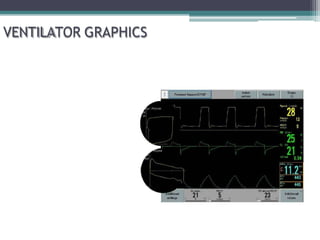
2. Purpose
• Graphics are waveforms that reflect the patient-
ventilator system and their interaction.
• Purposes of monitoring graphics:
• Allow users to interpret, evaluate, and troubleshoot
the ventilator and the patient’s response to the
ventilator.
• Monitor the patient’s disease status (C and Raw).
• Assess the patient’s response to therapy.
• Monitor proper ventilator function
• Allow fine tuning of ventilator to decrease WOB,
optimize ventilation, and maximize patient
comfort.
3. Purpose
• A skilled practitioner can use ventilator
graphics to assess the status of the patient’s
lungs in the same way a cardiologist uses an
EKG to view the condition of the heart.
• This is especially important for critical care
physicians & respiratory therapists to help
make appropriate recommendations and
to ensure proper functioning of the
ventilator.
4. To understand ventilator graphics, clinicians need to consider a model of the respiratory
system
Single-compartment model of the respiratory system
3 parameters: P, V, F
2 mechanical properties of interest:
Resistance - the ratio of pressure change to flow change
Elastance - the ratio of volume change to pressure change
6. Types of Waveforms
Scalars and Loops:
• Scalars: Plot pressure, volume, or flow against time.
Time is the x-axis.
• Loops: Plot pressure or flow against volume. (P/V or
F/V). There is no time component.
7. Types of Waveforms
Basic shapes of waveforms:
•Generally, the ramp waves are considered the same as exponential shapes, so you really
only need to remember three: square, ramp, and sine waves.
8. Types of Waveforms
Basic shapes of waveforms:
• Square wave:
▫ Represents a constant or set parameter.
▫ For example, pressure setting in PC mode
or flowrate setting in VC mode.
• Ramp wave:
▫ Represents a variable parameter
▫ Will vary with changes in lung
characteristics
▫ Can be accelerating or decelerating
• Sine wave:
▫ Seen with spontaneous, unsupported
breathing
9. Pressure Modes
Types of Waveforms
Volume Modes
Volume Control
SIMV (Vol. Control)
Pressure Control
PRVC
SIMV (PRVC)
SIMV (Press. Control)
Pressure Support
Volume Support
Pressure
Flow
Volume
Pressure
Flow
Volume
10. Question: What are the three types of waveforms?
Types of Waveforms
Pressure
Flow
Volume
Answer:
11. • In Volume modes,
the shape of the
pressure wave will
be a ramp for
mandatory
breaths.
• In Pressure modes, the
shape of the pressure wave
will be a square shape.
• This means that pressure is
constant during inspiration or
pressure is a set parameter.
•In Volume modes, adding an inspiratory pause (or hold) will add a small plateau to
the waveform.
•This is thought to improve distribution of ventilation.
Pressure Waveform
12. Can be used to assess:
•Air trapping (auto-PEEP)
•Airway Obstruction
•Bronchodilator Response
•Respiratory Mechanics (C/Raw)
•Active Exhalation
•Breath Type (Pressure vs. Volume)
•PIP, Pplat
•CPAP, PEEP
•Asynchrony
•Triggering Effort
Pressure Waveform
13. Pressure Waveform
•The baseline for the pressure waveform will be higher, when PEEP is added.
•There will be a negative deflection just before the waveform with patient
triggered breaths.
5
15
No patient effort Patient effort
PEEP +5
14. A
B
1
2
Inspiratory hold
= MAP
1 = Peak Inspiratory Pressure (PIP)
2 = Plateau Pressure (Pplat)
A = Airway Resistance (Raw)
B = Alveolar Distending Pressure
• The area under the entire curve represents the mean airway pressure (MAP).
Pressure Waveform
15. Increased Airway Resistance
A.
PIP
Decreased Compliance
PIP
Raw
Pplat
B.
•A -Increase in airway resistance (Raw) causes the PIP to increase, but Pplat pressure
remains normal (PIP - Pplat is transairway pressure)
•B-A decrease in lung compliance causes the entire waveform to increase in size.
(More pressure is needed to achieve the same tidal volume). The difference between
PIP and Pplat remains normal.
Pressure Waveform
Raw
Pplat
17. Volume Waveform
• The Volume waveform will generally have a “mountain
peak” appearance at the top. It may also have a plateau,
or “flattened” area at the peak of the waveform.
•There will also be a plateau if an inspiratory pause time is set or inspiratory hold
maneuver is applied to the breath.
18. Can be used to assess:
•Air trapping (auto-PEEP)
•Leaks
•Tidal Volume
•Active Exhalation
•Asynchrony
Volume Waveform
20. Volume Waveform
Air-Trapping or Leak
•If the exhalation side of the waveform doesn’t return to baseline, it could be
from air-trapping or there could be a leak (ET tube, vent circuit, chest tube, etc.)
Loss of volume
21. Volume Waveform
Question: The volume waveform is most commonly used to
assess which two conditions?
Answer: Air trapping and leaks
22. Flow Waveform
• In Volume modes, the
shape of the flow wave
will be square.
• This means that flow
remains constant or
flowrate is a set
parameter.
• In Pressure modes,
the shape of the flow
waveform will have
ramp pattern.
•Some ventilators allow you to select the desired flow pattern in Volume Control mode.
23. Flow Waveform
Can be used to assess:
•Air trapping (auto-PEEP)
•Airway Obstruction
•Bronchodilator Response
•Active Exhalation
•Breath Type (Pressure vs. Volume)
•Inspiratory Flow
•Asynchrony
•Triggering Effort
25. Flow Waveform
•The decelerating flow pattern may be preferred over the constant flow pattern. The
same tidal volume can be delivered, but with a lower peak pressure.
26. Flow Waveform
Auto-Peep (air trapping)
•If the expiratory portion of the waveform doesn’t return to baseline before the start of
the next breath starts, there could be air trapping. (emphysema, improper I:E ratio)
Start of next breath
Expiratory flow
doesn’t return to
baseline
= Normal
27. Flow Waveform
Bronchodilator Response: Increase
in PEFR, and shorter expiratory
time
Pre-Bronchodilator Post-Bronchodilator
•To assess response to bronchodilator therapy, you should see an increase in peak
expiratory flow rate.
•The expiratory portion of the curve should return to baseline sooner.
Peak flow
Improved Peak Flow
shorter
exp. time
long
exp. time
28. Pressure Modes
Types of Waveforms
Volume Modes
•In Pressure Limited, control modes (time-cycled), inspiratory flow should return to baseline.
•In support modes (flow-cycled), flow does not return to baseline.
Pressure
Flow
Volume
Pressure
Flow
Volume
Volume Control
SIMV (Vol. Control)
Pressure Control
PRVC
SIMV (PRVC)
SIMV (Press. Control)
Pressure Support
Volume Support
29. •The area of no flow indicated by the red line is known as a “zero-flow state”.
•This indicates that inspiratory time is too long for this patient.
Types of Waveforms
30. Pressure Modes
Types of Waveforms
Volume Modes
Question: How can I tell what type of mode (or type of breath) is this? Is it Volume or Pressure?
Remember the letter “P”. In Pressure modes…The Pressure waveform…has a Plateau.
Pressure Control
PRVC
SIMV (PRVC)
SIMV (Press. control)
Volume Control
SIMV (Vol. control)
Pressure Support/
Volume Support
Pressure
Flow
Volume
Pressure
Flow
Volume
31. Is it a Volume or Pressure mode?
Is it a Control (rate) or Support mode?
Interpret the mode:
Types of Waveforms
The pressure waveform has a plateau
The flow waveform doesn't return to baseline
33. Pressure/Volume Loops
• Volume is plotted on the y-axis, Pressure on the x-
axis.
• Inspiratory curve is upward, Expiratory curve is
downward.
• Spontaneous breaths go clockwise and positive
pressure breaths go counterclockwise.
• The bottom of the loop will be at the set PEEP
level. It will be at 0 if there’s no PEEP set.
• If an imaginary line is drawn down the middle of
the loop, the area to the right represents
inspiratory resistance and the area to the left
represents expiratory resistance.
34. Pressure/Volume Loops
Can be used to assess:
•Lung Overdistention
•Airway Obstruction
•Bronchodilator Response
•Respiratory Mechanics (C/Raw)
•WOB
•Flow Starvation
•Leaks
•Triggering Effort
40. 15 30
5
Pressure/Volume Loops
A Leak
•The expiratory portion of the loop doesn’t return to baseline. This indicates a leak.
500
250
41. Pressure/Volume Loops
•The lower inflection point represents the point of alveolar opening (recruitment).
•Some lung protection strategies for treating ARDS, suggest setting PEEP just above the
lower inflection point.
Inflection Points
42. Pressure/Volume Loops
5 15 30
Question: What does this loop indicate?
Answer: Decreased lung compliance. (ARDS, CHF, Atelectasis)
500
250
43. Pressure/Volume Loops
5 15 30
Question: What is occurring when there is a bird beak appearance on the P/V loop?
Answer: Lung overdistention. Pressure continues to increase, while volume remains the same.
500
250
46. Flow/Volume Loops
• Flow is plotted on the y axis and volume on the x axis
• Flow volume loops used for ventilator graphics are the
same as ones used for Pulmonary Function Testing,
(usually upside down).
• Inspiration is above the horizontal line and expiration is
below.
• The shape of the inspiratory portion of the curve will
match the flow waveform.
• The shape of the exp flow curve represents passive
exhalation.
• Can be used to determine the PIF, PEF, and Vt
• Looks circular with spontaneous breaths
47. Flow/Volume Loops
Can be used to assess:
•Air trapping
•Airway Obstruction
•Airway Resistance
•Bronchodilator Response
•Insp/Exp Flow
•Flow Starvation
•Leaks
•Water or Secretion accumulation
•Asynchrony
50. Flow/Volume Loops
0
200 400 600
20
40
60
-20
-40
-60
Expiratory part
of loop does not
return to starting
point, indicating
a leak.
A Leak
•If there is a leak, the loop will not meet at the starting point where inhalation starts and
exhalation ends. It can also occur with air-trapping.
= Normal
51. 0 0
Reduced
Peak
Flow
“scooping”
Flow/Volume Loops
•The expiratory part of the curve “scoops” with diseases that cause small airway
obstruction (high expiratory resistance). e.g. asthma, emphysema.
Airway Obstruction
“normal
PFT view”
53. Flow/Volume Loops
0
200 400 600
20
40
60
-20
-40
-60
Question: When the expiratory side of the loop doesn’t return to baseline, this indicates what?
Answer: There is a leak. (ETT cuff, vent circuit)
54. 0 0
Flow/Volume Loops
Question: What is the term used for the part of the loop indicated by the arrow?
Answer: This is known as “scooping”. It’s caused by airway obstruction.
55. Air Trapping (auto-PEEP)
• Causes:
• Insufficient expiratory time
• Early collapse of unstable alveoli/airways during exhalation
• How to Identify it on the graphics
• Pressure wave: while performing an expiratory hold, the waveform rises
above baseline.
• Flow wave: the expiratory flow doesn’t return to baseline before the next
breath begins.
• Volume wave: the expiratory portion doesn’t return to baseline.
• Flow/Volume Loop: the loop doesn’t meet at the baseline
• Pressure/Volume Loop: the loop doesn’t meet at the baseline
• How to Fix:
• Give a treatment, adjust I-time, increase flow, add PEEP.
56. Airway Resistance Changes
• Causes:
• Bronchospasm
• ETT problems (too small, kinked, obstructed, patient biting)
• High flow rate
• Secretion build-up
• Damp or blocked expiratory valve/filter
• Water in the HME
• How to Identify it on the graphics
• Pressure wave: PIP increases, but the plateau stays the same
• Flow wave: it takes longer for the exp side to reach baseline/exp flow
rate is reduced
• Volume wave: it takes longer for the exp curve to reach the baseline
• Pressure/Volume loop: the loop will be wider. Increase Insp. Resistance
will cause it to bulge to the right. Exp resistance, bulges to the left.
• Flow/Volume loop: decreased exp flow with a scoop in the exp curve
• How to fix
• Give a treatment, suction patient, drain water, change HME, change
ETT, add a bite block, reduce PF rate, change exp filter.
57. Compliance Changes
• Decreased compliance
• Causes
🞄 ARDS
🞄 Atelectasis
🞄 Abdominal distension
🞄 CHF
🞄 Consolidation
🞄 Fibrosis
🞄 Hyperinflation
🞄 Pneumothorax
🞄 Pleural effusion
• How to Identify it on the graphics
🞄 Pressure wave: PIP and plateau
both increase
🞄 Pressure/Volume loop: lays
more horizontal
• Increased compliance
• Causes
🞄 Emphysema
🞄 Surfactant Therapy
• How to Identify it on the graphics
🞄 Pressure wave: PIP and plateau
both decrease
🞄 Pressure/Volume loop: Stands
more vertical (upright)
58. Leaks
• Causes
• Expiratory leak: ETT cuff leak , chest tube leak, BP fistula, NG
tube in trachea
• Inspiratory leak: loose connections, ventilator malfunction,
faulty flow sensor
• How to ID it
• Pressure wave: Decreased PIP
• Volume wave: Expiratory side of wave doesn’t return to baseline
• Flow wave: PEF decreased
• Pressure/Volume loop: exp side doesn’t return to the baseline
• Flow/Volume loop: exp side doesn’t return to baseline
• How to fix it
• Check possible causes listed above
• Do a leak test and make sure all connections are tight
59. Asynchrony
• Causes (Flow, Rate, or Triggering)
• Air hunger (flow starvation)
• Neurological Injury
• Improperly set sensitivity
• How to ID it
• Pressure wave: patient tries to inhale/exhale in the middle of the waveform,
causing a dip in the pressure
• Flow wave: patient tries to inhale/exhale in the middle of the waveform, causing
erratic flows/dips in the waveform
• Pressure/Volume loop: patient makes effort to breath causing dips in loop either
Insp/Exp.
• Flow/Volume loop: patient makes effort to breath causing dips in loop either
Insp/Exp.
• How to fix it:
• Try increasing the flow rate, decreasing the I-time, or increasing the set rate to
“capture” the patient.
• Change the mode - sometimes changing from partial to full support will solve the
problem
• If neurological, may need paralytic or sedative
• Adjust sensitivity
63. Rise Time
•The inspiratory rise time determines the amount of
time it takes to reach the desired airway pressure or
peak flow rate.
•Used to assess if ventilator is meeting patient’s demand in Pressure Support mode.
•In SIMV, rise time becomes a % of the breath cycle.
64. Rise Time
too fast too slow
• If rise time is too fast (less), you can get an overshoot in the
pressure wave, creating a pressure “spike”. If this occurs, you
need to increase the rise time. This makes the flow valve open a
bit more slowly.
• If rise time is too slow (more), the pressure wave becomes rounded
or slanted, when it should be more square. This will decrease Vt
delivery and may not meet the patient’s inspiratory demands. If this
occurs, you will need to decrease the rise time to open the valve
faster.
pressure spike
65. Inspiratory Cycle Off
•The inspiratory cycle off determines when the
ventilator flow cycles from inspiration to expiration, in
Pressure Support mode.
Also know as–
•Inspiratory flow termination,
•Expiratory flow sensitivity,
•Inspiratory flow cycle %,
•E-cycle, etc…
•The flow-cycling variable is given different names depending on the brand of ventilator.
66. Inspiratory Cycle Off
•The breath ends when inspiratory flow has dropped to a specific flow value.
Inspiration ends
pressure
flow
67. Inspiratory Cycle Off
•In the above example, the machine is set to cycle inspiration off at 30% of the patient’s
peak inspiratory flow.
100% of Patient’s
Peak Inspiratory Flow
Flow
30%
100%
75%
50%
68. Inspiratory Cycle Off
•A –The cycle off percentage is too high, cycling off too soon. This makes the breath too
small. (not enough Vt.)
•B – The cycle off percentage is too low, making the breath too long. This forces the
patient to actively exhale (increase WOB), creating an exhalation “spike”.
60%
10%
Exhalation
spike
A B
100% 100%
69. Rise Time
Question: The red portion of the waveform indicates
that rise time is what?
Answer: It indicates that the rise time is too slow
71. Sources:
• Rapid Interpretation of Ventilator
Waveforms
• Ventilator Waveform Analysis –
Susan Pearson
• Golden Moments in Mechanical
Ventilation – Maquet, inc.
• Servo-I Graphics – Maquet, inc.