
Recommended
More Related Content
What's hot
What's hot (19)
Similar to Ushupo.v6
Similar to Ushupo.v6 (20)
Ushupo.v6
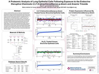
- 1. A Proteomic Analysis of Lung Epithelial Cells Following Exposure to the Endocrine Disruptive Chemicals 2,3,7,8-tetrachlorodibenzo-p-dioxin and Arsenic Trioxide Ryan P. Lynch & David M. Smalley Maine Institute for Human Genetics & Health, 246 Sylvan Road, Bangor ME 04401 (rlynch@emh.org) Abstract 2,3,7,8-tetrachlorodibenzo-p-dioxin Protein Expression Influenced By TCDD (2,3,7,8-tetrachlorodibenzo-p-dioxin) and arsenic are environmental endocrine disruptive chemicals (EDCs) that increase risk of lung cancer. While many studies Treated Human Bronchial Epithelial Cells 2,3,7,8-tetrachlorodibenzo-p-dioxin have attempted to examine the pathways associated with the transformation of normal epithelial cells into malignant cells, the mechanisms remain unclear. The goal of the Peptides from 2 altered Peptides from 2 proteins present study was to examine changes in the proteome of lung epithelial cells following proteins with no change exposure to low doses of these environmental EDCs. Immortalized human lung epithelial cells (NuLi-1) were treated with TCDD (2, 10, 50 nM) or As2O3 (0.5, 2, 10 µM), or vehicle for 24 hrs, and then lysed with 6M urea/PBS. The proteins were reduced, alkylated, and digested, and the peptides were isolated. The peptides were 3 SD then differentially labeled with an isobaric tag (iTRAQ) and the samples were mixed. Following 2D separation (isoelectric focusing and reverse phase-LC), the peptides were analyzed by MALDI-TOF/TOF using an Applied Biosystems 4800 Plus mass spectrometer. The spectra were analyzed using Protein Pilot software with the Paragon algorithm. Each EDC treatment set generated approximately 1000 proteins. Relative quantitation was performed on proteins with at least 2 peptides identified. While most proteins remained unchanged, several were significantly altered following TCDD exposure, and none after arsenic. These included proteins previously reported to be altered, as well as others that we believe are novel. Proteins involved with the extracellular matrix, cell regulation, and a number of mitochondrial associated proteins are shown to be altered. The results of this study are currently being validated using alternative strategies. 3 SD Materials & Methods Downregulated Proteins Upregulated Proteins (>3 SD from mean; >4 peptides) (>3 SD from mean; >4 peptides) Protein Biological Function Subcellular Location Protein Biological Function Subcellular Location Beta-actin-like protein 2 Cell motility Cytoplasm; cytoskeleton Blocks the elongation and Tropomodulin-3 depolymerization of the actin filaments Cytoplasm; cytoskeleton Non-histone chromosomal protein HMG-17 Chromatin organization Nucleus; cytoplasm Tenascin Signal transduction Extracellular matrix 40S ribosomal protein S21 Translational elongation Transcriptional regulation; cell cycle High mobility group protein HMGI-C regulation Nucleus Mitochondrial ATP synthesis coupled Mitochondria; mitochondria Elongation factor 1-beta Translational elongation ATP synthase subunit b, mitochondrial proton transport inner membrane L-lactate dehydrogenase B chain Anaerobic glycolysis; oxidation reduction Cytoplasm Histone H2A type 1-C Nucleosome assembly Nucleus Cell redox homeostasis; oxidation Endoplasmic reticulum Thioredoxin domain-containing protein 12 reduction lumen Hydroxymethylglutaryl-CoA synthase, Cholesterol/isoprenoid biosynthetic 60S acidic ribosomal protein P1 Translational elongation cytoplasmic process Cytoplasm 26S proteasome non-ATPase regulatory Microtubule-based movement; protein subunit 2 Regulation of protein catabolic process As2O3 Treated Human Tubulin beta-3 chain polymerization Tyrosine-protein phosphatase non- Negative regulation of insulin receptor Endoplasmic reticulum Intermediate filament organization; skin receptor type 1 signaling pathway membrane Keratin, type I cytoskeletal 9 development Rho protein signal transduction; actin PHF3 Isoform 1 of PHD finger protein 3 Transcription Nucleus Myosin IXB isoform 1 filament-based movement Cytoplasm Bronchial Epithelial Cells Isoform Beta of Tripartite motif-containing Transcription from RNA polymerase II Methionine adenosyltransferase 2 subunit Extracellular polysaccharide biosynthetic protein 29 promoter Cytoplasm beta process; one-carbon metabolic process DNA replication; double-strand break Putative RNA-binding protein 3 (RBM3) Positive regulation of translation Cytoplasm; nucleus Flap endonuclease 1 repair Nucleus Nucleus envelope; Transport; lipid metabolic process; Protein S100-A6 Signal transduction cytoplasm Fatty acid-binding protein, epidermal epidermis development Cytoplasm Actin, cytoplasmic 1 Cellular component movement Cytoplasm; cytoskeleton Hemoglobin subunit epsilon Oxygen transport Ras GTPase-activating protein-binding Neurotransmitter catabolic process; Mitochondria outer protein 2 Transport; Ras protein signal transduction Cytoplasm Monoamine oxidase A oxidation reduction membrane YWHAH 14-3-3 protein eta Transport; cell signalling 40S ribosomal protein S25 Translational elongation Cytoplasm; mitochondria; Isoform 2 of Proteasome activator Protein ETHE1, mitochondrial Mitochondrial metabolic homeostasis nucleus complex Apoptosis regulation Cytoplasm; nucleus Histone H1.4 Nucleosome assembly Nucleus Mucin-16 Cell adhesion Cell membrane Hepatoma-derived growth factor Cell proliferation; transcription regulation; Cytoplasm; nucleus Eukaryotic translation elongation factor 1 epsilon-1 Translation regulation; DNA damage repair Cytoplasm; nucleus Copyright Maine Institute for Human Genetics & Health March 7, 2010 3 SD Summary/Conclusions •Low doses of 2,3,7,8-tetrachlorodibenzo-p-dioxin induced expression of multiple proteins in human bronchial epithelial cells, whereas no significant changes in protein expression were observed after treatment with arsenic trioxide. •Two-fold changes in relative protein levels from 4 samples can easily be detected for the 100 most abundant proteins using iTRAQ labeling with a Database Search Results minimum of 2 peptides per protein. As2O3 Treated Human Bronchial Epithelial Cells 3 SD Set 1 Set 2 Set 3 Cumulative Spectra (total) 11316 18442 18560 48318 Spectra (identified) 10058 12689 13861 39274 Peptides 5848 8360 8672 18862 Proteins 957 923 1009 1583