SlideShare a Scribd company logo
1 of 40
US ATRFACTS
Dr. Kamal Sayed / MSc US AAU
OK
What is the principle of ultrasound?
An electric current passes through a cable to the transducer and is applied
to the crystals, causing them to deform and vibrate. This vibration produces
the ultrasound beam. The frequency of the ultrasound waves produced is
predetermined by the crystals in the transducer.
•
Artefacts
•
US Artifacts are any alterations in the image which
do not represent an actual image of the examined area.
•
They may be produced by technical imaging errors or result
from the complex interaction of the US with biological tissues.
Reverberation artifacts appear as a series of equally spaced
lines. One can avoid artefacts by turninig off all electric
equipment so that artefact does not hinder cardiac anatomy
exam.
•
Cauterization artifact is another example of how external
electrical equipment can cause distorted ultrasound images.
•
What does artifact mean in medical terms?
•
In medical imaging, artifacts are misrepresentations of tissue
structures produced by imaging techniques such as
ultrasound, X-ray, CT scan, and magnetic resonance imaging
(MRI). ... Physicians typically learn to recognize some of
these artifacts to avoid mistaking them for actual pathology.
•
Examples of tools causing artefacts include :
•
include stone tools, pottery vessels, metal objects such as
weapons and items of personal adornment such as buttons,
jewelry and clothing. Bones that show signs of human
modification are also examples.
•
Artifacts are items, usually found at an archaeological dig, are
any things made by or used by humans.
Some examples would be whole pottery, pot shards, stone
tools, decorative and religious artworks, bones of the animals
that the group ate, and sometimes human remains. Things
such as shelters, firepits, etc
•
Artifacts
•
https://radiopaedia.org/articles/blooming-artifact-
ultrasound?lang=gb
•
acoustic enhancement
•
acoustic shadowing
•
aliasing artifact
•
anisotropy
•
beam width artifact
•
blooming artifact
•
comet tail artifact
–
colour comet tail artifact
•
double aorta artifact
•
electrical interference artifact
•
hardware-related artifacts
–
transducer-related artifact
•
mirror image artifact
•
multipath artifact
•
colour bruit artifact
•
colour flash artifact
•
•
reverberation artifact
•
refraction artifact
•
ring down artifact
•
side lobe artifact
•
speckle artifact
•
speed displacement artifact
•
side lobe artifact
•
twinkling artifact
•
An image artifact is any feature which appears in
an image which is not present in the original imaged object.
An image artifact is sometime the result of improper
operation of the imager, and other times a consequence of
natural processes or properties of the human body
•
•
•
. Mirror image artefact is one of the beam path artefacts.
These occur when an ultrasound beam is not reflected
directly back to the transducer after hitting a reflective
surface, but rather takes an indirect return journey.
•
The primary beam reflects from such a surface
(e.g. diaphragm) but instead of directly being received by
the transducer, it encounters another structure (e.g. a nodular
lesion) in its path and is reflected back to the highly reflective
surface (e.g. diaphragm).
•
Slide (13)
•
•
It then again reflects back towards the transducer
•
. To avoid this artifact, change the position and angle of scanning to
change the angel of insonation of the primary
•
ultrasound beam.
In rhematology, mirrors nearly always are bone surfaces.
•
The mirror artefact is easily seen as such when the true image as
well as the mirror and mirror image are all in the image.
•
The mirror image is slightly trickier when only the mirror and mirror
image are present
•
•
. The ultrasound machine makes a false assumption that the
returning echo has been reflected once and hence the
delayed echoes are judged as if being returned from a deeper
structure, thus giving a mirror artifact on the other side of the
•
reflective surface
•
•
. It is a friendly artifact that allows the sonographer to
exclude pleural effusion by the reflection of the liver image
through the diaphragm. Examples :
•
@ reflection of a liver lesion into the thorax (the
commonest example)
•
@ reflection of abdominal ascites mimicking pleural
effusion
•
@ duplication of gestational sac (either ghost twin or
heterotopic pregnancy) 3
•
@ duplication of the uterus
•
Images slides ()
Scrotum (upper image) /Liver lesion (lower image)
•
Acoustic enhancement
•
Acoustic enhancement also called posterior
enhancement or enhanced through transmission, refers to
the increased echoes deep to structures that transmit sound
exceptionally well.
•
This is characteristic of fluid-filled structures such as cysts,
the urinary bladder and the gallbladder.
•
The fluid only attenuates the sound less than the surrounding
tissue.
•
Slide (15)
Acoustic enhancement: upper (hep cyst + GB)/ lower (epidermal inclusion cyst)
•
The time gain compensation (TGC) overcompensates through
the fluid-filled structure causing deeper tissues to be brighter.
•
Simply it is seen as increased echogenicity (whiteness)
posterior to the cystic area.
•
The presence of acoustic enhancement aids in the
identification of cystic masses but some solid masses,
especially lymphoma, may also show acoustic enhancement
posteriorly.
•
Acoustic shadowing (posterior acoustic shadowing)
•
is characterised by a signal void (dark or black area) behind
structures that strongly absorb or reflect ultrasonic waves.
•
it is a form of imaging artifact.
•
This happens most frequently with solid structures, as sound
conducts most rapidly in areas where molecules are closely packed,
such as in bone or stones.
•
Slide (18)
•
•
•
LT image (cholelithiasis)/ MID image (renal calculus)/
RT image (gas in colon diverticulum)
•
Beam width artifact
•
Occurs when a reflective object located beyond the widened
ultrasound beam, after the focal zone, creates false
detectable echoes that are displayed as overlapping the
structure of interest.
•
it occurs when scanning an anechoic structure and some
peripheral echoes are identified, i.e. gas bubbles in the
duodenum simulating small gallstones and peripheric echoes
in the bladder.
•
It is possible to avoid this beam width artifact by 1- adjusting
the focal zone to the depth level of interest
•
2- and by placing the transducer at the centre of the object
being studied.
•
Mirror image artifact is seen when there is a highly reflective
•
surface (e.g. diaphragm) in the path of the primary beam.
•
The primary beam reflects from such a surface
(e.g. diaphragm) but instead of directly being received by
the transducer, it encounters another structure (e.g. a nodular
lesion) in its path and is reflected back to the highly reflective
surface (e.g. diaphragm). It then again reflects back towards
the transducer
•
. To avoid this artifact, change the position and angle of
scanning to change the angel of insonation of the primary
ultrasound beam.
•
In rheumatology, mirrors are nearly always bone surfaces.
•
The mirror artefact is easily seen as such when the true image
as well as the mirror and mirror image are all in the image.
•
The mirror image is slightly trickier when only the mirror and
mirror image are present
•
. The ultrasound machine makes a false assumption that the
returning echo has been reflected once and hence the
delayed echoes are judged as if being returned from a deeper
structure, thus giving a mirror artifact on the other side of the
reflective surface.
•
•
It is a friendly artifact that allows the sonographer to exclude
pleural effusion by the reflection of the liver image through
the diaphragm.
•
Examples:
•
@ reflection of a liver lesion into the thorax (the commonest
example)
•
@ reflection of abdominal ascites mimicking pleural effusion
•
@ duplication of gestational sac (either ghost twin or
heterotopic pregnancy) 3
•
@ duplication of the uterus
•
Images slides (24)
Liver lesion (upper image) Scrotum / scrotum (lower image)
•
reverberation artifact
•
Reverberation artifact occurs when an ultrasound beam
encounters two strong parallel reflectors.
•
When the ultrasound beam reflects back and forth between
the reflectors ("reverberates"), the ultrasound transducer
interprets the sound waves returning as deeper structures
since it took longer for the wave to return to the transducer.
•
when the ultrasound beam reflects back and forth between
the reflectors ("reverberates"),
•
Slides (29/30)
•
the ultrasound transducer interprets the sound waves
returning as deeper structures since it took longer for the
wave to return to the transducer.
•
Reverberation artifacts can be improved by changing the
angle of insonation so that reverberation between strong
parallel reflectors cannot occur.
•
Comet-tail artifact is a specific type of reverberation artifact.
This results a short train of reverberations from an echogenic
focus which has strong parallel reflectors within it (e.g.
cholesterol crystals in adenomyomatosis).
•
It is advisable always to let the colour box go to the top of the
image to be aware of possible reverberation sources
•
Slide (31).
•
•
With comet tail artifact, the space between the two strong
parallel reflectors may be less than 1/2 the space pulse
length, causing the echoes to be displayed as triangular lines
(the later echoes get attenuated and have a decreased
amplitude, manifesting on the display as decreased width).
•
Anisotropy artefact is an angle-generated artifact.
•
It is produced in tissue that contains multiple, parallel linear
sound interfaces (e.g., tendons, ligaments) that lead to the
preferential reflection of the beam in one direction
•
. When the ultrasound beam is incident on a fibrillar structure
as a tendon or a ligament, the organised fibrils may reflect a
majority of the insonating sound beam in a direction away
from the transducer. When this occurs, the transducer does
not receive the returning echo and assumes that the
insonated area should be hypoechoic.
•
This anisotropic effect is dependant on the angle of the
insonating beam. The maximum return echo occurs when the
ultrasound beam is perpendicular to the tendon.
•
Decreasing the insonating angle on a normal tendon will
cause it to change from brightly hyperechoic (the actual echo
from tightly bound tendon fibres) to darkly hypoechoic. If the
angle is then increased, the tendon will again appear
hyperechoic
•
Slide (35).
•
•
If the artefact causes a normal tendon to appear hypoechoic,
it may falsely lead to a diagnosis of tendinosis or tear
•
. In some situations, anisotropy may be useful in diagnosis. If a
tendon is surrounded by other brightly hyperechoic structures
(e.g. fat), then altering the angle of the transducer will cause
the tendon to become hypoechoic, differentiating it from the
other structures.
•
Anistropy : upper: TXR NOT perp. To volar (palmar) wrist
lower : TXR perpendicular to volar wrist
Grating lobes Artefacts are the maxima of the main beam.
Side lobes and grating lobes are both unwanted parts of
the US beam emitted off axis that produce image artifacts due
to error in positioning the returning echo.
•
POINT SPREAD ARTEFACT
•
This artifact occurs when two reflectors are perpendicular to
the beam's main axis create one reflection on the image.
•
It is also called point spread artifact.
•
Lateral resolution is determined by beam width.
•
Point spread artifact is another term of describing suboptimal
lateral resolution.
•
Speed displacement artifact, also known as propagation
velocity artifact,
•
is a gray scale ultrasound finding that can be identified as an
area of focal discontinuity and displacement of an echo
deeper than that its actual position in an imaged structure.
Depth determination by an US machine is based on the
principle that the average propagation velocity of sound in
human tissue is 1540 m/s, and as such the {go return} time
between transmission and detecting the returned sound wave
to the transducer is multiplied by this number and halved to
determine distance, regardless of tissue type
•
As a result, if the true propagation velocity of a tissue falls
significantly below or above 1540 m/s, such as fat or bone,
then the distance calculated by the machine will be false,
displaying an inaccurate depth measurement.
•
By this same principle, if there is differential variation in tissue
composition of the tissues under the same ultrasound beam,
then different return times to the transducer will be
processed as different depths of tissue as opposed to
differences in propagation velocity between the tissues.
•
This may result in discontinuity in the displayed ultrasound
image, and as such is referred to as a propagation velocity
misrepresentation.
•
A commonly encountered scenario is speed displacement
artifact due to slowing of the US beam by focal fat, such as in
focal fatty sparing in case of hepatic steatosis.
•

More Related Content

What's hot

CT ITS BASIC PHYSICS
CT ITS BASIC PHYSICSCT ITS BASIC PHYSICS
CT ITS BASIC PHYSICSDEEPAK
 
radiological anatomy of retroperitoneum powerpoint
radiological anatomy of  retroperitoneum powerpointradiological anatomy of  retroperitoneum powerpoint
radiological anatomy of retroperitoneum powerpointDactarAdhikari
 
Ultrasound transducer doppler ppt pdf pk
Ultrasound transducer doppler ppt pdf pkUltrasound transducer doppler ppt pdf pk
Ultrasound transducer doppler ppt pdf pkDr pradeep Kumar
 
Principle of usg imaging, construction of transducers
Principle of usg imaging, construction of transducersPrinciple of usg imaging, construction of transducers
Principle of usg imaging, construction of transducersDev Lakhera
 
Pancreas ultrasound
Pancreas ultrasoundPancreas ultrasound
Pancreas ultrasoundSafi. Khan
 
Basic physics of Ultrasound
Basic physics of UltrasoundBasic physics of Ultrasound
Basic physics of UltrasoundNouman Ahmed
 
Chapter 6 image quality in ct
Chapter 6 image quality in ct Chapter 6 image quality in ct
Chapter 6 image quality in ct Muntaser S.Ahmad
 
Radiology Spots PPT- 2 by Dr Chandni Wadhwani
Radiology Spots PPT- 2 by Dr Chandni WadhwaniRadiology Spots PPT- 2 by Dr Chandni Wadhwani
Radiology Spots PPT- 2 by Dr Chandni WadhwaniChandni Wadhwani
 
Basic physics of ultrasound.JH
Basic physics of ultrasound.JHBasic physics of ultrasound.JH
Basic physics of ultrasound.JHhari baskar
 
Ultrasound contrast agents
Ultrasound contrast agentsUltrasound contrast agents
Ultrasound contrast agentsNaseem Abu hawash
 
Physic Of Ultrasound
Physic Of UltrasoundPhysic Of Ultrasound
Physic Of UltrasoundKhalis Karim
 
Ct image quality artifacts and it remedy
Ct image quality artifacts and it remedyCt image quality artifacts and it remedy
Ct image quality artifacts and it remedyYashawant Yadav
 
Doppler physics ppt
Doppler physics pptDoppler physics ppt
Doppler physics pptAbhilasha Singh
 
MR ENTEROGRAPHY
MR ENTEROGRAPHYMR ENTEROGRAPHY
MR ENTEROGRAPHYNihal Ahmed
 
Artifacts usg, ct, mri radiology ppt,pdf pk
Artifacts usg, ct, mri radiology  ppt,pdf pkArtifacts usg, ct, mri radiology  ppt,pdf pk
Artifacts usg, ct, mri radiology ppt,pdf pkDr pradeep Kumar
 
CT ARTIFACTS.pptx
CT ARTIFACTS.pptxCT ARTIFACTS.pptx
CT ARTIFACTS.pptxdypradio
 

What's hot (20)

CT ITS BASIC PHYSICS
CT ITS BASIC PHYSICSCT ITS BASIC PHYSICS
CT ITS BASIC PHYSICS
 
radiological anatomy of retroperitoneum powerpoint
radiological anatomy of  retroperitoneum powerpointradiological anatomy of  retroperitoneum powerpoint
radiological anatomy of retroperitoneum powerpoint
 
Ultrasound transducer doppler ppt pdf pk
Ultrasound transducer doppler ppt pdf pkUltrasound transducer doppler ppt pdf pk
Ultrasound transducer doppler ppt pdf pk
 
Principle of usg imaging, construction of transducers
Principle of usg imaging, construction of transducersPrinciple of usg imaging, construction of transducers
Principle of usg imaging, construction of transducers
 
Pancreas ultrasound
Pancreas ultrasoundPancreas ultrasound
Pancreas ultrasound
 
Ultrasound artifacts
Ultrasound artifactsUltrasound artifacts
Ultrasound artifacts
 
Basic physics of Ultrasound
Basic physics of UltrasoundBasic physics of Ultrasound
Basic physics of Ultrasound
 
Chapter 6 image quality in ct
Chapter 6 image quality in ct Chapter 6 image quality in ct
Chapter 6 image quality in ct
 
Radiology Spots PPT- 2 by Dr Chandni Wadhwani
Radiology Spots PPT- 2 by Dr Chandni WadhwaniRadiology Spots PPT- 2 by Dr Chandni Wadhwani
Radiology Spots PPT- 2 by Dr Chandni Wadhwani
 
Doppler Physics
Doppler PhysicsDoppler Physics
Doppler Physics
 
Basic physics of ultrasound.JH
Basic physics of ultrasound.JHBasic physics of ultrasound.JH
Basic physics of ultrasound.JH
 
Ultrasound contrast agents
Ultrasound contrast agentsUltrasound contrast agents
Ultrasound contrast agents
 
Physic Of Ultrasound
Physic Of UltrasoundPhysic Of Ultrasound
Physic Of Ultrasound
 
Ct image quality artifacts and it remedy
Ct image quality artifacts and it remedyCt image quality artifacts and it remedy
Ct image quality artifacts and it remedy
 
Doppler physics ppt
Doppler physics pptDoppler physics ppt
Doppler physics ppt
 
Defecography (1).pptx
Defecography (1).pptxDefecography (1).pptx
Defecography (1).pptx
 
MR ENTEROGRAPHY
MR ENTEROGRAPHYMR ENTEROGRAPHY
MR ENTEROGRAPHY
 
Artifacts usg, ct, mri radiology ppt,pdf pk
Artifacts usg, ct, mri radiology  ppt,pdf pkArtifacts usg, ct, mri radiology  ppt,pdf pk
Artifacts usg, ct, mri radiology ppt,pdf pk
 
CT ARTIFACTS.pptx
CT ARTIFACTS.pptxCT ARTIFACTS.pptx
CT ARTIFACTS.pptx
 
Mri physics
Mri physicsMri physics
Mri physics
 

Similar to Us artefacts

ARTIFACTS IN ECHO-1.pptx
ARTIFACTS IN ECHO-1.pptxARTIFACTS IN ECHO-1.pptx
ARTIFACTS IN ECHO-1.pptxAnayaAnaya14
 
Ultrasound artifacts and contrast enhanced ultrasound
Ultrasound artifacts and contrast enhanced ultrasoundUltrasound artifacts and contrast enhanced ultrasound
Ultrasound artifacts and contrast enhanced ultrasoundArjun Reddy
 
ultrasoundartifacts-200113150706.pdf
ultrasoundartifacts-200113150706.pdfultrasoundartifacts-200113150706.pdf
ultrasoundartifacts-200113150706.pdfGenesisGeronaga
 
Ultrasound Artifact
Ultrasound ArtifactUltrasound Artifact
Ultrasound ArtifactAmer Sohail
 
general Radiological anatomy
general Radiological anatomygeneral Radiological anatomy
general Radiological anatomyDr Sandeep k Sharma
 
Basics of sonography and anatomy of chest wall
Basics of sonography and anatomy of chest wallBasics of sonography and anatomy of chest wall
Basics of sonography and anatomy of chest wallGamal Agmy
 
Sudanese Chest Sonography Workshop (Basics of sonography and anatomy of chest...
Sudanese Chest Sonography Workshop (Basics of sonography and anatomy of chest...Sudanese Chest Sonography Workshop (Basics of sonography and anatomy of chest...
Sudanese Chest Sonography Workshop (Basics of sonography and anatomy of chest...Gamal Agmy
 
Lecture 24 Ultrasonography in animal reproduction
Lecture 24 Ultrasonography in animal reproductionLecture 24 Ultrasonography in animal reproduction
Lecture 24 Ultrasonography in animal reproductionDrGovindNarayanPuroh
 
ULTRASOUND - ARTIFACTS.pptx
ULTRASOUND - ARTIFACTS.pptxULTRASOUND - ARTIFACTS.pptx
ULTRASOUND - ARTIFACTS.pptxShubhamRai502947
 
artefacts in ultrasound imaging: physics .pptx
artefacts in ultrasound imaging: physics .pptxartefacts in ultrasound imaging: physics .pptx
artefacts in ultrasound imaging: physics .pptxWangjuSumnyan
 
M-2 2018-ag-7688 Diagnostic ultrasonography in domestic animal reproduction.ppt
M-2 2018-ag-7688 Diagnostic ultrasonography in domestic animal reproduction.pptM-2 2018-ag-7688 Diagnostic ultrasonography in domestic animal reproduction.ppt
M-2 2018-ag-7688 Diagnostic ultrasonography in domestic animal reproduction.pptAliHussain4701
 
Probe construction, types and usg artefacts
Probe construction, types and usg artefactsProbe construction, types and usg artefacts
Probe construction, types and usg artefactsVishakha Mittal
 
Usg artefacts
Usg artefacts Usg artefacts
Usg artefacts Prajwith Rai
 
usg artifacts.pptx
usg artifacts.pptxusg artifacts.pptx
usg artifacts.pptxrohanjohnjacob
 
ULTRASOUND BY DR.ABHIJEET MAJHI,MS .pptx
ULTRASOUND BY DR.ABHIJEET MAJHI,MS .pptxULTRASOUND BY DR.ABHIJEET MAJHI,MS .pptx
ULTRASOUND BY DR.ABHIJEET MAJHI,MS .pptxAbhijeet Majhi
 
DIFFERENT TYPES OF RADIOGRAPHY CT_ARTIFACTS
DIFFERENT TYPES OF RADIOGRAPHY CT_ARTIFACTSDIFFERENT TYPES OF RADIOGRAPHY CT_ARTIFACTS
DIFFERENT TYPES OF RADIOGRAPHY CT_ARTIFACTSMichaelAkortsu1
 
Physics of ultrasound imaging
Physics of ultrasound imagingPhysics of ultrasound imaging
Physics of ultrasound imagingmtuzer
 
Ultrasound imaging
Ultrasound imagingUltrasound imaging
Ultrasound imagingAnjan Dangal
 

Similar to Us artefacts (20)

ARTIFACTS IN ECHO-1.pptx
ARTIFACTS IN ECHO-1.pptxARTIFACTS IN ECHO-1.pptx
ARTIFACTS IN ECHO-1.pptx
 
Us physics 3
Us physics 3Us physics 3
Us physics 3
 
Ultrasound artifacts and contrast enhanced ultrasound
Ultrasound artifacts and contrast enhanced ultrasoundUltrasound artifacts and contrast enhanced ultrasound
Ultrasound artifacts and contrast enhanced ultrasound
 
ultrasoundartifacts-200113150706.pdf
ultrasoundartifacts-200113150706.pdfultrasoundartifacts-200113150706.pdf
ultrasoundartifacts-200113150706.pdf
 
Ultrasound Artifact
Ultrasound ArtifactUltrasound Artifact
Ultrasound Artifact
 
general Radiological anatomy
general Radiological anatomygeneral Radiological anatomy
general Radiological anatomy
 
Basics of sonography and anatomy of chest wall
Basics of sonography and anatomy of chest wallBasics of sonography and anatomy of chest wall
Basics of sonography and anatomy of chest wall
 
Sudanese Chest Sonography Workshop (Basics of sonography and anatomy of chest...
Sudanese Chest Sonography Workshop (Basics of sonography and anatomy of chest...Sudanese Chest Sonography Workshop (Basics of sonography and anatomy of chest...
Sudanese Chest Sonography Workshop (Basics of sonography and anatomy of chest...
 
Lecture 24 Ultrasonography in animal reproduction
Lecture 24 Ultrasonography in animal reproductionLecture 24 Ultrasonography in animal reproduction
Lecture 24 Ultrasonography in animal reproduction
 
ULTRASOUND - ARTIFACTS.pptx
ULTRASOUND - ARTIFACTS.pptxULTRASOUND - ARTIFACTS.pptx
ULTRASOUND - ARTIFACTS.pptx
 
artefacts in ultrasound imaging: physics .pptx
artefacts in ultrasound imaging: physics .pptxartefacts in ultrasound imaging: physics .pptx
artefacts in ultrasound imaging: physics .pptx
 
M-2 2018-ag-7688 Diagnostic ultrasonography in domestic animal reproduction.ppt
M-2 2018-ag-7688 Diagnostic ultrasonography in domestic animal reproduction.pptM-2 2018-ag-7688 Diagnostic ultrasonography in domestic animal reproduction.ppt
M-2 2018-ag-7688 Diagnostic ultrasonography in domestic animal reproduction.ppt
 
Probe construction, types and usg artefacts
Probe construction, types and usg artefactsProbe construction, types and usg artefacts
Probe construction, types and usg artefacts
 
Usg artefacts
Usg artefacts Usg artefacts
Usg artefacts
 
Ct artiact
Ct artiactCt artiact
Ct artiact
 
usg artifacts.pptx
usg artifacts.pptxusg artifacts.pptx
usg artifacts.pptx
 
ULTRASOUND BY DR.ABHIJEET MAJHI,MS .pptx
ULTRASOUND BY DR.ABHIJEET MAJHI,MS .pptxULTRASOUND BY DR.ABHIJEET MAJHI,MS .pptx
ULTRASOUND BY DR.ABHIJEET MAJHI,MS .pptx
 
DIFFERENT TYPES OF RADIOGRAPHY CT_ARTIFACTS
DIFFERENT TYPES OF RADIOGRAPHY CT_ARTIFACTSDIFFERENT TYPES OF RADIOGRAPHY CT_ARTIFACTS
DIFFERENT TYPES OF RADIOGRAPHY CT_ARTIFACTS
 
Physics of ultrasound imaging
Physics of ultrasound imagingPhysics of ultrasound imaging
Physics of ultrasound imaging
 
Ultrasound imaging
Ultrasound imagingUltrasound imaging
Ultrasound imaging
 

More from KamalEldirawi

Hepatic doppler us [2]
Hepatic doppler us [2]Hepatic doppler us [2]
Hepatic doppler us [2]KamalEldirawi
 
Hepatic doppler us [3]
Hepatic doppler us [3]Hepatic doppler us [3]
Hepatic doppler us [3]KamalEldirawi
 
Hepatic dopp us [1]
Hepatic dopp  us [1]Hepatic dopp  us [1]
Hepatic dopp us [1]KamalEldirawi
 
Us physics (11)
Us physics (11)Us physics (11)
Us physics (11)KamalEldirawi
 
Introductory us
Introductory usIntroductory us
Introductory usKamalEldirawi
 
Illustrated carotid doppler 3
Illustrated carotid doppler 3Illustrated carotid doppler 3
Illustrated carotid doppler 3KamalEldirawi
 
Illustrated carotid doppler 2
Illustrated carotid doppler 2Illustrated carotid doppler 2
Illustrated carotid doppler 2KamalEldirawi
 
Illustrated carotid doppler 1
Illustrated carotid doppler 1Illustrated carotid doppler 1
Illustrated carotid doppler 1KamalEldirawi
 
Umbilical artery doppler [1]
Umbilical artery doppler [1]Umbilical artery doppler [1]
Umbilical artery doppler [1]KamalEldirawi
 
Doppler principles [2]
Doppler principles [2]Doppler principles [2]
Doppler principles [2]KamalEldirawi
 
Doppler principles [1]
Doppler principles [1]Doppler principles [1]
Doppler principles [1]KamalEldirawi
 
Hepatic doppler us [3]
Hepatic doppler us [3]Hepatic doppler us [3]
Hepatic doppler us [3]KamalEldirawi
 
Hepatic doppler us [2]
Hepatic doppler us [2]Hepatic doppler us [2]
Hepatic doppler us [2]KamalEldirawi
 
Hepatic dopp us [1]
Hepatic dopp  us [1]Hepatic dopp  us [1]
Hepatic dopp us [1]KamalEldirawi
 
Doppler ultrasound (4)
Doppler ultrasound (4)Doppler ultrasound (4)
Doppler ultrasound (4)KamalEldirawi
 
Doppler us (2)
Doppler us (2)Doppler us (2)
Doppler us (2)KamalEldirawi
 
Doppler artefact (1)
Doppler artefact (1)Doppler artefact (1)
Doppler artefact (1)KamalEldirawi
 
Doppler us (3)
Doppler us (3)Doppler us (3)
Doppler us (3)KamalEldirawi
 
Doppler ultrasounds (1)
Doppler ultrasounds (1)Doppler ultrasounds (1)
Doppler ultrasounds (1)KamalEldirawi
 
Doppler Physics (3)
Doppler Physics (3)Doppler Physics (3)
Doppler Physics (3)KamalEldirawi
 

More from KamalEldirawi (20)

Hepatic doppler us [2]
Hepatic doppler us [2]Hepatic doppler us [2]
Hepatic doppler us [2]
 
Hepatic doppler us [3]
Hepatic doppler us [3]Hepatic doppler us [3]
Hepatic doppler us [3]
 
Hepatic dopp us [1]
Hepatic dopp  us [1]Hepatic dopp  us [1]
Hepatic dopp us [1]
 
Us physics (11)
Us physics (11)Us physics (11)
Us physics (11)
 
Introductory us
Introductory usIntroductory us
Introductory us
 
Illustrated carotid doppler 3
Illustrated carotid doppler 3Illustrated carotid doppler 3
Illustrated carotid doppler 3
 
Illustrated carotid doppler 2
Illustrated carotid doppler 2Illustrated carotid doppler 2
Illustrated carotid doppler 2
 
Illustrated carotid doppler 1
Illustrated carotid doppler 1Illustrated carotid doppler 1
Illustrated carotid doppler 1
 
Umbilical artery doppler [1]
Umbilical artery doppler [1]Umbilical artery doppler [1]
Umbilical artery doppler [1]
 
Doppler principles [2]
Doppler principles [2]Doppler principles [2]
Doppler principles [2]
 
Doppler principles [1]
Doppler principles [1]Doppler principles [1]
Doppler principles [1]
 
Hepatic doppler us [3]
Hepatic doppler us [3]Hepatic doppler us [3]
Hepatic doppler us [3]
 
Hepatic doppler us [2]
Hepatic doppler us [2]Hepatic doppler us [2]
Hepatic doppler us [2]
 
Hepatic dopp us [1]
Hepatic dopp  us [1]Hepatic dopp  us [1]
Hepatic dopp us [1]
 
Doppler ultrasound (4)
Doppler ultrasound (4)Doppler ultrasound (4)
Doppler ultrasound (4)
 
Doppler us (2)
Doppler us (2)Doppler us (2)
Doppler us (2)
 
Doppler artefact (1)
Doppler artefact (1)Doppler artefact (1)
Doppler artefact (1)
 
Doppler us (3)
Doppler us (3)Doppler us (3)
Doppler us (3)
 
Doppler ultrasounds (1)
Doppler ultrasounds (1)Doppler ultrasounds (1)
Doppler ultrasounds (1)
 
Doppler Physics (3)
Doppler Physics (3)Doppler Physics (3)
Doppler Physics (3)
 

Recently uploaded

VIP Service Call Girls Sindhi Colony đź“ł 7877925207 For 18+ VIP Call Girl At Th...
VIP Service Call Girls Sindhi Colony đź“ł 7877925207 For 18+ VIP Call Girl At Th...VIP Service Call Girls Sindhi Colony đź“ł 7877925207 For 18+ VIP Call Girl At Th...
VIP Service Call Girls Sindhi Colony đź“ł 7877925207 For 18+ VIP Call Girl At Th...jageshsingh5554
 
Call Girls Horamavu WhatsApp Number 7001035870 Meeting With Bangalore Escorts
Call Girls Horamavu WhatsApp Number 7001035870 Meeting With Bangalore EscortsCall Girls Horamavu WhatsApp Number 7001035870 Meeting With Bangalore Escorts
Call Girls Horamavu WhatsApp Number 7001035870 Meeting With Bangalore Escortsvidya singh
 
Call Girls Service Navi Mumbai Samaira 8617697112 Independent Escort Service ...
Call Girls Service Navi Mumbai Samaira 8617697112 Independent Escort Service ...Call Girls Service Navi Mumbai Samaira 8617697112 Independent Escort Service ...
Call Girls Service Navi Mumbai Samaira 8617697112 Independent Escort Service ...Call girls in Ahmedabad High profile
 
CALL ON ➥9907093804 🔝 Call Girls Baramati ( Pune) Girls Service
CALL ON ➥9907093804 🔝 Call Girls Baramati ( Pune)  Girls ServiceCALL ON ➥9907093804 🔝 Call Girls Baramati ( Pune)  Girls Service
CALL ON ➥9907093804 🔝 Call Girls Baramati ( Pune) Girls ServiceMiss joya
 
Premium Call Girls Cottonpet Whatsapp 7001035870 Independent Escort Service
Premium Call Girls Cottonpet Whatsapp 7001035870 Independent Escort ServicePremium Call Girls Cottonpet Whatsapp 7001035870 Independent Escort Service
Premium Call Girls Cottonpet Whatsapp 7001035870 Independent Escort Servicevidya singh
 
Call Girl Number in Vashi Mumbai📲 9833363713 💞 Full Night Enjoy
Call Girl Number in Vashi Mumbai📲 9833363713 💞 Full Night EnjoyCall Girl Number in Vashi Mumbai📲 9833363713 💞 Full Night Enjoy
Call Girl Number in Vashi Mumbai📲 9833363713 💞 Full Night Enjoybabeytanya
 
Russian Call Girls in Pune Tanvi 9907093804 Short 1500 Night 6000 Best call g...
Russian Call Girls in Pune Tanvi 9907093804 Short 1500 Night 6000 Best call g...Russian Call Girls in Pune Tanvi 9907093804 Short 1500 Night 6000 Best call g...
Russian Call Girls in Pune Tanvi 9907093804 Short 1500 Night 6000 Best call g...Miss joya
 
Best Rate (Hyderabad) Call Girls Jahanuma âźź 8250192130 âźź High Class Call Girl...
Best Rate (Hyderabad) Call Girls Jahanuma âźź 8250192130 âźź High Class Call Girl...Best Rate (Hyderabad) Call Girls Jahanuma âźź 8250192130 âźź High Class Call Girl...
Best Rate (Hyderabad) Call Girls Jahanuma âźź 8250192130 âźź High Class Call Girl...astropune
 
â™›VVIP Hyderabad Call Girls Chintalkuntađź–•7001035870đź–•Riya Kappor Top Call Girl ...
â™›VVIP Hyderabad Call Girls Chintalkuntađź–•7001035870đź–•Riya Kappor Top Call Girl ...â™›VVIP Hyderabad Call Girls Chintalkuntađź–•7001035870đź–•Riya Kappor Top Call Girl ...
â™›VVIP Hyderabad Call Girls Chintalkuntađź–•7001035870đź–•Riya Kappor Top Call Girl ...astropune
 
Call Girl Number in Panvel Mumbai📲 9833363713 💞 Full Night Enjoy
Call Girl Number in Panvel Mumbai📲 9833363713 💞 Full Night EnjoyCall Girl Number in Panvel Mumbai📲 9833363713 💞 Full Night Enjoy
Call Girl Number in Panvel Mumbai📲 9833363713 💞 Full Night Enjoybabeytanya
 
Call Girls Service Jaipur Grishma WhatsApp ❤8445551418 VIP Call Girls Jaipur
Call Girls Service Jaipur Grishma WhatsApp ❤8445551418 VIP Call Girls JaipurCall Girls Service Jaipur Grishma WhatsApp ❤8445551418 VIP Call Girls Jaipur
Call Girls Service Jaipur Grishma WhatsApp ❤8445551418 VIP Call Girls Jaipurparulsinha
 
VIP Russian Call Girls in Varanasi Samaira 8250192130 Independent Escort Serv...
VIP Russian Call Girls in Varanasi Samaira 8250192130 Independent Escort Serv...VIP Russian Call Girls in Varanasi Samaira 8250192130 Independent Escort Serv...
VIP Russian Call Girls in Varanasi Samaira 8250192130 Independent Escort Serv...Neha Kaur
 
Call Girls Service In Shyam Nagar Whatsapp 8445551418 Independent Escort Service
Call Girls Service In Shyam Nagar Whatsapp 8445551418 Independent Escort ServiceCall Girls Service In Shyam Nagar Whatsapp 8445551418 Independent Escort Service
Call Girls Service In Shyam Nagar Whatsapp 8445551418 Independent Escort Serviceparulsinha
 
(Rocky) Jaipur Call Girl - 9521753030 Escorts Service 50% Off with Cash ON De...
(Rocky) Jaipur Call Girl - 9521753030 Escorts Service 50% Off with Cash ON De...(Rocky) Jaipur Call Girl - 9521753030 Escorts Service 50% Off with Cash ON De...
(Rocky) Jaipur Call Girl - 9521753030 Escorts Service 50% Off with Cash ON De...indiancallgirl4rent
 
Book Paid Powai Call Girls Mumbai đ– ‹ 9930245274 đ– ‹Low Budget Full Independent H...
Book Paid Powai Call Girls Mumbai đ– ‹ 9930245274 đ– ‹Low Budget Full Independent H...Book Paid Powai Call Girls Mumbai đ– ‹ 9930245274 đ– ‹Low Budget Full Independent H...
Book Paid Powai Call Girls Mumbai đ– ‹ 9930245274 đ– ‹Low Budget Full Independent H...Call Girls in Nagpur High Profile
 
Kesar Bagh Call Girl Price 9548273370 , Lucknow Call Girls Service
Kesar Bagh Call Girl Price 9548273370 , Lucknow Call Girls ServiceKesar Bagh Call Girl Price 9548273370 , Lucknow Call Girls Service
Kesar Bagh Call Girl Price 9548273370 , Lucknow Call Girls Servicemakika9823
 
Bangalore Call Girl Whatsapp Number 100% Complete Your Sexual Needs
Bangalore Call Girl Whatsapp Number 100% Complete Your Sexual NeedsBangalore Call Girl Whatsapp Number 100% Complete Your Sexual Needs
Bangalore Call Girl Whatsapp Number 100% Complete Your Sexual NeedsGfnyt
 
CALL ON ➥9907093804 🔝 Call Girls Hadapsar ( Pune) Girls Service
CALL ON ➥9907093804 🔝 Call Girls Hadapsar ( Pune)  Girls ServiceCALL ON ➥9907093804 🔝 Call Girls Hadapsar ( Pune)  Girls Service
CALL ON ➥9907093804 🔝 Call Girls Hadapsar ( Pune) Girls ServiceMiss joya
 
VIP Call Girls Pune Vani 9907093804 Short 1500 Night 6000 Best call girls Ser...
VIP Call Girls Pune Vani 9907093804 Short 1500 Night 6000 Best call girls Ser...VIP Call Girls Pune Vani 9907093804 Short 1500 Night 6000 Best call girls Ser...
VIP Call Girls Pune Vani 9907093804 Short 1500 Night 6000 Best call girls Ser...Miss joya
 
Low Rate Call Girls Pune Esha 9907093804 Short 1500 Night 6000 Best call girl...
Low Rate Call Girls Pune Esha 9907093804 Short 1500 Night 6000 Best call girl...Low Rate Call Girls Pune Esha 9907093804 Short 1500 Night 6000 Best call girl...
Low Rate Call Girls Pune Esha 9907093804 Short 1500 Night 6000 Best call girl...Miss joya
 

Recently uploaded (20)

VIP Service Call Girls Sindhi Colony đź“ł 7877925207 For 18+ VIP Call Girl At Th...
VIP Service Call Girls Sindhi Colony đź“ł 7877925207 For 18+ VIP Call Girl At Th...VIP Service Call Girls Sindhi Colony đź“ł 7877925207 For 18+ VIP Call Girl At Th...
VIP Service Call Girls Sindhi Colony đź“ł 7877925207 For 18+ VIP Call Girl At Th...
 
Call Girls Horamavu WhatsApp Number 7001035870 Meeting With Bangalore Escorts
Call Girls Horamavu WhatsApp Number 7001035870 Meeting With Bangalore EscortsCall Girls Horamavu WhatsApp Number 7001035870 Meeting With Bangalore Escorts
Call Girls Horamavu WhatsApp Number 7001035870 Meeting With Bangalore Escorts
 
Call Girls Service Navi Mumbai Samaira 8617697112 Independent Escort Service ...
Call Girls Service Navi Mumbai Samaira 8617697112 Independent Escort Service ...Call Girls Service Navi Mumbai Samaira 8617697112 Independent Escort Service ...
Call Girls Service Navi Mumbai Samaira 8617697112 Independent Escort Service ...
 
CALL ON ➥9907093804 🔝 Call Girls Baramati ( Pune) Girls Service
CALL ON ➥9907093804 🔝 Call Girls Baramati ( Pune)  Girls ServiceCALL ON ➥9907093804 🔝 Call Girls Baramati ( Pune)  Girls Service
CALL ON ➥9907093804 🔝 Call Girls Baramati ( Pune) Girls Service
 
Premium Call Girls Cottonpet Whatsapp 7001035870 Independent Escort Service
Premium Call Girls Cottonpet Whatsapp 7001035870 Independent Escort ServicePremium Call Girls Cottonpet Whatsapp 7001035870 Independent Escort Service
Premium Call Girls Cottonpet Whatsapp 7001035870 Independent Escort Service
 
Call Girl Number in Vashi Mumbai📲 9833363713 💞 Full Night Enjoy
Call Girl Number in Vashi Mumbai📲 9833363713 💞 Full Night EnjoyCall Girl Number in Vashi Mumbai📲 9833363713 💞 Full Night Enjoy
Call Girl Number in Vashi Mumbai📲 9833363713 💞 Full Night Enjoy
 
Russian Call Girls in Pune Tanvi 9907093804 Short 1500 Night 6000 Best call g...
Russian Call Girls in Pune Tanvi 9907093804 Short 1500 Night 6000 Best call g...Russian Call Girls in Pune Tanvi 9907093804 Short 1500 Night 6000 Best call g...
Russian Call Girls in Pune Tanvi 9907093804 Short 1500 Night 6000 Best call g...
 
Best Rate (Hyderabad) Call Girls Jahanuma âźź 8250192130 âźź High Class Call Girl...
Best Rate (Hyderabad) Call Girls Jahanuma âźź 8250192130 âźź High Class Call Girl...Best Rate (Hyderabad) Call Girls Jahanuma âźź 8250192130 âźź High Class Call Girl...
Best Rate (Hyderabad) Call Girls Jahanuma âźź 8250192130 âźź High Class Call Girl...
 
â™›VVIP Hyderabad Call Girls Chintalkuntađź–•7001035870đź–•Riya Kappor Top Call Girl ...
â™›VVIP Hyderabad Call Girls Chintalkuntađź–•7001035870đź–•Riya Kappor Top Call Girl ...â™›VVIP Hyderabad Call Girls Chintalkuntađź–•7001035870đź–•Riya Kappor Top Call Girl ...
â™›VVIP Hyderabad Call Girls Chintalkuntađź–•7001035870đź–•Riya Kappor Top Call Girl ...
 
Call Girl Number in Panvel Mumbai📲 9833363713 💞 Full Night Enjoy
Call Girl Number in Panvel Mumbai📲 9833363713 💞 Full Night EnjoyCall Girl Number in Panvel Mumbai📲 9833363713 💞 Full Night Enjoy
Call Girl Number in Panvel Mumbai📲 9833363713 💞 Full Night Enjoy
 
Call Girls Service Jaipur Grishma WhatsApp ❤8445551418 VIP Call Girls Jaipur
Call Girls Service Jaipur Grishma WhatsApp ❤8445551418 VIP Call Girls JaipurCall Girls Service Jaipur Grishma WhatsApp ❤8445551418 VIP Call Girls Jaipur
Call Girls Service Jaipur Grishma WhatsApp ❤8445551418 VIP Call Girls Jaipur
 
VIP Russian Call Girls in Varanasi Samaira 8250192130 Independent Escort Serv...
VIP Russian Call Girls in Varanasi Samaira 8250192130 Independent Escort Serv...VIP Russian Call Girls in Varanasi Samaira 8250192130 Independent Escort Serv...
VIP Russian Call Girls in Varanasi Samaira 8250192130 Independent Escort Serv...
 
Call Girls Service In Shyam Nagar Whatsapp 8445551418 Independent Escort Service
Call Girls Service In Shyam Nagar Whatsapp 8445551418 Independent Escort ServiceCall Girls Service In Shyam Nagar Whatsapp 8445551418 Independent Escort Service
Call Girls Service In Shyam Nagar Whatsapp 8445551418 Independent Escort Service
 
(Rocky) Jaipur Call Girl - 9521753030 Escorts Service 50% Off with Cash ON De...
(Rocky) Jaipur Call Girl - 9521753030 Escorts Service 50% Off with Cash ON De...(Rocky) Jaipur Call Girl - 9521753030 Escorts Service 50% Off with Cash ON De...
(Rocky) Jaipur Call Girl - 9521753030 Escorts Service 50% Off with Cash ON De...
 
Book Paid Powai Call Girls Mumbai đ– ‹ 9930245274 đ– ‹Low Budget Full Independent H...
Book Paid Powai Call Girls Mumbai đ– ‹ 9930245274 đ– ‹Low Budget Full Independent H...Book Paid Powai Call Girls Mumbai đ– ‹ 9930245274 đ– ‹Low Budget Full Independent H...
Book Paid Powai Call Girls Mumbai đ– ‹ 9930245274 đ– ‹Low Budget Full Independent H...
 
Kesar Bagh Call Girl Price 9548273370 , Lucknow Call Girls Service
Kesar Bagh Call Girl Price 9548273370 , Lucknow Call Girls ServiceKesar Bagh Call Girl Price 9548273370 , Lucknow Call Girls Service
Kesar Bagh Call Girl Price 9548273370 , Lucknow Call Girls Service
 
Bangalore Call Girl Whatsapp Number 100% Complete Your Sexual Needs
Bangalore Call Girl Whatsapp Number 100% Complete Your Sexual NeedsBangalore Call Girl Whatsapp Number 100% Complete Your Sexual Needs
Bangalore Call Girl Whatsapp Number 100% Complete Your Sexual Needs
 
CALL ON ➥9907093804 🔝 Call Girls Hadapsar ( Pune) Girls Service
CALL ON ➥9907093804 🔝 Call Girls Hadapsar ( Pune)  Girls ServiceCALL ON ➥9907093804 🔝 Call Girls Hadapsar ( Pune)  Girls Service
CALL ON ➥9907093804 🔝 Call Girls Hadapsar ( Pune) Girls Service
 
VIP Call Girls Pune Vani 9907093804 Short 1500 Night 6000 Best call girls Ser...
VIP Call Girls Pune Vani 9907093804 Short 1500 Night 6000 Best call girls Ser...VIP Call Girls Pune Vani 9907093804 Short 1500 Night 6000 Best call girls Ser...
VIP Call Girls Pune Vani 9907093804 Short 1500 Night 6000 Best call girls Ser...
 
Low Rate Call Girls Pune Esha 9907093804 Short 1500 Night 6000 Best call girl...
Low Rate Call Girls Pune Esha 9907093804 Short 1500 Night 6000 Best call girl...Low Rate Call Girls Pune Esha 9907093804 Short 1500 Night 6000 Best call girl...
Low Rate Call Girls Pune Esha 9907093804 Short 1500 Night 6000 Best call girl...
 

Us artefacts

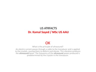
  • 1. US ATRFACTS Dr. Kamal Sayed / MSc US AAU OK What is the principle of ultrasound? An electric current passes through a cable to the transducer and is applied to the crystals, causing them to deform and vibrate. This vibration produces the ultrasound beam. The frequency of the ultrasound waves produced is predetermined by the crystals in the transducer.
  • 2. • Artefacts • US Artifacts are any alterations in the image which do not represent an actual image of the examined area. • They may be produced by technical imaging errors or result from the complex interaction of the US with biological tissues. Reverberation artifacts appear as a series of equally spaced lines. One can avoid artefacts by turninig off all electric equipment so that artefact does not hinder cardiac anatomy exam. • Cauterization artifact is another example of how external electrical equipment can cause distorted ultrasound images.
  • 3. • What does artifact mean in medical terms? • In medical imaging, artifacts are misrepresentations of tissue structures produced by imaging techniques such as ultrasound, X-ray, CT scan, and magnetic resonance imaging (MRI). ... Physicians typically learn to recognize some of these artifacts to avoid mistaking them for actual pathology.
  • 4. • Examples of tools causing artefacts include : • include stone tools, pottery vessels, metal objects such as weapons and items of personal adornment such as buttons, jewelry and clothing. Bones that show signs of human modification are also examples. • Artifacts are items, usually found at an archaeological dig, are any things made by or used by humans. Some examples would be whole pottery, pot shards, stone tools, decorative and religious artworks, bones of the animals that the group ate, and sometimes human remains. Things such as shelters, firepits, etc
  • 6. • comet tail artifact – colour comet tail artifact • double aorta artifact • electrical interference artifact • hardware-related artifacts – transducer-related artifact • mirror image artifact • multipath artifact • colour bruit artifact • colour flash artifact •
  • 7. • reverberation artifact • refraction artifact • ring down artifact • side lobe artifact • speckle artifact • speed displacement artifact • side lobe artifact • twinkling artifact
  • 8. • An image artifact is any feature which appears in an image which is not present in the original imaged object. An image artifact is sometime the result of improper operation of the imager, and other times a consequence of natural processes or properties of the human body • •
  • 9. • . Mirror image artefact is one of the beam path artefacts. These occur when an ultrasound beam is not reflected directly back to the transducer after hitting a reflective surface, but rather takes an indirect return journey. • The primary beam reflects from such a surface (e.g. diaphragm) but instead of directly being received by the transducer, it encounters another structure (e.g. a nodular lesion) in its path and is reflected back to the highly reflective surface (e.g. diaphragm). • Slide (13) •
  • 10. • It then again reflects back towards the transducer • . To avoid this artifact, change the position and angle of scanning to change the angel of insonation of the primary • ultrasound beam. In rhematology, mirrors nearly always are bone surfaces. • The mirror artefact is easily seen as such when the true image as well as the mirror and mirror image are all in the image. • The mirror image is slightly trickier when only the mirror and mirror image are present •
  • 11. • . The ultrasound machine makes a false assumption that the returning echo has been reflected once and hence the delayed echoes are judged as if being returned from a deeper structure, thus giving a mirror artifact on the other side of the • reflective surface •
  • 12. • . It is a friendly artifact that allows the sonographer to exclude pleural effusion by the reflection of the liver image through the diaphragm. Examples : • @ reflection of a liver lesion into the thorax (the commonest example) • @ reflection of abdominal ascites mimicking pleural effusion • @ duplication of gestational sac (either ghost twin or heterotopic pregnancy) 3 • @ duplication of the uterus • Images slides ()
  • 13. Scrotum (upper image) /Liver lesion (lower image)
  • 14. • Acoustic enhancement • Acoustic enhancement also called posterior enhancement or enhanced through transmission, refers to the increased echoes deep to structures that transmit sound exceptionally well. • This is characteristic of fluid-filled structures such as cysts, the urinary bladder and the gallbladder. • The fluid only attenuates the sound less than the surrounding tissue. • Slide (15)
  • 15. Acoustic enhancement: upper (hep cyst + GB)/ lower (epidermal inclusion cyst)
  • 16. • The time gain compensation (TGC) overcompensates through the fluid-filled structure causing deeper tissues to be brighter. • Simply it is seen as increased echogenicity (whiteness) posterior to the cystic area. • The presence of acoustic enhancement aids in the identification of cystic masses but some solid masses, especially lymphoma, may also show acoustic enhancement posteriorly.
  • 17. • Acoustic shadowing (posterior acoustic shadowing) • is characterised by a signal void (dark or black area) behind structures that strongly absorb or reflect ultrasonic waves. • it is a form of imaging artifact. • This happens most frequently with solid structures, as sound conducts most rapidly in areas where molecules are closely packed, such as in bone or stones. • Slide (18) • • •
  • 18. LT image (cholelithiasis)/ MID image (renal calculus)/ RT image (gas in colon diverticulum)
  • 19. • Beam width artifact • Occurs when a reflective object located beyond the widened ultrasound beam, after the focal zone, creates false detectable echoes that are displayed as overlapping the structure of interest. • it occurs when scanning an anechoic structure and some peripheral echoes are identified, i.e. gas bubbles in the duodenum simulating small gallstones and peripheric echoes in the bladder.
  • 20. • It is possible to avoid this beam width artifact by 1- adjusting the focal zone to the depth level of interest • 2- and by placing the transducer at the centre of the object being studied.
  • 21. • Mirror image artifact is seen when there is a highly reflective • surface (e.g. diaphragm) in the path of the primary beam. • The primary beam reflects from such a surface (e.g. diaphragm) but instead of directly being received by the transducer, it encounters another structure (e.g. a nodular lesion) in its path and is reflected back to the highly reflective surface (e.g. diaphragm). It then again reflects back towards the transducer • . To avoid this artifact, change the position and angle of scanning to change the angel of insonation of the primary ultrasound beam. •
  • 22. In rheumatology, mirrors are nearly always bone surfaces. • The mirror artefact is easily seen as such when the true image as well as the mirror and mirror image are all in the image. • The mirror image is slightly trickier when only the mirror and mirror image are present • . The ultrasound machine makes a false assumption that the returning echo has been reflected once and hence the delayed echoes are judged as if being returned from a deeper structure, thus giving a mirror artifact on the other side of the reflective surface. •
  • 23. • It is a friendly artifact that allows the sonographer to exclude pleural effusion by the reflection of the liver image through the diaphragm. • Examples: • @ reflection of a liver lesion into the thorax (the commonest example) • @ reflection of abdominal ascites mimicking pleural effusion • @ duplication of gestational sac (either ghost twin or heterotopic pregnancy) 3 • @ duplication of the uterus • Images slides (24)
  • 24. Liver lesion (upper image) Scrotum / scrotum (lower image)
  • 25. • reverberation artifact • Reverberation artifact occurs when an ultrasound beam encounters two strong parallel reflectors. • When the ultrasound beam reflects back and forth between the reflectors ("reverberates"), the ultrasound transducer interprets the sound waves returning as deeper structures since it took longer for the wave to return to the transducer. • when the ultrasound beam reflects back and forth between the reflectors ("reverberates"), • Slides (29/30)
  • 26. • the ultrasound transducer interprets the sound waves returning as deeper structures since it took longer for the wave to return to the transducer. • Reverberation artifacts can be improved by changing the angle of insonation so that reverberation between strong parallel reflectors cannot occur.
  • 27. • Comet-tail artifact is a specific type of reverberation artifact. This results a short train of reverberations from an echogenic focus which has strong parallel reflectors within it (e.g. cholesterol crystals in adenomyomatosis). • It is advisable always to let the colour box go to the top of the image to be aware of possible reverberation sources • Slide (31). •
  • 28. • With comet tail artifact, the space between the two strong parallel reflectors may be less than 1/2 the space pulse length, causing the echoes to be displayed as triangular lines (the later echoes get attenuated and have a decreased amplitude, manifesting on the display as decreased width).
  • 29.
  • 30.
  • 31.
  • 32. • Anisotropy artefact is an angle-generated artifact. • It is produced in tissue that contains multiple, parallel linear sound interfaces (e.g., tendons, ligaments) that lead to the preferential reflection of the beam in one direction • . When the ultrasound beam is incident on a fibrillar structure as a tendon or a ligament, the organised fibrils may reflect a majority of the insonating sound beam in a direction away from the transducer. When this occurs, the transducer does not receive the returning echo and assumes that the insonated area should be hypoechoic.
  • 33. • This anisotropic effect is dependant on the angle of the insonating beam. The maximum return echo occurs when the ultrasound beam is perpendicular to the tendon. • Decreasing the insonating angle on a normal tendon will cause it to change from brightly hyperechoic (the actual echo from tightly bound tendon fibres) to darkly hypoechoic. If the angle is then increased, the tendon will again appear hyperechoic • Slide (35). •
  • 34. • If the artefact causes a normal tendon to appear hypoechoic, it may falsely lead to a diagnosis of tendinosis or tear • . In some situations, anisotropy may be useful in diagnosis. If a tendon is surrounded by other brightly hyperechoic structures (e.g. fat), then altering the angle of the transducer will cause the tendon to become hypoechoic, differentiating it from the other structures. •
  • 35. Anistropy : upper: TXR NOT perp. To volar (palmar) wrist lower : TXR perpendicular to volar wrist
  • 36. Grating lobes Artefacts are the maxima of the main beam. Side lobes and grating lobes are both unwanted parts of the US beam emitted off axis that produce image artifacts due to error in positioning the returning echo.
  • 37. • POINT SPREAD ARTEFACT • This artifact occurs when two reflectors are perpendicular to the beam's main axis create one reflection on the image. • It is also called point spread artifact. • Lateral resolution is determined by beam width. • Point spread artifact is another term of describing suboptimal lateral resolution.
  • 38. • Speed displacement artifact, also known as propagation velocity artifact, • is a gray scale ultrasound finding that can be identified as an area of focal discontinuity and displacement of an echo deeper than that its actual position in an imaged structure. Depth determination by an US machine is based on the principle that the average propagation velocity of sound in human tissue is 1540 m/s, and as such the {go return} time between transmission and detecting the returned sound wave to the transducer is multiplied by this number and halved to determine distance, regardless of tissue type
  • 39. • As a result, if the true propagation velocity of a tissue falls significantly below or above 1540 m/s, such as fat or bone, then the distance calculated by the machine will be false, displaying an inaccurate depth measurement. • By this same principle, if there is differential variation in tissue composition of the tissues under the same ultrasound beam, then different return times to the transducer will be processed as different depths of tissue as opposed to differences in propagation velocity between the tissues.
  • 40. • This may result in discontinuity in the displayed ultrasound image, and as such is referred to as a propagation velocity misrepresentation. • A commonly encountered scenario is speed displacement artifact due to slowing of the US beam by focal fat, such as in focal fatty sparing in case of hepatic steatosis. •