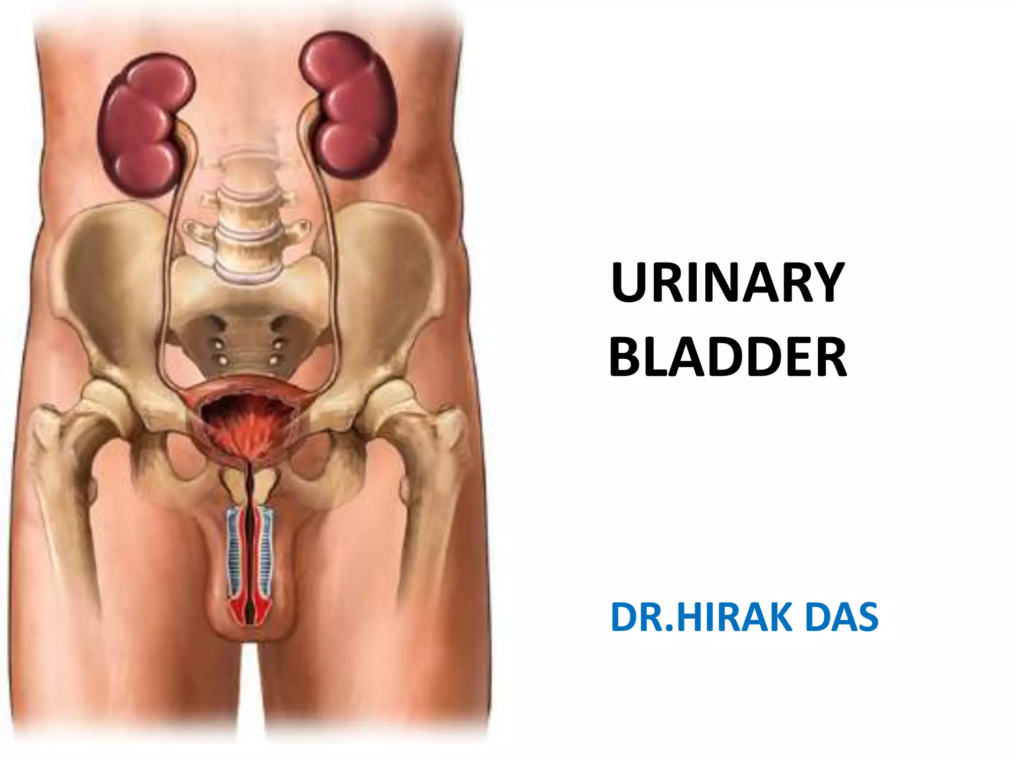
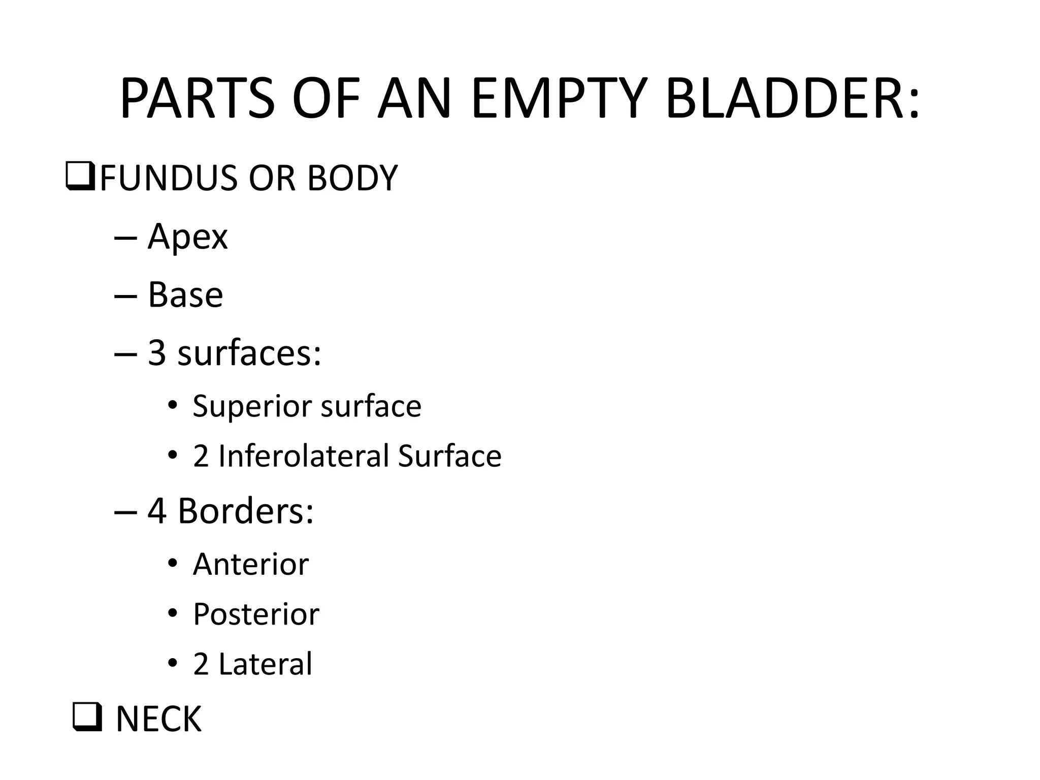
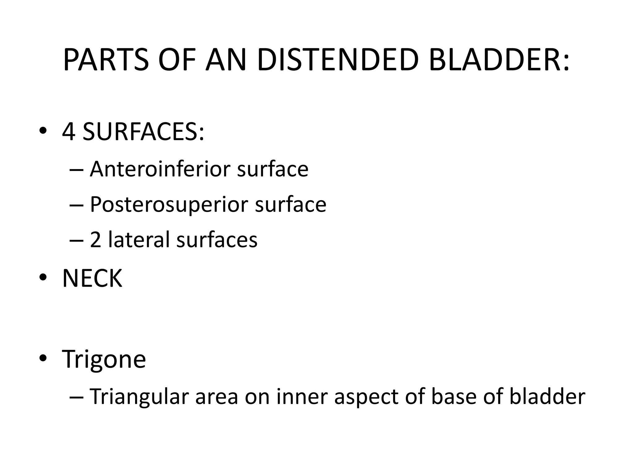
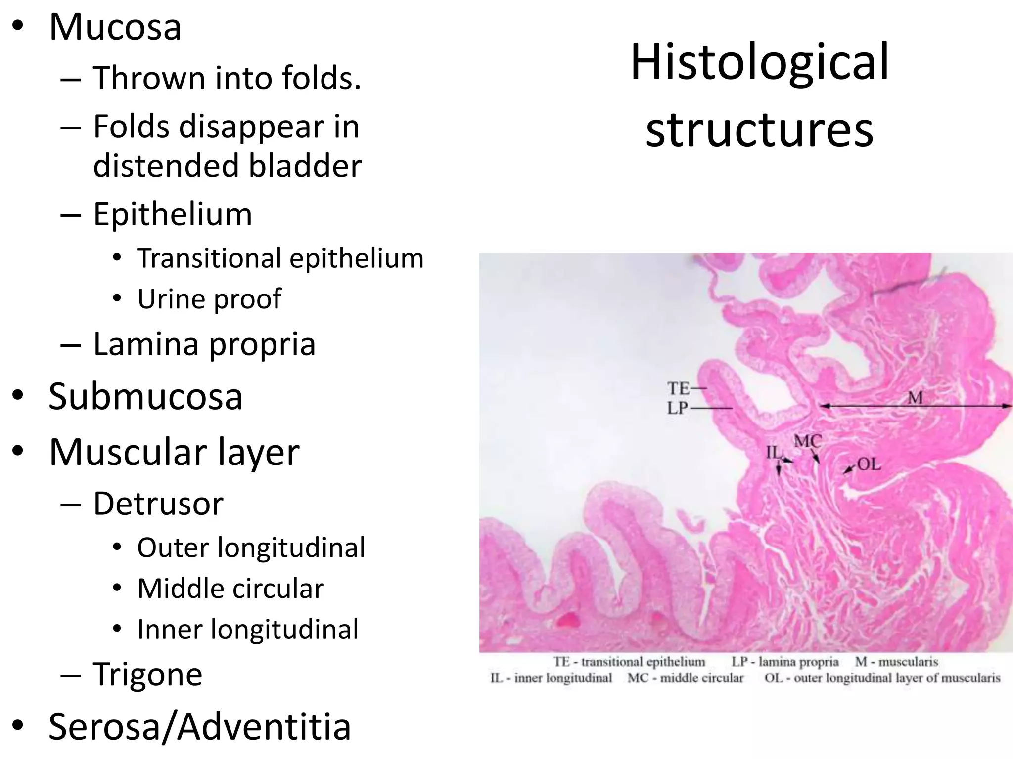
URINARY
BLADDER
DR.HIRAK DAS
URINARY
BLADDER
• A hollow organ that stores urine
• Location:
– Empty bladder: Pelvis
– Distended bladder:
Abdominopelvic
• Shape:
– Empty bladder: Tetrahedral
– Distended bladder: Ovoid
• Capacity
– 220 ml (average)
– Painful sensation start when
amount reach above 450 ml
– Above 800 ml, micturition goes
beyond involuntary control.
– Beyond 1 litre (approx): Bladder
will rupture Anatomical
capacity.
PARTS OF AN EMPTY BLADDER:
FUNDUS OR BODY
– Apex
– Base
– 3 surfaces:
• Superior surface
• 2 Inferolateral Surface
– 4 Borders:
• Anterior
• Posterior
• 2 Lateral
 NECK
PARTS OF AN DISTENDED BLADDER:
• 4 SURFACES:
– Anteroinferior surface
– Posterosuperior surface
– 2 lateral surfaces
• NECK
• Trigone
– Triangular area on inner aspect of base of bladder
Histological
structures
• Mucosa
– Thrown into folds.
– Folds disappear in
distended bladder
– Epithelium
• Transitional epithelium
• Urine proof
– Lamina propria
• Submucosa
• Muscular layer
– Detrusor
• Outer longitudinal
• Middle circular
• Inner longitudinal
– Trigone
• Serosa/Adventitia

Urinary bladder.pptx

  • 1.
  • 2.
    URINARY BLADDER • A holloworgan that stores urine • Location: – Empty bladder: Pelvis – Distended bladder: Abdominopelvic • Shape: – Empty bladder: Tetrahedral – Distended bladder: Ovoid • Capacity – 220 ml (average) – Painful sensation start when amount reach above 450 ml – Above 800 ml, micturition goes beyond involuntary control. – Beyond 1 litre (approx): Bladder will rupture Anatomical capacity.
  • 3.
    PARTS OF ANEMPTY BLADDER: FUNDUS OR BODY – Apex – Base – 3 surfaces: • Superior surface • 2 Inferolateral Surface – 4 Borders: • Anterior • Posterior • 2 Lateral  NECK
  • 4.
    PARTS OF ANDISTENDED BLADDER: • 4 SURFACES: – Anteroinferior surface – Posterosuperior surface – 2 lateral surfaces • NECK • Trigone – Triangular area on inner aspect of base of bladder
  • 5.
    Histological structures • Mucosa – Throwninto folds. – Folds disappear in distended bladder – Epithelium • Transitional epithelium • Urine proof – Lamina propria • Submucosa • Muscular layer – Detrusor • Outer longitudinal • Middle circular • Inner longitudinal – Trigone • Serosa/Adventitia