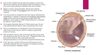
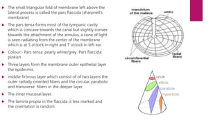
Tympanic membrane
development
 The first brancial arch
 Cleft forming the external auditory canal
 pouch forming the mucosal layer.
 The mesenchyme in between these two layers grow to form the
middle layer of tympanic membrane.
 Formed by 28th -30th week of gestation.
 Lies at the medial end of external auditory canal and
forms the majority of the lateral wall of tympanic cavity
thus forming a partition between the two cavities.
 Oval in shape, being broader above than below,
forming an angle of 55 degree with the floor of the
meatus.
 It is horizontal in newborns but the reason behind this
angulation in adults is because the anterior and inferior
walls of the external canal grows longer than the
posterior and superior walls. Adult position is reached
by about 5 years of age.
 Its longest diameter being 9-10mm (post.sup. To ant
inf) and shortest 8-9mm with a thickness of 0.1mm.
 The circumference is thickened to form the annulus
which sits in the sulcus, which is absent superiorly,
notch of rivinus.
 The tympanic bone is the one housing the sulcus and
the squamous part forms the roof . Therefore there are
two suture lines in the canal wall with the
tympanosquamous ant and the tympanomastoid post.
 From the sup. Limits of the sulcus the annulus runs
centrally towards the ant. Process of malleus forming
the ant and post malleolar folds.
 The small triangular fold of membrane left above the
lateral process is called the pars flaccida (sharpnell’s
membrane)
 The pars tensa forms most of the tympanic cavity
which is concave towards the canal but slightly convex
towards the attachment of the annulus, a cone of light
is seen radiating from the center of the membrane
which is at 5 o’clock in right and 7 o’clock in left ear.
 Colour:- Pars tensa: pearly white/grey Pars flaccida:
pinkish
 Three layers form the membrane outer epithelial layer
the epidermis.
 middle firbrous layer which consist of of two layers the
outer radially oriented fibers and the circular, parabolic
and transverse fibers in the deeper layer.
 The inner mucosal layer.
 The lamina propia in the flaccida is less marked and
the orientation is random.
 Blood supply :-
 deep auricular artery branch of maxillary artery- external side
 mucosal side :-
1. ant. Tympanic artery,
2. stylomastoid branch of the post auricular artery
3. middle meningeal artery (probable)
 Nerve supply :-
1. auriculotemporal nerve V3 (ant half of lateral surface),
2. auricular branch of vagus(post half of lateral surface),
3. tympanic branch of glossopharyngeal
 Both the vascular and nerve supply have anastomosis in the
lamina propia and both are relatively sparse in the middle part
of the post half of the tympanic membrane.
 The chorda tympani nerve which enter the middle ear through
the ant. canaliculus runs between the fibrous and mucosal layer
of the tympanic membrane across upper part of the handle of
malleus above the attachment of TT and then continues within
the membrane but below the level of the post mallelor fold.
 Prussak space is found between the pars flaccida and the
neck of the malleus, bounded by the lateral malleolar fold.
There are two openings in it called the ant and post pouch
of von troltsch
 the epithelium of the external canal is diff from the
epidermis of rest of the body where epidermis grows from
the basal layer to surface , here they grow outward and
obliquely so that the layer migrates outward towards the
canal opening with rate of 0.1mm/day.
physiology
 The realtively large area of the tympanic membrane to the stapes footplate 18:1 contributes to overcoming the
acoustic impedence mismatch at the air fluid interface between the middle and inner ear.
 The incident sound from the EAC hits the membrane and sets up a travelling wave, which is mainly collected at the
rim of the membrane.
 This is then conducted to the umbo and coupled to the manubrium of the malleus.
 Note:- hearing is better preserved with central perforation than marginal as energy transfer from the canal to the
membrane is via the rim of the membrane.
 The reflectance of the membrane is high at the lower frequencies below 1KHZ while it is lowest between 1-4 khz,
indicating that between these frequencies maximum energy is delivered to the cochlea and the middle ear is most
efficient.
 At higher frequencies the reflectance rises again with near total at 15khz.
 Provides sonic shielding of the round window membrane

Tympanic membrane.pptx

  • 1.
  • 2.
    development  The firstbrancial arch  Cleft forming the external auditory canal  pouch forming the mucosal layer.  The mesenchyme in between these two layers grow to form the middle layer of tympanic membrane.  Formed by 28th -30th week of gestation.
  • 3.
     Lies atthe medial end of external auditory canal and forms the majority of the lateral wall of tympanic cavity thus forming a partition between the two cavities.  Oval in shape, being broader above than below, forming an angle of 55 degree with the floor of the meatus.  It is horizontal in newborns but the reason behind this angulation in adults is because the anterior and inferior walls of the external canal grows longer than the posterior and superior walls. Adult position is reached by about 5 years of age.  Its longest diameter being 9-10mm (post.sup. To ant inf) and shortest 8-9mm with a thickness of 0.1mm.  The circumference is thickened to form the annulus which sits in the sulcus, which is absent superiorly, notch of rivinus.  The tympanic bone is the one housing the sulcus and the squamous part forms the roof . Therefore there are two suture lines in the canal wall with the tympanosquamous ant and the tympanomastoid post.  From the sup. Limits of the sulcus the annulus runs centrally towards the ant. Process of malleus forming the ant and post malleolar folds.
  • 4.
     The smalltriangular fold of membrane left above the lateral process is called the pars flaccida (sharpnell’s membrane)  The pars tensa forms most of the tympanic cavity which is concave towards the canal but slightly convex towards the attachment of the annulus, a cone of light is seen radiating from the center of the membrane which is at 5 o’clock in right and 7 o’clock in left ear.  Colour:- Pars tensa: pearly white/grey Pars flaccida: pinkish  Three layers form the membrane outer epithelial layer the epidermis.  middle firbrous layer which consist of of two layers the outer radially oriented fibers and the circular, parabolic and transverse fibers in the deeper layer.  The inner mucosal layer.  The lamina propia in the flaccida is less marked and the orientation is random.
  • 5.
     Blood supply:-  deep auricular artery branch of maxillary artery- external side  mucosal side :- 1. ant. Tympanic artery, 2. stylomastoid branch of the post auricular artery 3. middle meningeal artery (probable)  Nerve supply :- 1. auriculotemporal nerve V3 (ant half of lateral surface), 2. auricular branch of vagus(post half of lateral surface), 3. tympanic branch of glossopharyngeal  Both the vascular and nerve supply have anastomosis in the lamina propia and both are relatively sparse in the middle part of the post half of the tympanic membrane.  The chorda tympani nerve which enter the middle ear through the ant. canaliculus runs between the fibrous and mucosal layer of the tympanic membrane across upper part of the handle of malleus above the attachment of TT and then continues within the membrane but below the level of the post mallelor fold.
  • 6.
     Prussak spaceis found between the pars flaccida and the neck of the malleus, bounded by the lateral malleolar fold. There are two openings in it called the ant and post pouch of von troltsch  the epithelium of the external canal is diff from the epidermis of rest of the body where epidermis grows from the basal layer to surface , here they grow outward and obliquely so that the layer migrates outward towards the canal opening with rate of 0.1mm/day.
  • 7.
    physiology  The realtivelylarge area of the tympanic membrane to the stapes footplate 18:1 contributes to overcoming the acoustic impedence mismatch at the air fluid interface between the middle and inner ear.  The incident sound from the EAC hits the membrane and sets up a travelling wave, which is mainly collected at the rim of the membrane.  This is then conducted to the umbo and coupled to the manubrium of the malleus.  Note:- hearing is better preserved with central perforation than marginal as energy transfer from the canal to the membrane is via the rim of the membrane.  The reflectance of the membrane is high at the lower frequencies below 1KHZ while it is lowest between 1-4 khz, indicating that between these frequencies maximum energy is delivered to the cochlea and the middle ear is most efficient.  At higher frequencies the reflectance rises again with near total at 15khz.  Provides sonic shielding of the round window membrane