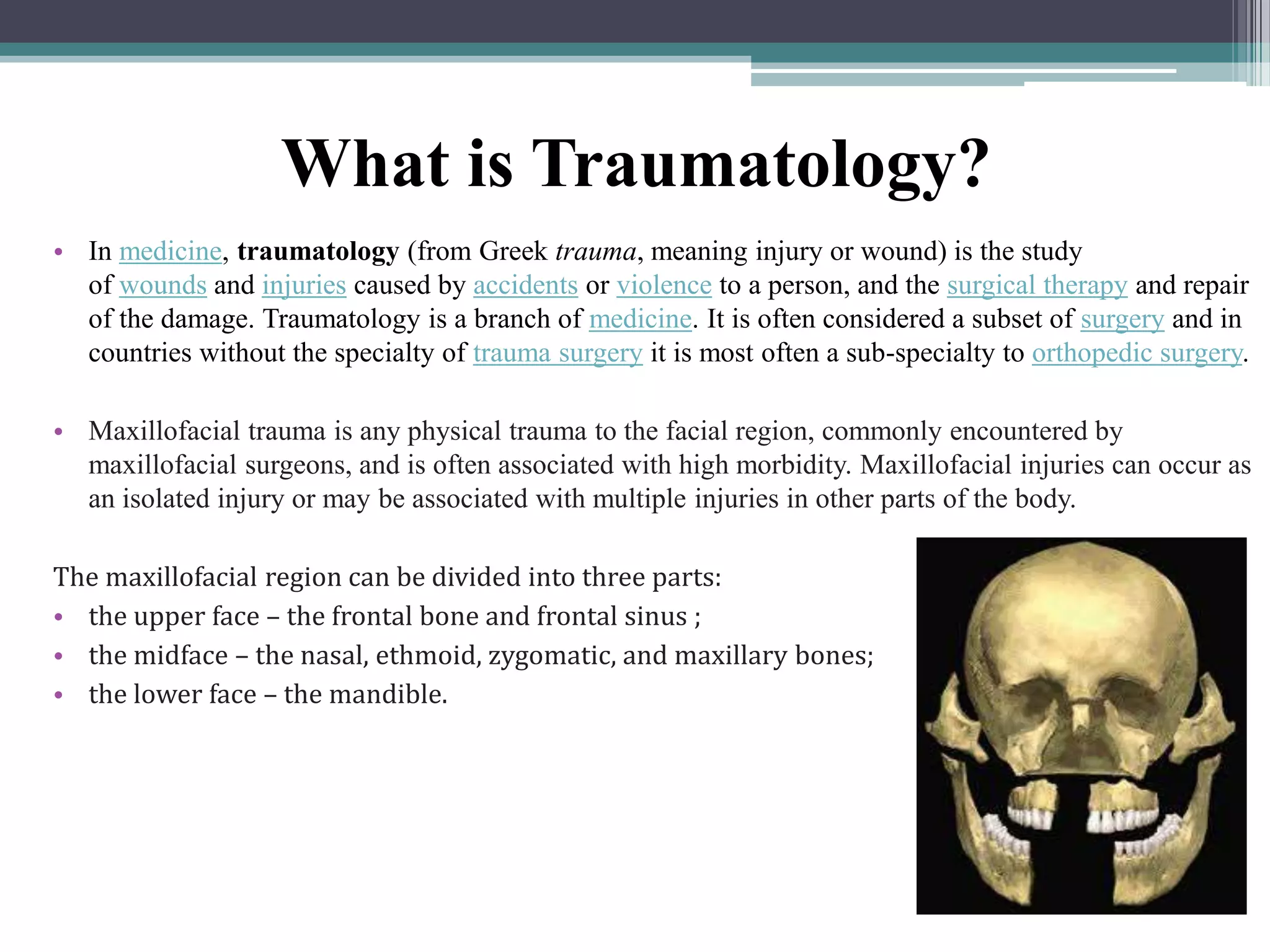
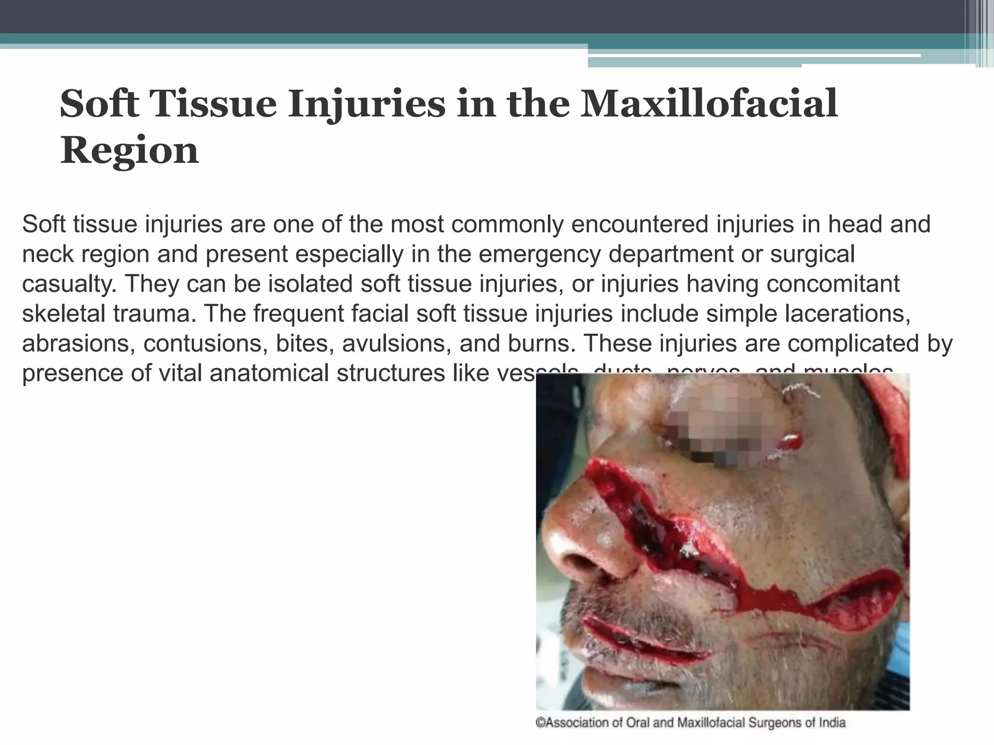
Traumatology of the maxillofacial region. Soft
tissue injuries of the face. Fractures of the bones
of the face. Trauma of the teeth and alveolar
processes of the jaws.
Lecturer: ADYLBEK uulu ISKENDER
CONTENT PLAN
• Definition of Traumatology;
• Soft tissue injuries of the face;
• Classification of soft tissue injuries of the face
• Fractures of the bones of the face
• Dislocations and fractures of teeth
What is Traumatology?
• In medicine, traumatology (from Greek trauma, meaning injury or wound) is the study
of wounds and injuries caused by accidents or violence to a person, and the surgical therapy and repair
of the damage. Traumatology is a branch of medicine. It is often considered a subset of surgery and in
countries without the specialty of trauma surgery it is most often a sub-specialty to orthopedic surgery.
• Maxillofacial trauma is any physical trauma to the facial region, commonly encountered by
maxillofacial surgeons, and is often associated with high morbidity. Maxillofacial injuries can occur as
an isolated injury or may be associated with multiple injuries in other parts of the body.
The maxillofacial region can be divided into three parts:
• the upper face – the frontal bone and frontal sinus ;
• the midface – the nasal, ethmoid, zygomatic, and maxillary bones;
• the lower face – the mandible.
Soft Tissue Injuries in the Maxillofacial
Region
Soft tissue injuries are one of the most commonly encountered injuries in head and
neck region and present especially in the emergency department or surgical
casualty. They can be isolated soft tissue injuries, or injuries having concomitant
skeletal trauma. The frequent facial soft tissue injuries include simple lacerations,
abrasions, contusions, bites, avulsions, and burns. These injuries are complicated by
presence of vital anatomical structures like vessels, ducts, nerves, and muscles.
The most common etiology of facial soft tissue injuries varies
according to the age, sex, and geographical distribution of the
population. Facial soft tissue trauma tends to occur in certain areas of
the head depending on the causative mechanism. It typically includes
the T-shaped area that includes forehead, nose, lips, and chin,
followed by the occiput and anterior
temporal areas.
• Falls are by far the most common cause, accounting for 48–51% of the
injuries;
• Non-fall impacts. Approximately 16% of facial soft tissue trauma is the
result of a non-fall impact with structural element such a door, wall, or
window frame;
• Assaults account for 16–32% of facial soft tissue injuries;
• Road traffic accidents cause 6–13% of facial trauma where soft tissue
injuries may occur alone or in combination with other fractures;
• Sporting injuries account for about 8% of facial soft tissue trauma;
• Others include occupational injuries, bites from humans or animals,
(A)Based on mechanism of injuries
(I)Mechanical or physical injuries:
1. When injury is caused due to blunt force:
a) Abrasions
b) Contusions
c) Lacerations
2. When injury is caused due to sharp force:
a) Incised wounds
b) Chop wounds
c) Stab/punctured wounds
(II)Thermal injuries:
1. Due to excessive cold: e.g., frostbite
2. Due to moist heat: e.g. burns and scald wounds
(III)Chemical injuries: Due to corrosive acids and alkalis
(IV)Miscellaneous: Electricity, lightning, etc.
(V)Explosions: Blast injuries
(B)Legal classification
1.Simple—soft tissue injuries without much
tissue loss and can be managed conservatively.
Heals rapidly without any permanent deformity.
2.Grievous injuries - injuries that endanger
life and cause severe disfigurement or
deformities like permanent hearing loss, vision
loss, and severe head injuries.
3.Dangerous—the grievous injuries that
endanger life immediately after impact
(C)Based on the communication of the injury
with the external environment:
• Closed wound: Only the underlying tissue and/or structures are
damaged without breaking the skin. Examples of closed wounds include
hematomas, contusions, and crush injuries. These types of wounds are
not contaminated and, hence, heal on their own without any sequelae.
• Open wounds: There is a break in the skin, which exposes the
underlying structures to the external environment. Open wounds include
simple and complex lacerations, avulsions, punctures, abrasions,
accidental tattooing, and retained foreign body with a tendency to heal
with scarring. These injuries require extensive exploration and
debridement followed by a course of antibiotic regimen for uneventful
healing
(D)According to the facial subunit(s) involved. The
major esthetic subunits on the face are the scalp, forehead,
nose, periorbital region, cheek, perioral region, auricle, and
neck
(E)Based on additional injuries to the related
structures;
(a)Injuries to nerve—The nerve injuries are most commonly encountered in
cases of open wounds. The nerve injuries are further classified into neuropraxia,
axonotmesis, and neurotmesis (Seddon’s classification of nerve injuries). The
most commonly encountered nerve injuries in maxillofacial region are the facial
nerve and trigeminal nerve.
(b)Injuries to arteries and veins—The face is a highly vascular region where
even a small injury leads to significant bleeding. Any wound in maxillofacial
region should be attended in emergency setting at the earliest due to chances of
injury to the extensive vascular network which necessitates hemostasis.
(c)Injuries to parotid duct—Seen in cases of deep lacerations on cheek at the
region of parotid duct. If undiagnosed and untreated, this injury can lead to
cumbersome sequelae of parotid fistula. Parotid duct injury is suspected if the
laceration involves an imaginary line joining the tragus of the ear to the upper lip.
(F)Rank and Wakefield classification of wounds;
(a)Tidy wounds—The wounds that are inflicted by sharp instruments
like surgical blades and contain no devitalized tissue are called tidy.
These wounds are closed primarily. Examples are surgical incisions,
cuts from glass, and knife wounds.
(b)Untidy wounds—Untidy wounds result from crushing, tearing,
avulsion, vascular injury, or burns and contain devitalized tissue. Such
wounds must be managed by wound excision. The devitalized tissue is
excised, and the untidy wound is converted into the tidy wound before
proper closure is achieved. The chances of infection are high if
inadequately managed.
(G)CDC classification of surgical wounds;
Surgical wounds can be classified into four different categories depending
on the bacterial load, the risk of infection, and where the wound is located
on the body.
• Class I: Clean wounds. They show no signs of infection or inflammation.
They often involve the eye, skin, or vascular system. It is often due to
non-penetrating (blunt) trauma.
• Class II: Clean-contaminated wounds. Although the wound may not
show signs of infection, it is at an increased risk of becoming infected
because of its location. For example, surgical wounds in the respiratory
tract like oropharynx and gastrointestinal tract may be at a high risk of
becoming infected.
• Class III: Contaminated wound. A surgical wound in which an outside
object has come into contact with the skin and has a high risk of
infection. For example, a gunshot wound may contaminate the skin
around where the surgical repair occurs.
• Class IV: Dirty-contaminated or infected wounds. The wounds that
have been exposed to fecal material and have a high bacterial load.
Contusions of the face.
• The cause of this damage is a direct blow with a
solid object (during a fight, boxing, from a
moving vehicle) or a blow when falling.
Symptoms.
• Abrasions, scratches, superficial wounds,
swelling and hematoma, especially in the
eyelids, lips, cheeks. Often, swelling makes it
difficult to recognize more severe injuries -
fractures.
contusions
contusions
• First aid for bruises: to ensure rest, to apply cold to the
site of the bruise during the first day; abrasions, small
wounds should be washed with an aqueous antiseptic
solution, dried, lubricated with an alcohol-containing
antiseptic solution. Do not apply a bandage.
• Treatment of bruises, hematomas is conservative. From
the 2nd day on the bruised area of UHF therapy (6-7
procedures); for resorption of hematoma - paraffin or
ozokerite (10-12 procedures) in combination with
electrophoresis. Extensive hematomas are opened to
avoid scarring. The duration of treatment is 2-2.5 weeks.
Facial wounds.
• Facial wounds can be superficial, deep, penetrating.
Features of facial injuries: heavy bleeding with damage
to large arteries, veins; pronounced gaping of the edges
when dissecting the facial muscles of the face;
discrepancy between the type of victim and the severity
of the damage due to the contraction and displacement
of the skin-muscle flaps; masking significant blood loss
with damage to the tongue, soft palate, as the patient
swallows blood; aspiration asphyxia in victims in an
unconscious state.
• First aid to the unconscious victim is provided
according to the following algorithm:
• 1. Lay the victim on his stomach or on his side with the
injured side.
• 2. Turn your head towards the injury.
• 3. Apply an aseptic pressure bandage.
• 4. Check the patency of the respiratory tract by removing
blood clots, vomit, foreign bodies (broken teeth,
dentures) from the oral cavity.
•5. Remove the tongue (to the teeth!) to prevent asphyxia.
First aid to the victim
• Note. It is forbidden to remove the tongue from the mouth in order to
avoid its biting during transportation.
• 6. Fix the tongue with an air duct or a pin and bandage.
• 7. Carry out the simplest anti-shock measures.
• 8. Transport the victim to the hospital.
Treatment
PST(primary surgical treatment)of facial wounds are performed
during the first day.
• The operation should be performed by an maxillofacial surgeon to
prevent possible complications: deformation of the face, limitation of
mobility of the lower jaw, narrowing of the nasal openings, etc.
In the treatment of facial wounds, sulfonamides and antibiotics are used
topically.
• Suturing
Fractures of the nose.
• The causes of a broken nose are a direct blow with a
fist, a heavy object or a blow with a face on a hard
surface during a fall, accidents on transport. As a rule,
not only the bones of the nose are damaged, but also
the cartilage septum with a violation of the mucous
membrane, so fractures are considered open.
Symptoms.
• Change in the usual shape of the nose, difficulty
breathing, nosebleeds, pathological mobility, local
soreness. Rapidly increasing edema and hematoma
often mask the picture of a fracture.
• First aid includes: the application of an aseptic sling-like
bandage, cold on the bridge of the nose, transportation
of the victim in a sitting position with his head lowered to
the chest.
Treatment.
• PST of the wound and the adjustment of nasal fragments
is carried out in the first hours or a day after the injury.
The adjustment is performed under local anesthesia
from inside the nasal cavity. To fix the corrected
fragments and stop bleeding, tamponade of the upper
and middle nasal passages is performed with gauze
strips lubricated with penicillin ointment for 5-7 days.
Tampons are changed every other day. Inside,
sulfonamides are prescribed for 10-15 days. It is
forbidden to blow your nose.:
Fractures of the zygomatic arch.
• The cause of these fractures is a direct blow to the zygomatic region. By
localization, fractures of the anterior part of the arch, posterior and both
departments are distinguished.
Symptoms:
• - with a fracture of the anterior section, flattening of the zygomatic region,
numbness of the skin of the suborbital region and unevenness of the lower
edge of the orbit are observed. In case of damage to the walls of the maxillary
(maxillary) sinus
• - bleeding from the corresponding half of the nose. If the upper wall of the
maxillary sinus is injured (it is also the lower wall of the orbit), then the eyeball is
displaced, diplopia occurs,
• - fractures of the posterior part of the zygomatic arch with displacement are
characterized by a limitation of mobility of the lower jaw (it is impossible to open
the mouth);
• - with severe fractures of both parts of the zygomatic arch, cerebral symptoms
appear (loss of consciousness, vomiting, nausea)
• - signs of a combined craniocerebral injury.
• First aid for isolated fractures of the zygomatic arch
is limited to the creation of rest, the local application
of cold, the application of an aseptic dressing if
necessary, the victim is transported to the hospital
in a sitting position or on foot accompanied. In the
case of a combined craniofacial injury, assistance is
provided according to the algorithm of action at the
scene of the incident during TBI. Treatment is
carried out in a specialized maxillofacial hospital.
Correction of displaced areas of the zygomatic bone
is best carried out in the first hours or days after the
injury to eliminate facial deformities, restore mobility
of the lower jaw and release the suborbital nerve
from compression.
Fractures of the lower jaw
• They occur as a result of a direct blow to the face with a fist, a heavy
object or a blow to the face when falling.
• By localization, fractures of the body, chin, angle, branch and alveolar
part of the jaw are distinguished.
• They can be single, two-sided or multiple. If the mucous membrane is
damaged, then these are open fractures.
Symptoms.
• The main symptoms of a fracture of the lower jaw: hemorrhage, pain,
impaired chewing, closing of the teeth, speech, eating, the correct ratio
of the dentition. With compression of the jaws, pain increases at the site
of bone damage; on palpation, crepitation of fragments is felt. Bilateral
fractures of the chin section of the lower jaw are especially dangerous,
since the tongue sinks and asphyxia occurs. Complications of fractures
of the lower jaw: traumatic osteomyelitis, perimaxillary phlegmon, and
with improper fusion of fragments - a false joint or persistent cicatricial
contracture of the jaw.
• First aid. When providing first aid, it is necessary
to ensure the patency of the respiratory tract, to
stop bleeding.
• Immobilization is best carried out with a stationary
rigid chin sling or sling-like bandage.
• Treatment of fractures of the lower jaw is carried
out in a hospital by maxillofacial surgeon.
• The main method of treatment is conservative:
fixation with wire splints for 3-6 weeks, depending
on the location and nature of the fracture. The
nurse is obliged to teach the patient the elements
of oral care and food intake.
Fractures of the upper jaw
• They arise from a strong direct blow to the jaw
area or when falling from a height, transport
accidents.
• By localization, fractures are distinguished: in
the region of the alveolar process; at the
junction of the jaw with the frontal and zygomatic
bones; passing through the zygomatic bone, eye
socket and nasal root.
• The last type of fracture is the most severe, as it
is combined with a fracture of the base of the
skull.
Symptoms.
• Victims complain of general weakness, headaches and
pain when swallowing.
• Depending on the localization of the fracture,
deformation in the nose, flattening of the middle part of
the face are observed.
• When the mouth is opened, the fragments of the jaw fall
down, the face lengthens.
• The closing of the teeth (bite) is disturbed.
• There is bleeding from the nose, oral cavity.
• There is a symptom of "glasses" of different origin.
• If this is local damage to the bones of the upper
jaw and soft tissues, then this symptom occurs
immediately after the injury and the hemorrhage
spreads widely beyond the circular eye muscle.
• If this is local damage to the bones of the upper
jaw and soft tissues, then this symptom occurs
immediately after the injury and the hemorrhage
spreads widely beyond the circular eye muscle.
Applying bandages
• When providing first aid to the victim, it is necessary to stop
bleeding, prevent aspiration of blood and vomit.
• With the integrity of the lower jaw and the presence of a sufficient
number of teeth on both jaws, apply a sling-like bandage, pressing
the lower jaw to the upper, or perform immobilization with a rigid
chin sling.
• If the teeth are insufficient or both jaws are damaged, a slingshot
bandage cannot be used.
• In this case, the air duct should be inserted, then a standard rigid
sling should be applied.
• Treatment of fractures of the upper jaw, as well as the lower, is
carried out in a hospital by maxillofacial surgeon.
Dislocations of the lower jaw.
• The cause of this injury is a wide opening of the
mouth during yawning, screaming, singing,
dental treatment.
• Typical dislocations of the lower jaw anteriorly,
when the articular head of the
temporomandibular joint comes out of the
articular cavity and palpates under the
zygomatic arch. The peculiarity of these
dislocations is that the capsule of the joint does
not rupture, but only stretches.
Dislocations of the lower jaw.
Symptoms:
—with a bilateral anterior dislocation, the mouth
is wide open, closing the teeth is impossible,
speech is slurred; swallowing and eating are
difficult; chewing is not possible; there is
abundant salivation.
• On palpation, a deep fossa is determined in
front of the goat;
• —with a unilateral dislocation, the mouth is half-
open, the chin is shifted to a healthy side.
Dislocations of the lower jaw.
• First aid: to ensure the rest of the lower jaw in a forced
position with the help of a sling-like bandage, to provide
care for the separation of saliva (using an oilcloth bib, a
napkin) and to quickly deliver the victim to the hospital to
prevent dehydration due to excessive salivation.
Treatment is conservative.
• To ease the tension of the masticatory muscles, a 2%
solution of novocaine (2-3 ml) or another anesthetic is
injected.
• The victim is seated so that his lower jaw is at the level of
the doctor's elbow, the assistant fixes the head.
• The victim is seated so that his lower jaw is at the level of
the doctor's elbow, the assistant fixes the head.
• The patient is applied a sling-like bandage for 7-10 days,
food is prescribed crushed, semi-liquid food.
Fracture of the orbit.
Edema of the periocular region, subcutaneous emphysema.
Weakening of sensitivity at the site of innervation of the lower orbital
nerve.
This involves the back of the nose, cheek, upper and lower eyelid, as
well as the upper teeth and gums.
Inability to look up, due to the capture of the lower rectus muscle.
Diplopia. Hemorrhages and swelling create a compaction of the
tissues of the orbit between the lower rectum, lower oblique muscles
and periosteum.
This makes the eyeball less mobile.
Displacement of the eyeball deeper (enophthalmos) occurs with
serious fractures.
Crepitation (crisp sound) above the lower part of the eye socket.
• First aid: instillation into the affected eye of
antibacterial drugs (30% sodium sulfacil solution,
0.25% chloramphenicol solution, etc.) anesthesia,
any analgesic you have (1-2 tab. "Analgina",
"Pentalgina", "Revalgina", etc.) taking sedatives
(tincture of valerian, motherwort, etc.) by applying
an aseptic dressing.
• A small piece of clean cloth or bandage should
completely cover the eye area. It is fixed with a
patch or bandage without pressure.
Foreign bodies are not removed from the eye!
• Urgent hospitalization in an ophthalmological
institution or in a trauma center with an eye
department.
Dislocations and fractures of
teeth
• Trauma to the teeth is a fairly common dental problem, which is most
often found in children. According to statistics, traumatic tooth injuries are
detected in 41-50% of preschool children and up to 33% of school
children. Children under 6 years old learn to walk, fall from a height, from
slides, swings, etc. School-age children are engaged in sports (hockey,
karate, football), are engaged in active games on the street, ride skates
and roller skates. All these are situations in which the risk of tooth injury
increases.
• Acute injuries to the teeth arise from simultaneously acting causes. Often,
patients, especially children, seek help not immediately, but after a long
time, and therefore it is difficult to diagnose and treat such lesions. The
type of injury is associated with the force of the blow, its direction, the
place of application. An important role is played by the age of the patient,
the condition of the teeth and periodontium.
• Currently, there is more than one classification system for dental injuries,
but the most common is the WHO approach.
Classification of dislocations and
fractures
Currently, there is more than one classification system for dental injuries, but the
most common is the WHO approach. Based on it, dental injuries are represented
by 8 classes:
• I-class - bruises of the teeth, which are further accompanied by cracks in the
enamel;
• II-class - fractures of an uncomplicated form;
• III-class - complicated fracture of the apex of the tooth - damage to the crown;
• IV-class - a complete fracture of the apex of the tooth;
• V-class - longitudinal fractures that affect the root and crown of the tooth
• VI-class - fracture of the roots of the tooth;
• VII-class - incomplete dislocation;
• VIII-class - complete dislocation.
Dislocations of teeth
• Dislocation of the tooth is a traumatic injury to
the tooth, as a result of which its connection with
the socket is broken.
• Dislocation of the tooth occurs most often as a
result of a blow to the crown of the tooth. More
often than others, dislocations are subjected to
frontal teeth on the upper jaw and less often on
the lower. Dislocations of premolars and molars
occur most often with careless removal of
neighboring teeth using an elevator.
Incomplete dislocation
• With an incomplete dislocation, part of the periodontal
fibers is torn. Fibers that have retained continuity are
usually stretched to one degree or another
• Incomplete dislocation is characterized by a change in
the position of the crown of the tooth in the dentition and
the root in relation to the walls of the alveoli
Полный вывих
• With a complete dislocation, the tissues of the entire
periodontium and the circular ligament of the tooth rupture.
This leads to tooth loss from the alveoli under the action of
the applied force or the tooth's own weight. The
neurovascular bundle is always ruptured. A fracture of the
edge of the alveoli may occur. Sometimes the tooth is held in
the alveolus by single preserved fibers of the circular ligament
of the tooth or due to the adhesive properties of two wet
surfaces: the root of the tooth and the wall of the alveoli.
Immobilization with composite materials and fixing arcs
Bus-bracket with fixing ligature wire
Treatment of dislocations
Treatment of incomplete dislocation includes:
• Repositioning of teeth;
• Immobilization with fiberglass tape in combination
with light-curing composite material for 4 weeks or
fixation with a mouthguard or smooth splint-bracket;
• Cavity hygiene and a sparing diet;
• Examination after 1 month;
• when determining the death of the pulp - its
extirpation and filling of the root canal.
Treatment for a complete dislocation includes:
• With a complete dislocation of the tooth, its
replantation is possible (no later than three days
after the injury),
• pulp extirpation and canal filling;
• replantation;
• Immobilization for 4 weeks with a mouthguard or
a smooth staple splint, fiberglass tape in
combination with a light-curing composite
material;
• a sparing diet.
Treatment of an injected dislocation includes:
• Reposition and immobilization for 4 weeks with
a mouthguard or smooth splint-bracket,
fiberglass tape in combination with light-curing
composite material;
• If it is impossible to reposition - tooth extraction
Replantation
• Replantation is the return of a tooth to its own socket. There
are simultaneous and delayed replantation of the tooth.
• With a one-time visit, the tooth is prepared for replantation,
the canal of its root is filled and the actual replantation is
carried out with subsequent splinting.
• With delayed replantation, the dislocated tooth is washed,
immersed in a physiological solution with an antibiotic and
placed temporarily (until replantation) in the refrigerator. After
a few hours or days, the tooth is trepanated, filled and
replanted.
Переломы зубов
The causes of a tooth fracture are mainly mechanical
injuries resulting from a blow or fall. The frontal teeth
of the upper jaw are more prone to fractures than the
teeth of the lower jaw, often tooth fractures are
combined with their incomplete dislocations.
Tooth fractures are:
• by type: with the opening of the pulp chamber and
without opening;
• by type: transverse, longitudinal and splintered;
• by location: in the crown area, in the neck of the
tooth and the root.
Fracture with opening the pulp chamber
Fracture without opening the pulp chamber
Cracks in enamel and dentin
Fracture (chip) of the crown in the enamel area
Chipping of the crown in the enamel area
Fracture of the crown in the enamel and dentin zone
Fracture of the tooth neck
Fracture of the root or apex of the root
Schematic representation of individual types of tooth fracture: a - at the
level of enamel and dentin without and with the opening of the tooth cavity,
b - at the level of the neck of the tooth, c - transverse, at the level of the
upper third of the root, g - longitudinal, d - transverse, at the level of the
middle third of the root
Treatment of fractures of the teeth
• The tactics of treatment in relation to such teeth
are solved collectively by dentists: therapists,
surgeons, orthopedists.
Thank you for your attention!!!
With love your DENTISTRY teacher
– ADYLBEK uulu ISKENDER

Traumatology of the maxillofacial region. Soft tissue injuries of the face.

  • 1.
    Traumatology of themaxillofacial region. Soft tissue injuries of the face. Fractures of the bones of the face. Trauma of the teeth and alveolar processes of the jaws. Lecturer: ADYLBEK uulu ISKENDER
  • 2.
    CONTENT PLAN • Definitionof Traumatology; • Soft tissue injuries of the face; • Classification of soft tissue injuries of the face • Fractures of the bones of the face • Dislocations and fractures of teeth
  • 3.
    What is Traumatology? •In medicine, traumatology (from Greek trauma, meaning injury or wound) is the study of wounds and injuries caused by accidents or violence to a person, and the surgical therapy and repair of the damage. Traumatology is a branch of medicine. It is often considered a subset of surgery and in countries without the specialty of trauma surgery it is most often a sub-specialty to orthopedic surgery. • Maxillofacial trauma is any physical trauma to the facial region, commonly encountered by maxillofacial surgeons, and is often associated with high morbidity. Maxillofacial injuries can occur as an isolated injury or may be associated with multiple injuries in other parts of the body. The maxillofacial region can be divided into three parts: • the upper face – the frontal bone and frontal sinus ; • the midface – the nasal, ethmoid, zygomatic, and maxillary bones; • the lower face – the mandible.
  • 4.
    Soft Tissue Injuriesin the Maxillofacial Region Soft tissue injuries are one of the most commonly encountered injuries in head and neck region and present especially in the emergency department or surgical casualty. They can be isolated soft tissue injuries, or injuries having concomitant skeletal trauma. The frequent facial soft tissue injuries include simple lacerations, abrasions, contusions, bites, avulsions, and burns. These injuries are complicated by presence of vital anatomical structures like vessels, ducts, nerves, and muscles.
  • 5.
    The most commonetiology of facial soft tissue injuries varies according to the age, sex, and geographical distribution of the population. Facial soft tissue trauma tends to occur in certain areas of the head depending on the causative mechanism. It typically includes the T-shaped area that includes forehead, nose, lips, and chin, followed by the occiput and anterior temporal areas. • Falls are by far the most common cause, accounting for 48–51% of the injuries; • Non-fall impacts. Approximately 16% of facial soft tissue trauma is the result of a non-fall impact with structural element such a door, wall, or window frame; • Assaults account for 16–32% of facial soft tissue injuries; • Road traffic accidents cause 6–13% of facial trauma where soft tissue injuries may occur alone or in combination with other fractures; • Sporting injuries account for about 8% of facial soft tissue trauma; • Others include occupational injuries, bites from humans or animals,
  • 6.
    (A)Based on mechanismof injuries (I)Mechanical or physical injuries: 1. When injury is caused due to blunt force: a) Abrasions b) Contusions c) Lacerations 2. When injury is caused due to sharp force: a) Incised wounds b) Chop wounds c) Stab/punctured wounds (II)Thermal injuries: 1. Due to excessive cold: e.g., frostbite 2. Due to moist heat: e.g. burns and scald wounds (III)Chemical injuries: Due to corrosive acids and alkalis (IV)Miscellaneous: Electricity, lightning, etc. (V)Explosions: Blast injuries
  • 7.
    (B)Legal classification 1.Simple—soft tissueinjuries without much tissue loss and can be managed conservatively. Heals rapidly without any permanent deformity. 2.Grievous injuries - injuries that endanger life and cause severe disfigurement or deformities like permanent hearing loss, vision loss, and severe head injuries. 3.Dangerous—the grievous injuries that endanger life immediately after impact
  • 8.
    (C)Based on thecommunication of the injury with the external environment: • Closed wound: Only the underlying tissue and/or structures are damaged without breaking the skin. Examples of closed wounds include hematomas, contusions, and crush injuries. These types of wounds are not contaminated and, hence, heal on their own without any sequelae. • Open wounds: There is a break in the skin, which exposes the underlying structures to the external environment. Open wounds include simple and complex lacerations, avulsions, punctures, abrasions, accidental tattooing, and retained foreign body with a tendency to heal with scarring. These injuries require extensive exploration and debridement followed by a course of antibiotic regimen for uneventful healing
  • 9.
    (D)According to thefacial subunit(s) involved. The major esthetic subunits on the face are the scalp, forehead, nose, periorbital region, cheek, perioral region, auricle, and neck (E)Based on additional injuries to the related structures; (a)Injuries to nerve—The nerve injuries are most commonly encountered in cases of open wounds. The nerve injuries are further classified into neuropraxia, axonotmesis, and neurotmesis (Seddon’s classification of nerve injuries). The most commonly encountered nerve injuries in maxillofacial region are the facial nerve and trigeminal nerve. (b)Injuries to arteries and veins—The face is a highly vascular region where even a small injury leads to significant bleeding. Any wound in maxillofacial region should be attended in emergency setting at the earliest due to chances of injury to the extensive vascular network which necessitates hemostasis. (c)Injuries to parotid duct—Seen in cases of deep lacerations on cheek at the region of parotid duct. If undiagnosed and untreated, this injury can lead to cumbersome sequelae of parotid fistula. Parotid duct injury is suspected if the laceration involves an imaginary line joining the tragus of the ear to the upper lip.
  • 10.
    (F)Rank and Wakefieldclassification of wounds; (a)Tidy wounds—The wounds that are inflicted by sharp instruments like surgical blades and contain no devitalized tissue are called tidy. These wounds are closed primarily. Examples are surgical incisions, cuts from glass, and knife wounds. (b)Untidy wounds—Untidy wounds result from crushing, tearing, avulsion, vascular injury, or burns and contain devitalized tissue. Such wounds must be managed by wound excision. The devitalized tissue is excised, and the untidy wound is converted into the tidy wound before proper closure is achieved. The chances of infection are high if inadequately managed.
  • 11.
    (G)CDC classification ofsurgical wounds; Surgical wounds can be classified into four different categories depending on the bacterial load, the risk of infection, and where the wound is located on the body. • Class I: Clean wounds. They show no signs of infection or inflammation. They often involve the eye, skin, or vascular system. It is often due to non-penetrating (blunt) trauma. • Class II: Clean-contaminated wounds. Although the wound may not show signs of infection, it is at an increased risk of becoming infected because of its location. For example, surgical wounds in the respiratory tract like oropharynx and gastrointestinal tract may be at a high risk of becoming infected. • Class III: Contaminated wound. A surgical wound in which an outside object has come into contact with the skin and has a high risk of infection. For example, a gunshot wound may contaminate the skin around where the surgical repair occurs. • Class IV: Dirty-contaminated or infected wounds. The wounds that have been exposed to fecal material and have a high bacterial load.
  • 12.
    Contusions of theface. • The cause of this damage is a direct blow with a solid object (during a fight, boxing, from a moving vehicle) or a blow when falling. Symptoms. • Abrasions, scratches, superficial wounds, swelling and hematoma, especially in the eyelids, lips, cheeks. Often, swelling makes it difficult to recognize more severe injuries - fractures. contusions contusions
  • 14.
    • First aidfor bruises: to ensure rest, to apply cold to the site of the bruise during the first day; abrasions, small wounds should be washed with an aqueous antiseptic solution, dried, lubricated with an alcohol-containing antiseptic solution. Do not apply a bandage. • Treatment of bruises, hematomas is conservative. From the 2nd day on the bruised area of UHF therapy (6-7 procedures); for resorption of hematoma - paraffin or ozokerite (10-12 procedures) in combination with electrophoresis. Extensive hematomas are opened to avoid scarring. The duration of treatment is 2-2.5 weeks.
  • 15.
    Facial wounds. • Facialwounds can be superficial, deep, penetrating. Features of facial injuries: heavy bleeding with damage to large arteries, veins; pronounced gaping of the edges when dissecting the facial muscles of the face; discrepancy between the type of victim and the severity of the damage due to the contraction and displacement of the skin-muscle flaps; masking significant blood loss with damage to the tongue, soft palate, as the patient swallows blood; aspiration asphyxia in victims in an unconscious state.
  • 17.
    • First aidto the unconscious victim is provided according to the following algorithm: • 1. Lay the victim on his stomach or on his side with the injured side. • 2. Turn your head towards the injury. • 3. Apply an aseptic pressure bandage. • 4. Check the patency of the respiratory tract by removing blood clots, vomit, foreign bodies (broken teeth, dentures) from the oral cavity. •5. Remove the tongue (to the teeth!) to prevent asphyxia.
  • 18.
    First aid tothe victim • Note. It is forbidden to remove the tongue from the mouth in order to avoid its biting during transportation. • 6. Fix the tongue with an air duct or a pin and bandage. • 7. Carry out the simplest anti-shock measures. • 8. Transport the victim to the hospital. Treatment PST(primary surgical treatment)of facial wounds are performed during the first day. • The operation should be performed by an maxillofacial surgeon to prevent possible complications: deformation of the face, limitation of mobility of the lower jaw, narrowing of the nasal openings, etc. In the treatment of facial wounds, sulfonamides and antibiotics are used topically. • Suturing
  • 19.
    Fractures of thenose. • The causes of a broken nose are a direct blow with a fist, a heavy object or a blow with a face on a hard surface during a fall, accidents on transport. As a rule, not only the bones of the nose are damaged, but also the cartilage septum with a violation of the mucous membrane, so fractures are considered open. Symptoms. • Change in the usual shape of the nose, difficulty breathing, nosebleeds, pathological mobility, local soreness. Rapidly increasing edema and hematoma often mask the picture of a fracture.
  • 21.
    • First aidincludes: the application of an aseptic sling-like bandage, cold on the bridge of the nose, transportation of the victim in a sitting position with his head lowered to the chest. Treatment. • PST of the wound and the adjustment of nasal fragments is carried out in the first hours or a day after the injury. The adjustment is performed under local anesthesia from inside the nasal cavity. To fix the corrected fragments and stop bleeding, tamponade of the upper and middle nasal passages is performed with gauze strips lubricated with penicillin ointment for 5-7 days. Tampons are changed every other day. Inside, sulfonamides are prescribed for 10-15 days. It is forbidden to blow your nose.:
  • 22.
    Fractures of thezygomatic arch. • The cause of these fractures is a direct blow to the zygomatic region. By localization, fractures of the anterior part of the arch, posterior and both departments are distinguished. Symptoms: • - with a fracture of the anterior section, flattening of the zygomatic region, numbness of the skin of the suborbital region and unevenness of the lower edge of the orbit are observed. In case of damage to the walls of the maxillary (maxillary) sinus • - bleeding from the corresponding half of the nose. If the upper wall of the maxillary sinus is injured (it is also the lower wall of the orbit), then the eyeball is displaced, diplopia occurs, • - fractures of the posterior part of the zygomatic arch with displacement are characterized by a limitation of mobility of the lower jaw (it is impossible to open the mouth); • - with severe fractures of both parts of the zygomatic arch, cerebral symptoms appear (loss of consciousness, vomiting, nausea) • - signs of a combined craniocerebral injury.
  • 24.
    • First aidfor isolated fractures of the zygomatic arch is limited to the creation of rest, the local application of cold, the application of an aseptic dressing if necessary, the victim is transported to the hospital in a sitting position or on foot accompanied. In the case of a combined craniofacial injury, assistance is provided according to the algorithm of action at the scene of the incident during TBI. Treatment is carried out in a specialized maxillofacial hospital. Correction of displaced areas of the zygomatic bone is best carried out in the first hours or days after the injury to eliminate facial deformities, restore mobility of the lower jaw and release the suborbital nerve from compression.
  • 25.
    Fractures of thelower jaw • They occur as a result of a direct blow to the face with a fist, a heavy object or a blow to the face when falling. • By localization, fractures of the body, chin, angle, branch and alveolar part of the jaw are distinguished. • They can be single, two-sided or multiple. If the mucous membrane is damaged, then these are open fractures. Symptoms. • The main symptoms of a fracture of the lower jaw: hemorrhage, pain, impaired chewing, closing of the teeth, speech, eating, the correct ratio of the dentition. With compression of the jaws, pain increases at the site of bone damage; on palpation, crepitation of fragments is felt. Bilateral fractures of the chin section of the lower jaw are especially dangerous, since the tongue sinks and asphyxia occurs. Complications of fractures of the lower jaw: traumatic osteomyelitis, perimaxillary phlegmon, and with improper fusion of fragments - a false joint or persistent cicatricial contracture of the jaw.
  • 27.
    • First aid.When providing first aid, it is necessary to ensure the patency of the respiratory tract, to stop bleeding. • Immobilization is best carried out with a stationary rigid chin sling or sling-like bandage. • Treatment of fractures of the lower jaw is carried out in a hospital by maxillofacial surgeon. • The main method of treatment is conservative: fixation with wire splints for 3-6 weeks, depending on the location and nature of the fracture. The nurse is obliged to teach the patient the elements of oral care and food intake.
  • 28.
    Fractures of theupper jaw • They arise from a strong direct blow to the jaw area or when falling from a height, transport accidents. • By localization, fractures are distinguished: in the region of the alveolar process; at the junction of the jaw with the frontal and zygomatic bones; passing through the zygomatic bone, eye socket and nasal root. • The last type of fracture is the most severe, as it is combined with a fracture of the base of the skull.
  • 29.
    Symptoms. • Victims complainof general weakness, headaches and pain when swallowing. • Depending on the localization of the fracture, deformation in the nose, flattening of the middle part of the face are observed. • When the mouth is opened, the fragments of the jaw fall down, the face lengthens. • The closing of the teeth (bite) is disturbed. • There is bleeding from the nose, oral cavity. • There is a symptom of "glasses" of different origin.
  • 30.
    • If thisis local damage to the bones of the upper jaw and soft tissues, then this symptom occurs immediately after the injury and the hemorrhage spreads widely beyond the circular eye muscle. • If this is local damage to the bones of the upper jaw and soft tissues, then this symptom occurs immediately after the injury and the hemorrhage spreads widely beyond the circular eye muscle.
  • 34.
  • 35.
    • When providingfirst aid to the victim, it is necessary to stop bleeding, prevent aspiration of blood and vomit. • With the integrity of the lower jaw and the presence of a sufficient number of teeth on both jaws, apply a sling-like bandage, pressing the lower jaw to the upper, or perform immobilization with a rigid chin sling. • If the teeth are insufficient or both jaws are damaged, a slingshot bandage cannot be used. • In this case, the air duct should be inserted, then a standard rigid sling should be applied. • Treatment of fractures of the upper jaw, as well as the lower, is carried out in a hospital by maxillofacial surgeon.
  • 36.
    Dislocations of thelower jaw. • The cause of this injury is a wide opening of the mouth during yawning, screaming, singing, dental treatment. • Typical dislocations of the lower jaw anteriorly, when the articular head of the temporomandibular joint comes out of the articular cavity and palpates under the zygomatic arch. The peculiarity of these dislocations is that the capsule of the joint does not rupture, but only stretches.
  • 37.
    Dislocations of thelower jaw. Symptoms: —with a bilateral anterior dislocation, the mouth is wide open, closing the teeth is impossible, speech is slurred; swallowing and eating are difficult; chewing is not possible; there is abundant salivation. • On palpation, a deep fossa is determined in front of the goat; • —with a unilateral dislocation, the mouth is half- open, the chin is shifted to a healthy side.
  • 38.
  • 39.
    • First aid:to ensure the rest of the lower jaw in a forced position with the help of a sling-like bandage, to provide care for the separation of saliva (using an oilcloth bib, a napkin) and to quickly deliver the victim to the hospital to prevent dehydration due to excessive salivation. Treatment is conservative. • To ease the tension of the masticatory muscles, a 2% solution of novocaine (2-3 ml) or another anesthetic is injected. • The victim is seated so that his lower jaw is at the level of the doctor's elbow, the assistant fixes the head. • The victim is seated so that his lower jaw is at the level of the doctor's elbow, the assistant fixes the head. • The patient is applied a sling-like bandage for 7-10 days, food is prescribed crushed, semi-liquid food.
  • 40.
    Fracture of theorbit. Edema of the periocular region, subcutaneous emphysema. Weakening of sensitivity at the site of innervation of the lower orbital nerve. This involves the back of the nose, cheek, upper and lower eyelid, as well as the upper teeth and gums. Inability to look up, due to the capture of the lower rectus muscle. Diplopia. Hemorrhages and swelling create a compaction of the tissues of the orbit between the lower rectum, lower oblique muscles and periosteum. This makes the eyeball less mobile. Displacement of the eyeball deeper (enophthalmos) occurs with serious fractures. Crepitation (crisp sound) above the lower part of the eye socket.
  • 43.
    • First aid:instillation into the affected eye of antibacterial drugs (30% sodium sulfacil solution, 0.25% chloramphenicol solution, etc.) anesthesia, any analgesic you have (1-2 tab. "Analgina", "Pentalgina", "Revalgina", etc.) taking sedatives (tincture of valerian, motherwort, etc.) by applying an aseptic dressing. • A small piece of clean cloth or bandage should completely cover the eye area. It is fixed with a patch or bandage without pressure. Foreign bodies are not removed from the eye! • Urgent hospitalization in an ophthalmological institution or in a trauma center with an eye department.
  • 44.
  • 45.
    • Trauma tothe teeth is a fairly common dental problem, which is most often found in children. According to statistics, traumatic tooth injuries are detected in 41-50% of preschool children and up to 33% of school children. Children under 6 years old learn to walk, fall from a height, from slides, swings, etc. School-age children are engaged in sports (hockey, karate, football), are engaged in active games on the street, ride skates and roller skates. All these are situations in which the risk of tooth injury increases. • Acute injuries to the teeth arise from simultaneously acting causes. Often, patients, especially children, seek help not immediately, but after a long time, and therefore it is difficult to diagnose and treat such lesions. The type of injury is associated with the force of the blow, its direction, the place of application. An important role is played by the age of the patient, the condition of the teeth and periodontium. • Currently, there is more than one classification system for dental injuries, but the most common is the WHO approach.
  • 46.
    Classification of dislocationsand fractures Currently, there is more than one classification system for dental injuries, but the most common is the WHO approach. Based on it, dental injuries are represented by 8 classes: • I-class - bruises of the teeth, which are further accompanied by cracks in the enamel; • II-class - fractures of an uncomplicated form; • III-class - complicated fracture of the apex of the tooth - damage to the crown; • IV-class - a complete fracture of the apex of the tooth; • V-class - longitudinal fractures that affect the root and crown of the tooth • VI-class - fracture of the roots of the tooth; • VII-class - incomplete dislocation; • VIII-class - complete dislocation.
  • 47.
    Dislocations of teeth •Dislocation of the tooth is a traumatic injury to the tooth, as a result of which its connection with the socket is broken. • Dislocation of the tooth occurs most often as a result of a blow to the crown of the tooth. More often than others, dislocations are subjected to frontal teeth on the upper jaw and less often on the lower. Dislocations of premolars and molars occur most often with careless removal of neighboring teeth using an elevator.
  • 48.
    Incomplete dislocation • Withan incomplete dislocation, part of the periodontal fibers is torn. Fibers that have retained continuity are usually stretched to one degree or another • Incomplete dislocation is characterized by a change in the position of the crown of the tooth in the dentition and the root in relation to the walls of the alveoli
  • 49.
    Полный вывих • Witha complete dislocation, the tissues of the entire periodontium and the circular ligament of the tooth rupture. This leads to tooth loss from the alveoli under the action of the applied force or the tooth's own weight. The neurovascular bundle is always ruptured. A fracture of the edge of the alveoli may occur. Sometimes the tooth is held in the alveolus by single preserved fibers of the circular ligament of the tooth or due to the adhesive properties of two wet surfaces: the root of the tooth and the wall of the alveoli.
  • 50.
    Immobilization with compositematerials and fixing arcs
  • 51.
  • 52.
    Treatment of dislocations Treatmentof incomplete dislocation includes: • Repositioning of teeth; • Immobilization with fiberglass tape in combination with light-curing composite material for 4 weeks or fixation with a mouthguard or smooth splint-bracket; • Cavity hygiene and a sparing diet; • Examination after 1 month; • when determining the death of the pulp - its extirpation and filling of the root canal.
  • 53.
    Treatment for acomplete dislocation includes: • With a complete dislocation of the tooth, its replantation is possible (no later than three days after the injury), • pulp extirpation and canal filling; • replantation; • Immobilization for 4 weeks with a mouthguard or a smooth staple splint, fiberglass tape in combination with a light-curing composite material; • a sparing diet.
  • 54.
    Treatment of aninjected dislocation includes: • Reposition and immobilization for 4 weeks with a mouthguard or smooth splint-bracket, fiberglass tape in combination with light-curing composite material; • If it is impossible to reposition - tooth extraction
  • 55.
    Replantation • Replantation isthe return of a tooth to its own socket. There are simultaneous and delayed replantation of the tooth. • With a one-time visit, the tooth is prepared for replantation, the canal of its root is filled and the actual replantation is carried out with subsequent splinting. • With delayed replantation, the dislocated tooth is washed, immersed in a physiological solution with an antibiotic and placed temporarily (until replantation) in the refrigerator. After a few hours or days, the tooth is trepanated, filled and replanted.
  • 56.
    Переломы зубов The causesof a tooth fracture are mainly mechanical injuries resulting from a blow or fall. The frontal teeth of the upper jaw are more prone to fractures than the teeth of the lower jaw, often tooth fractures are combined with their incomplete dislocations. Tooth fractures are: • by type: with the opening of the pulp chamber and without opening; • by type: transverse, longitudinal and splintered; • by location: in the crown area, in the neck of the tooth and the root.
  • 57.
    Fracture with openingthe pulp chamber Fracture without opening the pulp chamber Cracks in enamel and dentin Fracture (chip) of the crown in the enamel area Chipping of the crown in the enamel area Fracture of the crown in the enamel and dentin zone
  • 58.
    Fracture of thetooth neck Fracture of the root or apex of the root
  • 59.
    Schematic representation ofindividual types of tooth fracture: a - at the level of enamel and dentin without and with the opening of the tooth cavity, b - at the level of the neck of the tooth, c - transverse, at the level of the upper third of the root, g - longitudinal, d - transverse, at the level of the middle third of the root
  • 60.
    Treatment of fracturesof the teeth • The tactics of treatment in relation to such teeth are solved collectively by dentists: therapists, surgeons, orthopedists.
  • 61.
    Thank you foryour attention!!! With love your DENTISTRY teacher – ADYLBEK uulu ISKENDER