Tractions in orthopaedics by dr. d. p. swami
•Download as PPTX, PDF•
4 likes•472 views

definition, types of traction, uses, care, precautions, application

Recommended
More Related Content
What's hot
What's hot (20)
Similar to Tractions in orthopaedics by dr. d. p. swami
Similar to Tractions in orthopaedics by dr. d. p. swami (20)
More from DR. D. P. SWAMI
More from DR. D. P. SWAMI (20)
Recently uploaded
Recently uploaded (20)
Tractions in orthopaedics by dr. d. p. swami
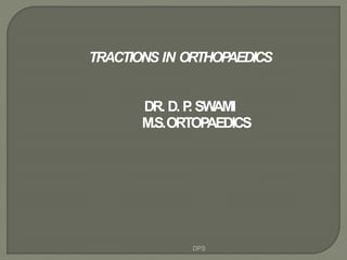
- 1. TRACTIONS IN ORTHOPAEDICS DR. D. P. SWAMI M.S.ORTOPAEDICS DPS
- 2. DEFINE TRACTION ? WHY TRACTIONS REQIRED? NAME ANY TRACTION ? DPS
- 3. DEFINITION: Traction is defined asan act of drawing or exerting apulling force applied to limbs, bones, or other tissues along thelongitudinal axis of the structure to pull the tissues apart, often for realignment. Traction when applied tothe injured limb can over come the effect of original deforming forces. DPS
- 4. DEFORMING FORCES IN # SUBTROCHANTERIC FEMUR DPS
- 5. • Reducea fracture • Reducedislocation of ajoint • Relieve pain • Restthe limb in functionalposition • Aid in healing of bone. • Overcome muscle spasmand deforming forces. • Correction of soft tissue contractures bypulling them gradually DPS
- 6. • Skin traction 1)Adhesive 2)Non-adhesive • Skeletal traction DPS
- 7. • Firm mattress or abed board. Facility to elevate thehead end and foot end of thebed. • An overhead frame, trapeze, monkey ropes and side rails to shift the position of thepatient. • Bars,pulleys, ropes, wt hangers, skeletal traction apparatus and plaster castmaterials. • Traction must always beopposed by counter traction. • Constant care and vigilance to avoid all the hazards ofprolonged bed rest DPS
- 8. Bohlers striuup with steinmannpin Bohlers Stiruup with steinmanpin Applied asskeletal tractionDPS
- 10. DPS
- 11. Usedasadefinitive method of treatment aswell asafirst aid or temporary measure. Mechanism: • Traction force is applied over alarge area. Load is spread and is more comfortable andefficient. • Forceapplied is transmitted from skin to the bones, via the superficial fascia, deep fasciaand intermuscular septa. • For better efficiency, the traction force isapplied only to the limb distal to the fracture. Maximum weight: Recommended is 6.7kg (depending on sizeand ageof patient ) (1/10th the bodyweight).DPS
- 13. • Prepare the skin by shaving washing and drying • Useadhesive strapping which canbe stretched only transversely • Avoid placing adhesive strapping over bony prominences • Leavealoop of 2 inches ( 5cm) projecting beyond the distal end of limb to allow the movement of finger / foot DPS
- 14. •Always leave afree skin between the straps •Must not be too tight or too loose •Leavethe heels free •Canbe safely used for 4-6 weeks •It may be pulled down day byday DPS
- 15. • Thisconsists of lengths of soft, ventilatedlatex foam rubber, laminated into astrong cloth backing. • Theseare useful in thin and atrophic skin orwhen there is sensitivity to adhesive strapping. It is applied in similar fashion asadhesive skin traction • Asthe grip is lesssecure, frequent reapplication may be necessary • Attached traction weight should notbe more than 4.5kg (10lbs) DPS
- 16. Non adhesive skin traction DPS
- 17. • Temporary management of femoral neck fractures and intertrochanteric fractures. • Management of femoral shaft fractures in olderand hefty children. • Undisplaced fracture of acetabulum. • After reduction of adislocation of the hip. • Prevent minor fixed flexion deformities of the hip or knee. • Management of low backache. DPS
- 18. 1. Abrasion & Laceration ofskin. 2. Dermatitis. 3. Anyfragile condition of skin. 4. Impairment of circulation-varicoseulcers, Impending gangrene. 5. Marked shortening of bony fragmentswhere more traction weight hasto beapplied. DPS
- 19. • Allergic reaction to adhesive. • Excoriation of skin from slipping ofadhesive strapping. • Pressuresoresaround malleoli & tendoachilles. • Common peroneal nerve palsy. DPS
- 20. Tractionforce isapplied directly to thebone bymeansof pinsor wiredriven through the bone DPS
- 21. Most commonly used pinsare 1) Steinman pin 2) Denhamspin 3) Kwire DPS
- 22. Steinman pin: • Are rigid stainless steel pins of varyinglength, 4 to 6 mmdiameter. • After insertion aspecial, stirrup (Bohler1929) is attached to thepin. • TheBohler stirrup allows the direction of the traction to be changed without turning the pin in the bone. DPS
- 23. DPS
- 24. Denham Pin: • short threaded length situated in the center • It engagesthe bony cortex and reducesthe risk of pinsliding. • Used in a) cancellous bones & b) osteporotic bones DPS
- 25. DPS
- 26. Kirschnerwire: • Isof small diameter and is insufficiently rigid untilpulled taut in aspecial stirrup, rotation of the stirrup is imparted tothe wire. • Though they are thin but if proper special stirrup is used they canwithstand alarge traction force becausethe stirrup provides longitudinal tension force which increasesthe rigidity of theK-wire. DPS
- 27. DPS
- 28. KwirestrainerDPS
- 29. DPS
- 30. Upper endoffemur: • Point of insertion is lateral surface offemur 2.5 cm below the most prominent part ofGT midway between ant and postsurface. • Usedin central fracture dislocation of hip • Cancellousscrew or screw eyeisused DPS
- 31. Lowerendof femur: • Point of insertion is determined by 2ways • Pin is passedasanteriorly aspossible to avoid neurovascular structures. • Avoid entering the kneejoint Disadvantages : Prolonged traction through lower endof femur predisposes to kneestifness DPS
- 32. DPS
- 33. Upper endof Tibia: • Point of insertion • Pin should be inserted from lateral tomedial side • In young patients avoid openepiphysis. DPS
- 34. f) Lowerendof Tibia: • Point of insertion 5 cm above the level of ankle joint g) Calcaneus: •Point of insertion •Avoid subtalar joint Advantages: Traction force directly in line of the calf muscles and couteract their pull Disadvantages: • Subtalar joint stiffness •Infection •Frequent looseing DPS
- 35. DPS
- 36. Olecranon: DPS
- 37. metacarpals: • Placedthrough diaphysis of 2nd and 3rd metacarpals. • It trasverses 2nd and 3rd metacarpal at right angle to longitudinal axisofradius. • USEDIN COMMINUTED#sOFBONESOF FOREARM-PARTICULARLY THATOFLOWER ENDOFRADIUS. DPS
- 38. DPS
- 39. DPS
- 40. • Introduction of infection into abone. • Incorrect placement of pin • -Allows pin to cut out of bone. • -Makes control of rotation of limbdifficult. • -Makes application of splintdifficult. • -Unequal pull causespin to move in the bonecausingischemic necrosis • Largetraction force. • -Distraction at fracturesite. • -Ligament damage. • Damageto epiphyseal growth plate inchildren. • Depressed scarand stiffness of joints.DPS
- 41. • Reasonfor applying Traction is to counteract deforming effect of muscle spasmand this tends todraw body in direction of traction. • Toprevent this, force is to be used inopposite direction calledCounter-traction. DPS
- 42. FIXEDTRACTION When counter traction acts through an appliance which obtains purchase on apart of the body, its called afixedtraction. DPS
- 43. • Definition: When the weight of all or part of the body acting under the influence ofgravity is utilized to provide counter traction, the arrangement is called sliding traction. • Principle: Thetraction force is applied by weight attached to adhesive strapping or asteel pinby a cord acting over apulley. Counter traction is obtained by raising one end of thebed by means of wooden blocks so that the body tends to slide in the oppositedirection. DPS
- 44. 1) In lower limb a. Bucksextension skin traction b. Perkins traction c. Russeltraction d. Tulloch- Brown Traction e. 90-90 Traction f. Gallows/ BryantsTraction g. Bohler – Braun frame h. Lateral upper femoraltraction i. Pelvic tracton DPS
- 45. 2) In upper limb a. Dunlop traction b. Olecronon pin traction c. Metacarpal pin traction 3) Spinal traction a. Cervical traction • Halter or non skeletaltraction ▪ Canvasor Chamoishead halter ▪ Crile head halter • Skull or skeletaltraction b. Halopelvic traction DPS
- 46. DPS
- 47. • Originally used in management of #sofpelvis, femur, tibia. • Skeletal traction being applied to injured leg, while the well leg was employed for counter traction. • But this method is valuable in correcting either abduction and adduction deformityat the hip. DPS
- 48. PRINCIPLE: • With abduction deformity at the hip,the affected limb appears to be longer. When Traction is applied to the well limb andAffected limb is simultaneously pushed Up (counter traction), the abduction deformity is reduced. DPS
- 49. DPS
- 50. USEDIN THETEMPORARYMANAGEMENTOF • Femoral neck fractures, • Femoral shaft fractures in older andlarger children, • Undisplaced #sof acetabulum, • In place of pelvictraction, • Correction of minor fixed flexiondeformites of hip • After reduction of dislocation of hip. DPS
- 51. DPS
- 52. • APPLYADHESIVESTRAPPINGTOABOVEKNEEORIN ELDERLYVENTOFOAMSKINTRACTION • SUPPORTTHELEGWITHPILLOW. • PASSTHECORDFROMSPREADEROVERPULLEY. • ATTACH2.3-3.2kgs (5 – 7 lbs) TOTHECORD. • ELEVATETHEFOOTENDOFBED. DPS
- 53. USEIN TREATMENTOF •Fracture tibia •# femur from subtrochanteric region distally in all ages •fracture Trochanter in <50yrs PRNCIPLE: • It is the useof Skeletal traction without any externalsplintage coupled with active movementsof injured limb • Perkins showed that by encouraging early muscular activity stiffness of jointwasprevented by extensibility of muscles by reciprocalinnervation. DPS
- 54. DPS
- 55. Indications: • Management of thefracture shaft of femur • After arthroplasty operations on the hip Application: • Below knee skin traction • Pulley attached to spreader • Soft sling placed under knee Weight adults – 3.6 kg chidren – 0.28- 1.8kg DPS
- 56. Advantage: Basedon law of parallelogram of forces that- the 2 pulley blocks at the foot of the bed theoretically doubles the pull on the limb and the resultant traction is in axisof 30° to the horizontal i.e. in line of shaft of femur DPS
- 57. • Devised by Obletz (1946) • Usedin # femur with wounds over postaspect of thigh (operative &post op management) • Subtrochanteric and proximal third #femur • Usedin both children andadults • Here both hip and knee are flexed to90 degree. • Skeletal traction is applied through lower femur or uppertibia • 3 methods of supporting leg in 90/90traction DPS
- 58. DPS
- 59. DPS
- 60. DPS
- 61. • Varus /valgus angulation at fracture site is controlled by moving the pulley,over which the traction cord passes,in a plane across the width of the bed. • Rotation is controlled by the knee being flexed. • As the union of fracture occurs, encourage active hip and knee exercise-extension , gradually lower the limb into a more horizontal position. DPS
- 62. 1. Those of skeletal traction. 2.Stiffness and loss of extension of the knee. 3.Flexion contracture of hip. 4.Injury to the lower femoral or upper tibial epiphyseal growth plates in children. 5. Neuro vascular damage. DPS
- 63. DPS
- 64. • Usedin # Shaft of femur in children <2yrs • Apply adhesive strapping to both lowerlimbs • Tie traction cords to an over headbeam • Tighten the traction cord to raise thebuttocks just clear the mattress • Counter traction obtained by weight ofpelvis DPS
- 65. • Vascular complication of Bryants traction may occur in either the injured or normal limb. • Acareful check must be done in bothlimbs during first 24-72hrs. -Bychecking color and temp of limbs. -Dorsiflexion of both anklepassively. • Bryants traction in children: under 2yrs - safe 2-4yrs - vascular complications more(can be prevented by using posterior splint). Over 4yrs - absolutely contraindicated. DPS
- 66. DPS
- 67. • In the initial management of CDHwhen diagnosed over the ageof 1year. • After 5 daysabduction of hip isstarted • Abduction is increased by 10* onalternate days • By 3wks hips should be fully abducted DPS
- 68. COMPLICATIONS: • The child will become restless and scream repeatedly with pain. • The pain is due to stretching of capsule and impingement of femoral head on superior lip of acetabulam. DPS
- 69. • In management of tibia and femoralfractures. DPS
- 70. • Most proximal pulley-to prevent footdrop. • 2 nd pulley-to apply traction in line of Femur. • 3 rd pulley-to apply traction in line of supracondylar area of femur and hightibial traction. • 4 th pulley-to apply traction in line of leg asin low tibial or calcanealtraction. DPS
- 71. DPS
- 72. • In pelvic traction special canvasharnessis buckled around the patientspelvis. • Long cords attach the harness to the footof the bed. • Foot end of the bed raised-providessliding traction. • Usedin conservative management of IVDP.To ensure that the pt lies quietly in bed rather than to distract the vertebralbodies. • Buck`straction may also be employed DPS
- 73. DPS
- 74. • Cervical spine -skeletal traction(skull traction) Crutchfield tongs Cone/Barton tongs Halo splint -non skeletal traction(halter traction) DPS
- 75. Indications: • Treatment of Cervical Spondylosis asan out patient • Maximum weight is 1.4 to 2.3kgs • Twotypes – Canvas& Crile head halter • Head end of bed should raised to providecounter- traction DPS
- 76. DPS
- 77. DPS
- 78. DPS
- 79. Applied by gaining purchase on the outertable of the skull withmetal pins Usedin the serious injuries of cervical spinelike • Toreduce adislocation or fracturedislocation - in both casewith traction the dislocation is under control and injury to spine does not occur • Tomaintain the position of c- Spinebefore and after operative fusion • For the treatment of cervical spondylosiswith severe nerve root compression • Maximum applicable weight is 9.1 to 18.2kg DPS
- 80. • For skull traction use a) Crutch field tongs b) Coneor Bartontongs c) Halo splint DPS
- 81. CRUTCHFIELDTONGS: • Fits in to parietalbone • Aspecial drill with ashoulder is used to enable an accurate depth of hole tobe drilled DPS
- 82. CONEORBARTONTONGS • Adrill is not required for theirinsertion. The threaded steel points are screwed into the parietal bones behind the ears DPS
- 83. • Sedatethe patient. • Shavethe scalponly locally. • Draw aline on the scalp,bisecting the skullfront to back. • Draw asecond line joining the tips ofmastoid process, it crossesthe 1st line at rightangle. • Fully open out thetongs. • With the fully open tongs lying equally on each side of Anteroposterior line, pressthe tongsinto the scalp making dimples on the 2ndline. • Infiltrate the areasof dimples down to and including the periosteum, with localanaesthesia. • Make small stab wound in scalp atdimples. • Using special drill point, drill the outer table of the skull in adirection parallel to the points of the tongs. Thedrill point is inserted to adepth of 3mm in children and 4mm inadults. DPS
- 84. • Torealignspine • Toprevent loss of function of undamaged neurological tissue • Toimprove neurologicalrecovery • Toobtain and maintain spinalinstability • Toobtain early functionalrecovery DPS
- 85. Level Minimum weight Maximum weight C1 2.3 kg 4.5 kg C2 2.7 kg 4.5 to 5.4kg C3 3.6 kg 4.5 to 6.7kg C4 4.5 kg 6.7 to 9.0kg C5 5.4 kg 9.0 to 11.3kg C6 6.7 kg 9.0 to 13.5kg C7 8.2 kg 11.3 to 15.8kg DPS
- 86. DPS
- 87. DPS
- 88. • Tongsmay pull out of skull • Tongsmay penetrate inner table • Osteomyelitis • Extradural hematoma • Extradural abscess • Subdural abscess • Cerebral abscess DPS