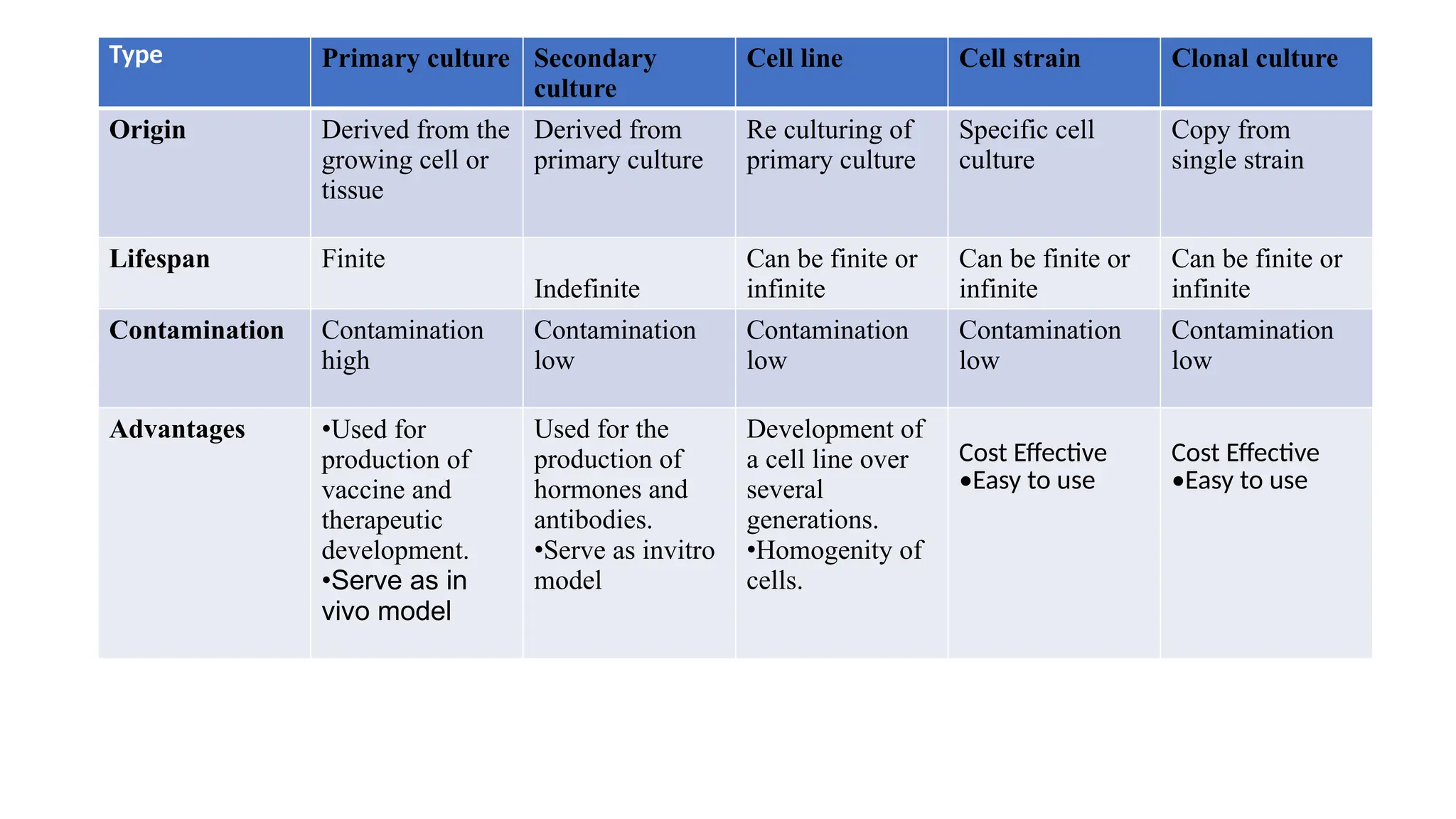
Type Primary cultureSecondary
culture
Cell line Cell strain Clonal culture
Origin Derived from the
growing cell or
tissue
Derived from
primary culture
Re culturing of
primary culture
Specific cell
culture
Copy from
single strain
Lifespan Finite
Indefinite
Can be finite or
infinite
Can be finite or
infinite
Can be finite or
infinite
Contamination Contamination
high
Contamination
low
Contamination
low
Contamination
low
Contamination
low
Advantages •Used for
production of
vaccine and
therapeutic
development.
•Serve as in
vivo model
Used for the
production of
hormones and
antibodies.
•Serve as invitro
model
Development of
a cell line over
several
generations.
•Homogenity of
cells.
Cost Effective
•Easy to use
Cost Effective
•Easy to use
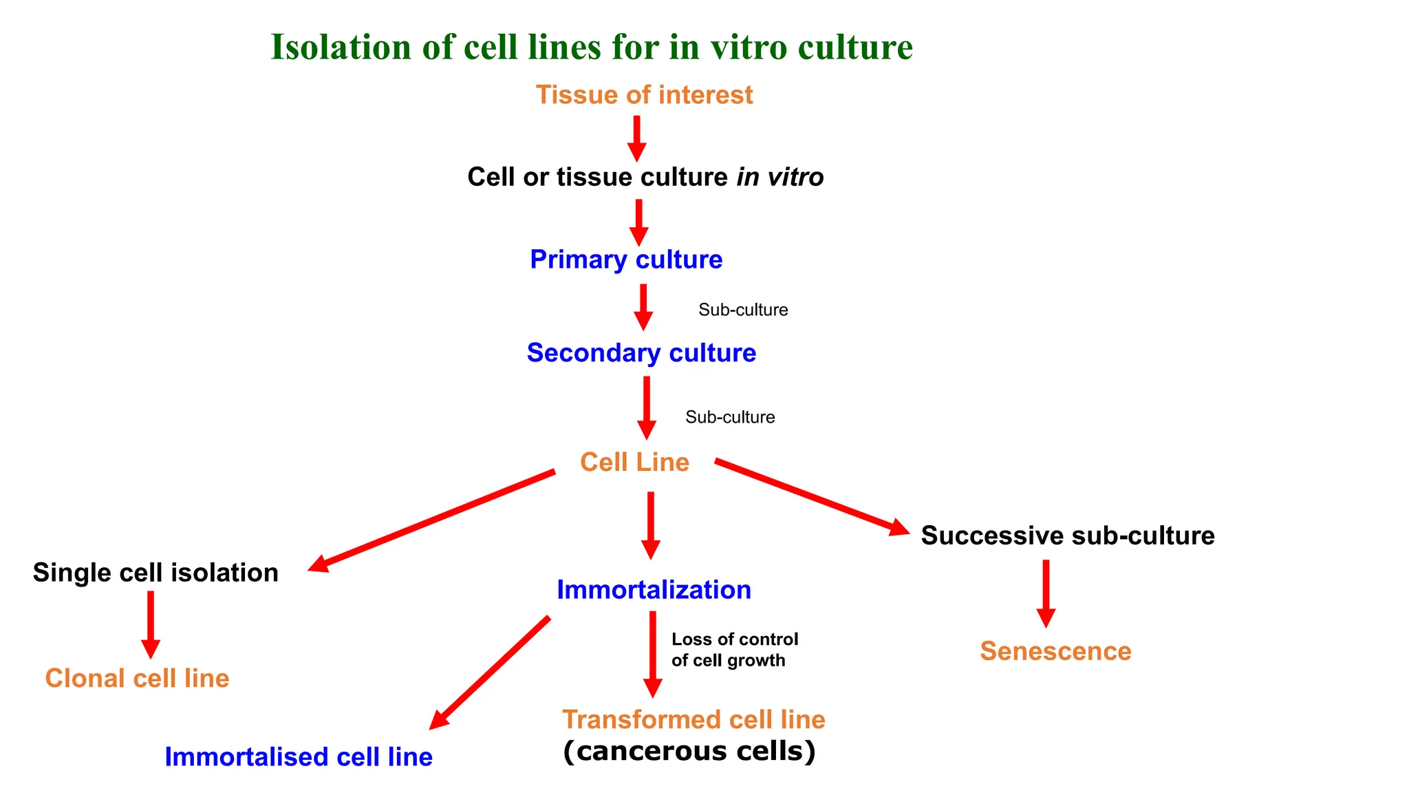
7.
Isolation of celllines for in vitro culture
Tissue of interest
Cell or tissue culture in vitro
Primary culture
Secondary culture
Cell Line
Immortalization
Transformed cell line
(cancerous cells)
Immortalised cell line
Successive sub-culture
Senescence
Single cell isolation
Clonal cell line
Loss of control
of cell growth
Sub-culture
Sub-culture
11.
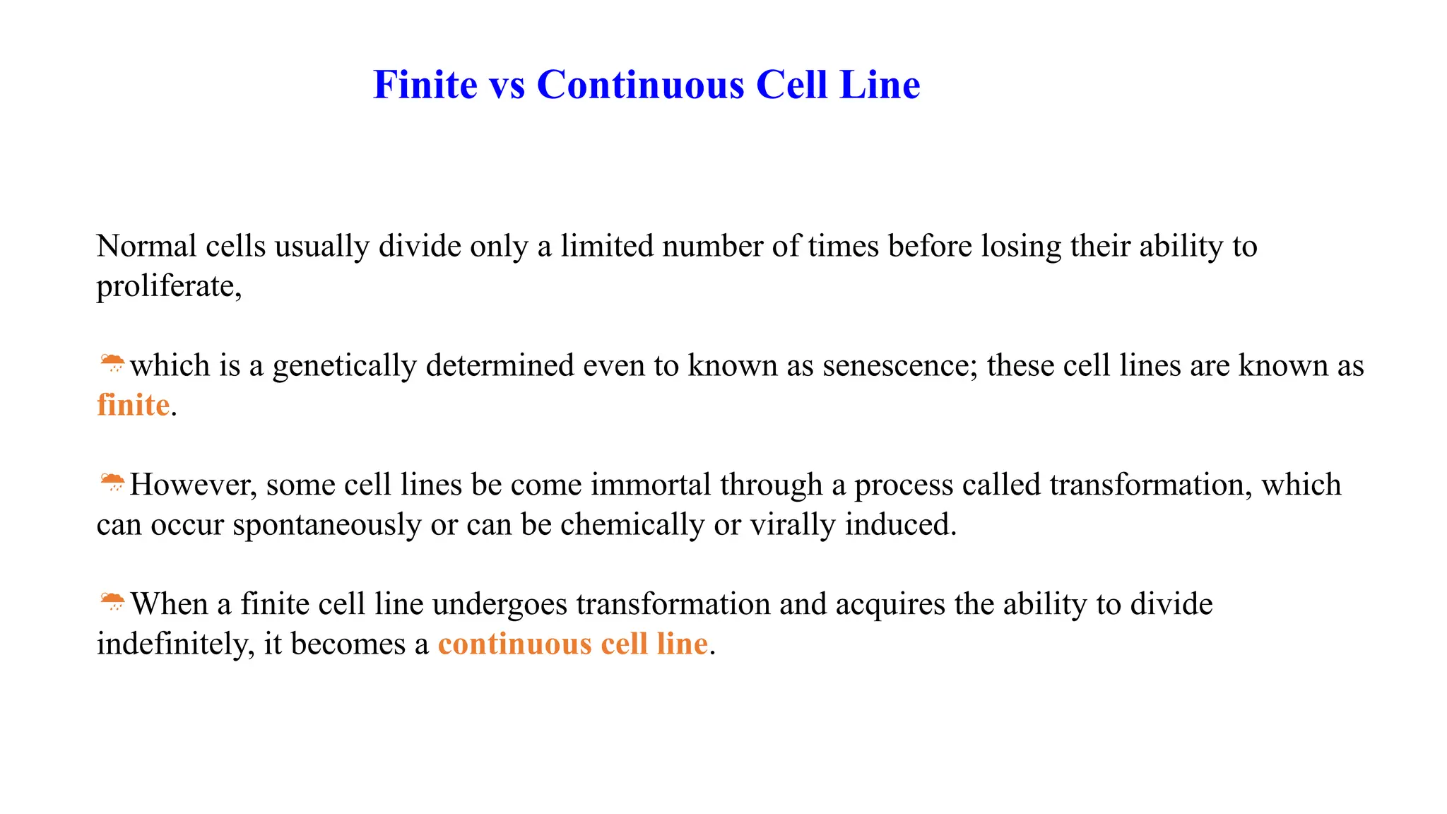
Normal cells usuallydivide only a limited number of times before losing their ability to
proliferate,
which is a genetically determined even to known as senescence; these cell lines are known as
finite.
However, some cell lines be come immortal through a process called transformation, which
can occur spontaneously or can be chemically or virally induced.
When a finite cell line undergoes transformation and acquires the ability to divide
indefinitely, it becomes a continuous cell line.
Finite vs Continuous Cell Line
12.
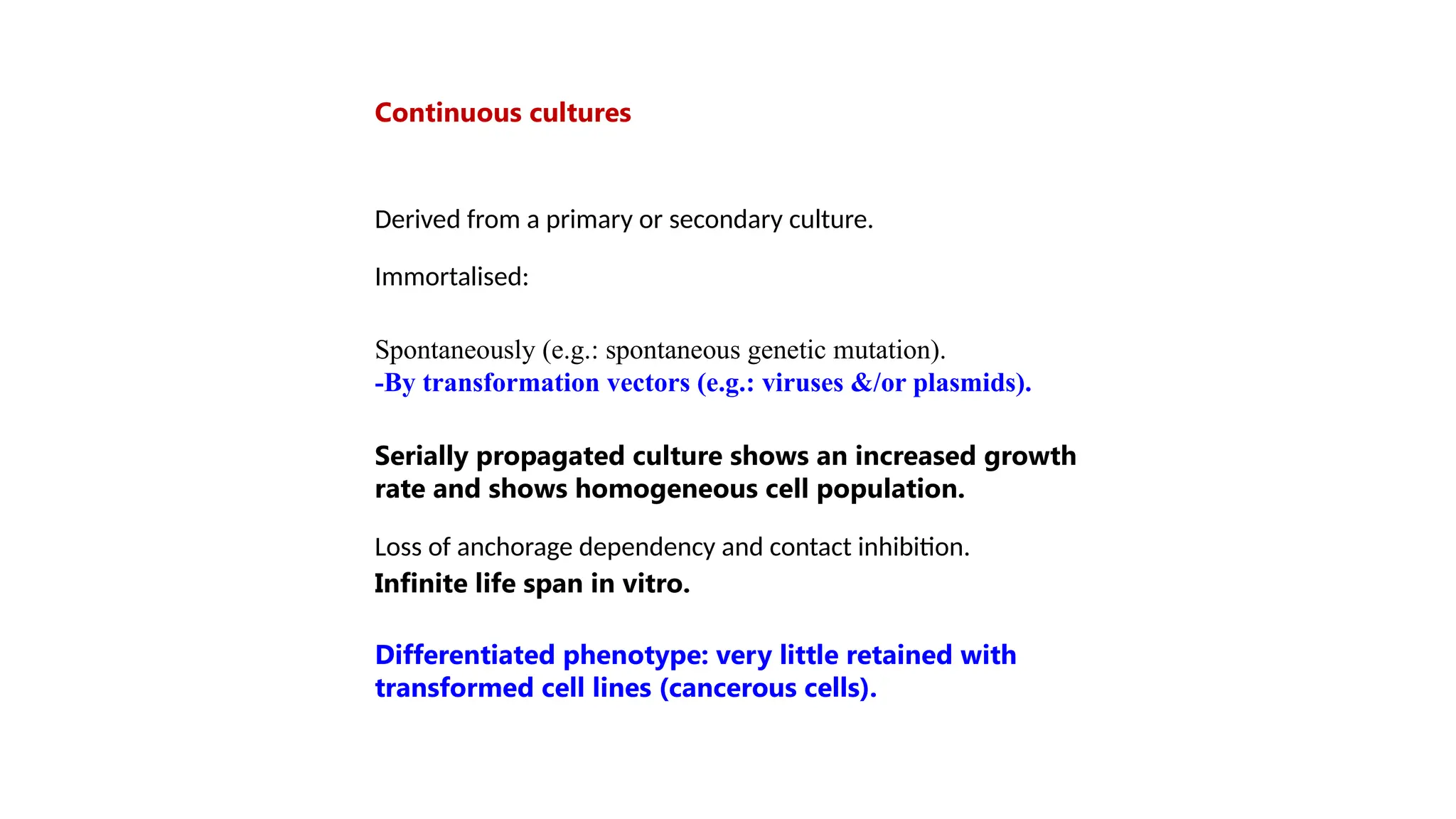
Continuous cultures
Derived froma primary or secondary culture.
Immortalised:
Spontaneously (e.g.: spontaneous genetic mutation).
-By transformation vectors (e.g.: viruses &/or plasmids).
Serially propagated culture shows an increased growth
rate and shows homogeneous cell population.
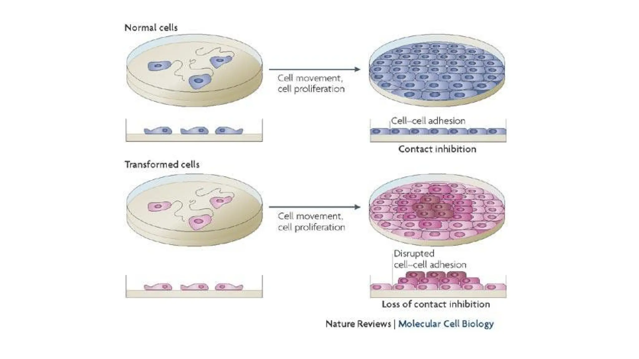
Loss of anchorage dependency and contact inhibition.
Infinite life span in vitro.
Differentiated phenotype: very little retained with
transformed cell lines (cancerous cells).
13.
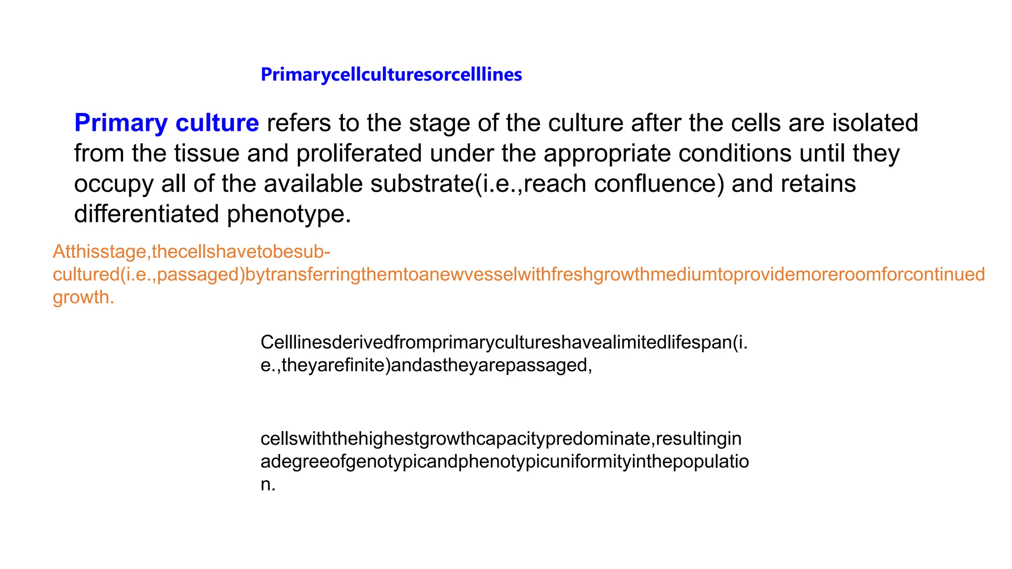
Primarycellculturesorcelllines
Primary culture refersto the stage of the culture after the cells are isolated
from the tissue and proliferated under the appropriate conditions until they
occupy all of the available substrate(i.e.,reach confluence) and retains
differentiated phenotype.
Atthisstage,thecellshavetobesub-
cultured(i.e.,passaged)bytransferringthemtoanewvesselwithfreshgrowthmediumtoprovidemoreroomforcontinued
growth.
Celllinesderivedfromprimarycultureshavealimitedlifespan(i.
e.,theyarefinite)andastheyarepassaged,
cellswiththehighestgrowthcapacitypredominate,resultingin
adegreeofgenotypicandphenotypicuniformityinthepopulatio
n.
15.
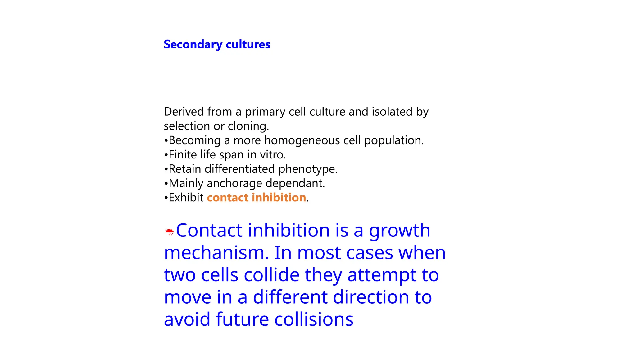
Derived from aprimary cell culture and isolated by
selection or cloning.
•Becoming a more homogeneous cell population.
•Finite life span in vitro.
•Retain differentiated phenotype.
•Mainly anchorage dependant.
•Exhibit contact inhibition.
Contact inhibition is a growth
mechanism. In most cases when
two cells collide they attempt to
move in a different direction to
avoid future collisions
Secondary cultures
16.
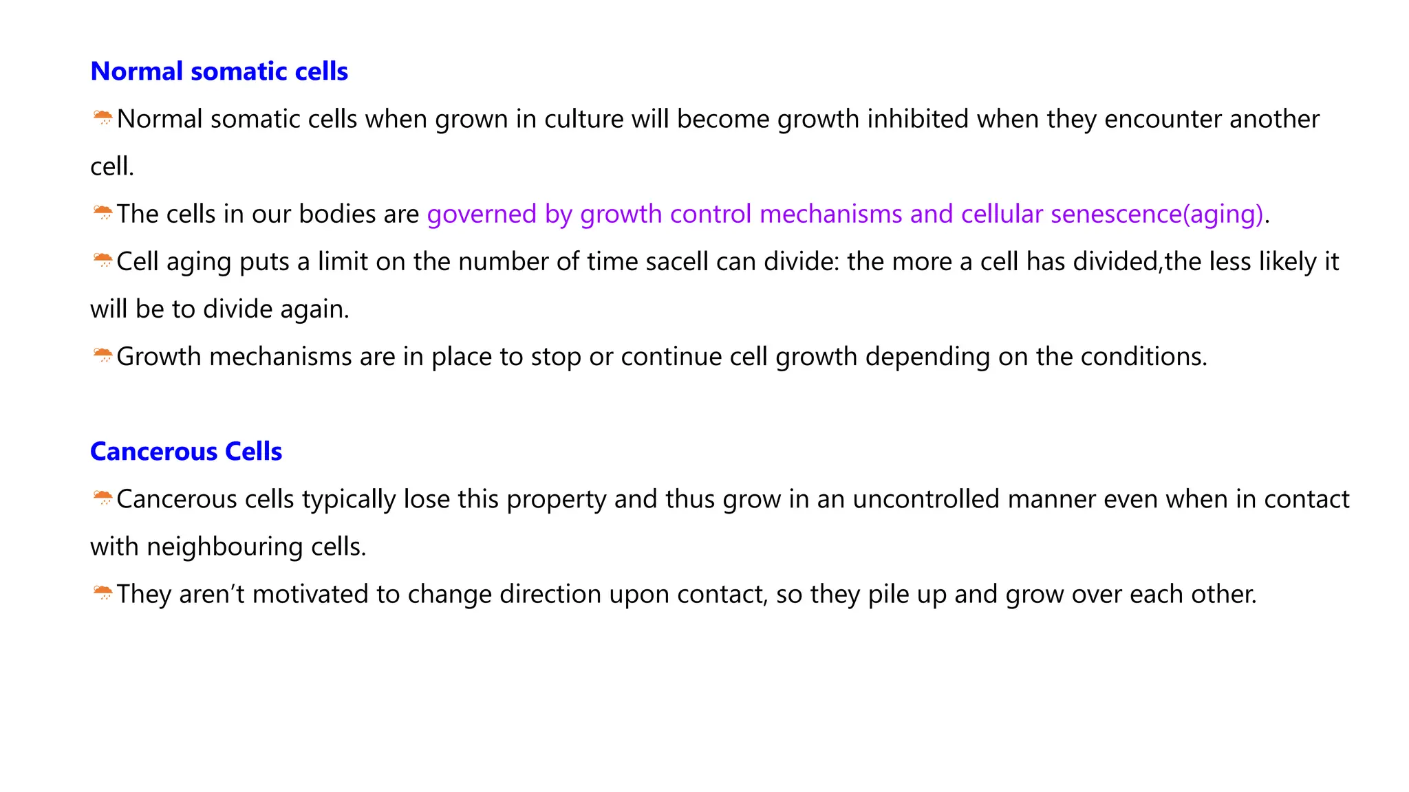
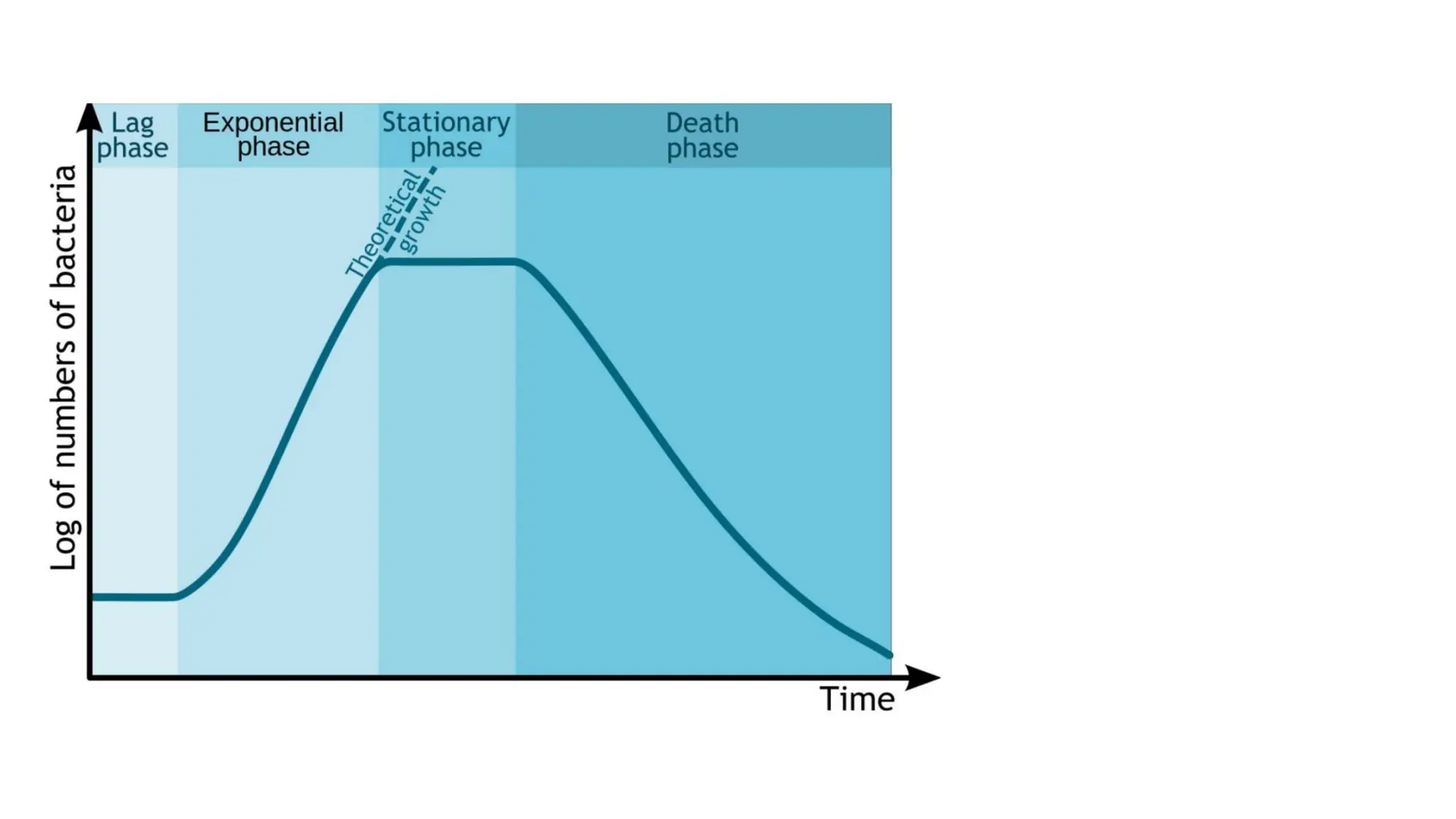
Normal somatic cells
Normalsomatic cells when grown in culture will become growth inhibited when they encounter another
cell.
The cells in our bodies are governed by growth control mechanisms and cellular senescence(aging).
Cell aging puts a limit on the number of time sacell can divide: the more a cell has divided,the less likely it
will be to divide again.
Growth mechanisms are in place to stop or continue cell growth depending on the conditions.
Cancerous Cells
Cancerous cells typically lose this property and thus grow in an uncontrolled manner even when in contact
with neighbouring cells.
They aren’t motivated to change direction upon contact, so they pile up and grow over each other.
20.
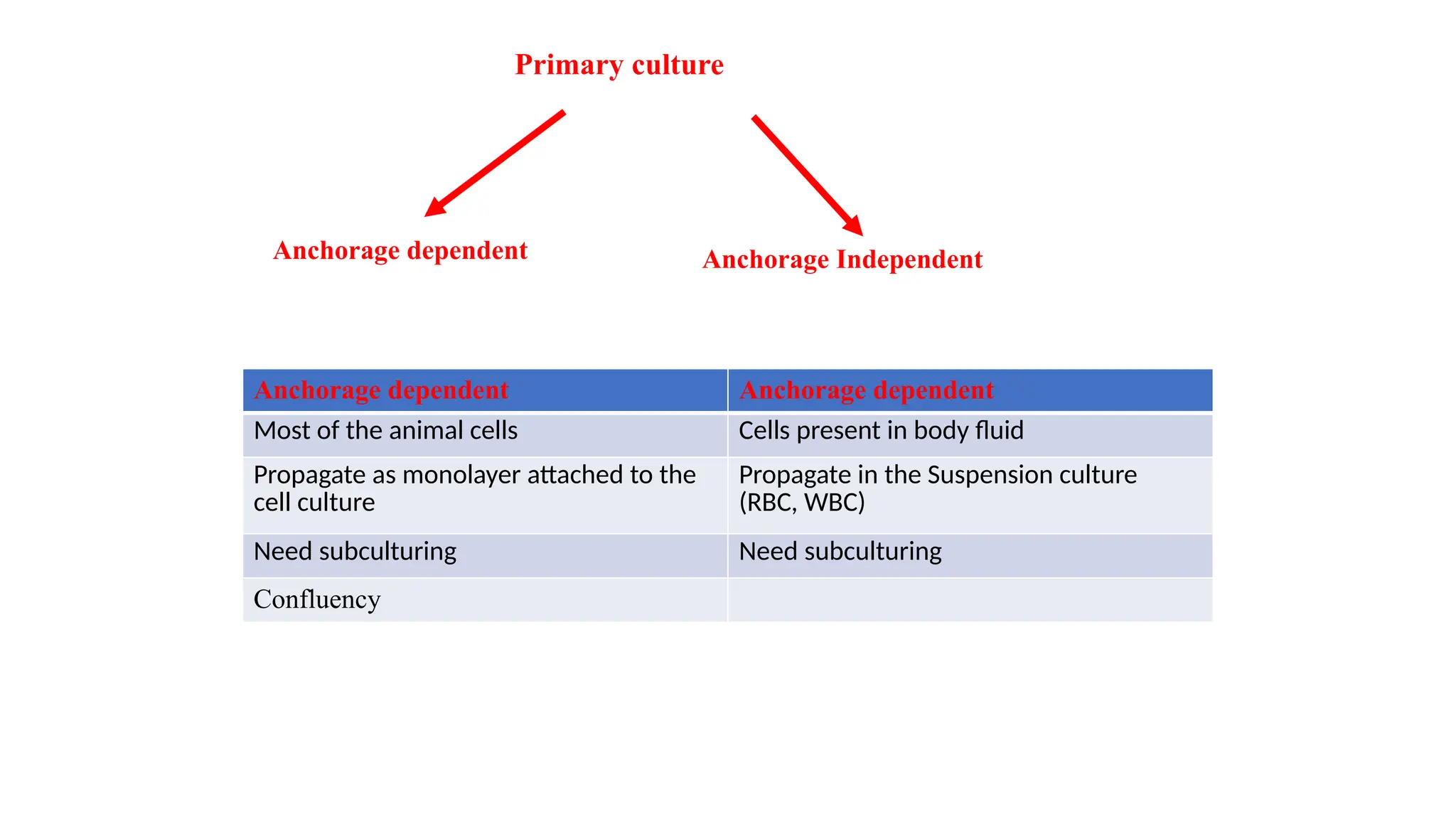
Primary culture
Anchorage dependentAnchorage Independent
Anchorage dependent Anchorage dependent
Most of the animal cells Cells present in body fluid
Propagate as monolayer attached to the
cell culture
Propagate in the Suspension culture
(RBC, WBC)
Need subculturing Need subculturing
Confluency
22.
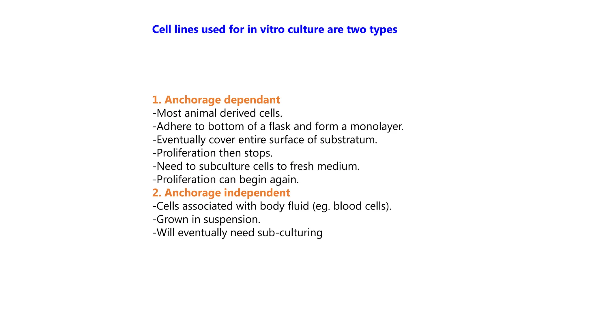
1. Anchorage dependant
-Mostanimal derived cells.
-Adhere to bottom of a flask and form a monolayer.
-Eventually cover entire surface of substratum.
-Proliferation then stops.
-Need to subculture cells to fresh medium.
-Proliferation can begin again.
2. Anchorage independent
-Cells associated with body fluid (eg. blood cells).
-Grown in suspension.
-Will eventually need sub-culturing
Cell lines used for in vitro culture are two types
24.
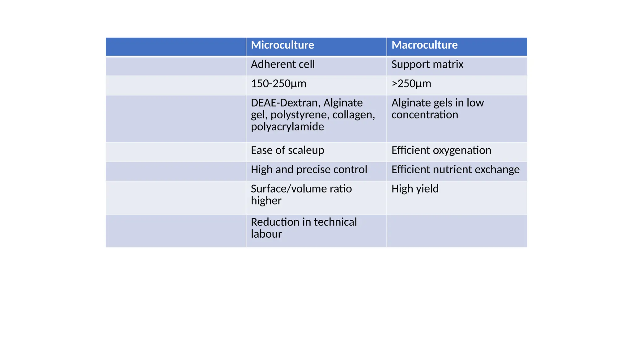
Microculture Macroculture
Carrier Adherentcell Support matrix
Size 150-250μm >250μm
Example DEAE-Dextran, Alginate gel, polystyrene, collagen,
polyacrylamide Alginate gels in lowconcentration
Advantages Ease of scaleup Efficient oxygenation
High and precise control Efficient nutrient exchange
Surface/volume ratio higher High yield
Reduction in technical labour
Microculture Macroculture
Adherent cell Support matrix
150-250μm >250μm
DEAE-Dextran, Alginate
gel, polystyrene, collagen,
polyacrylamide
Alginate gels in low
concentration
Ease of scaleup Efficient oxygenation
High and precise control Efficient nutrient exchange
Surface/volume ratio
higher
High yield
Reduction in technical
labour
26.
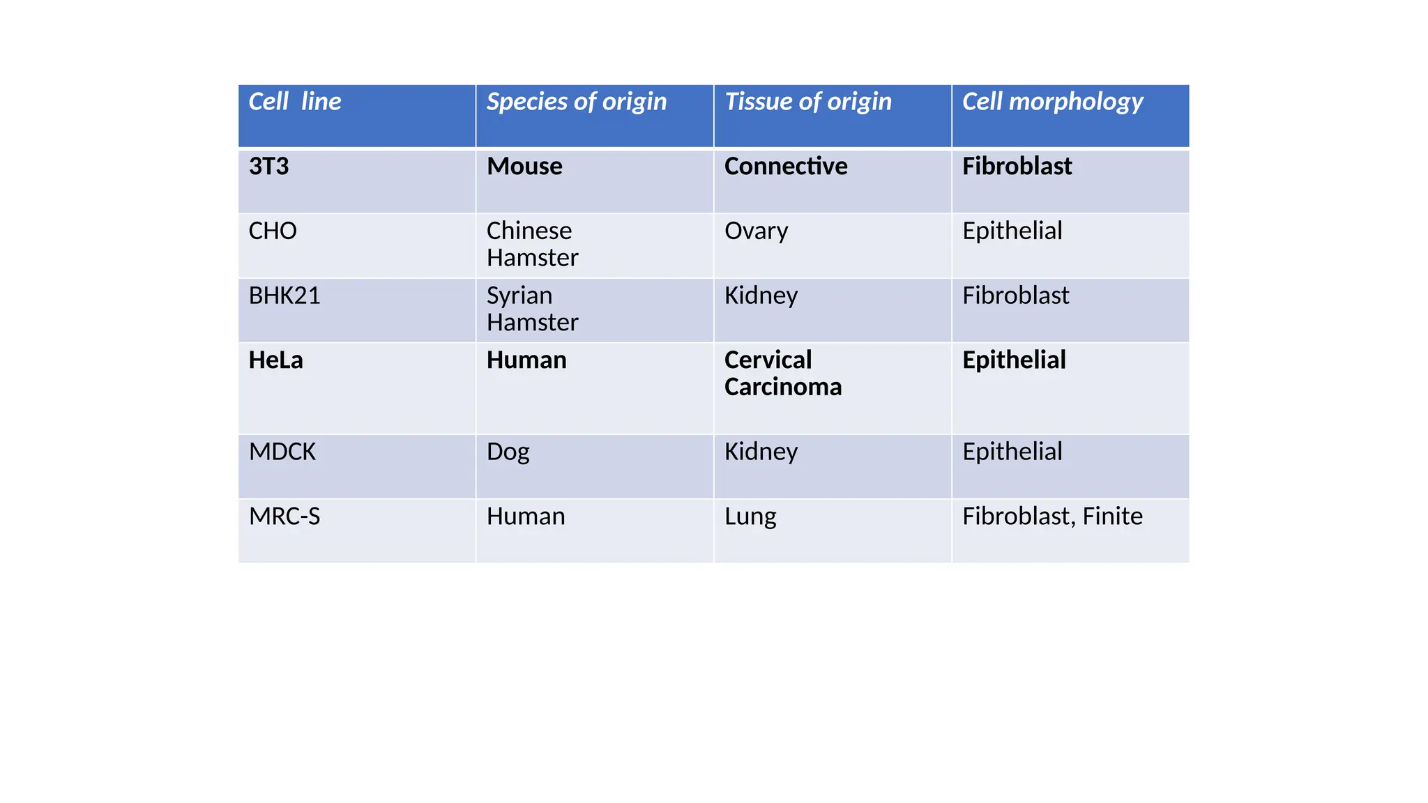
Cell line Speciesof origin Tissue of origin Cell morphology
3T3 Mouse Connective Fibroblast
CHO Chinese
Hamster
Ovary Epithelial
BHK21 Syrian
Hamster
Kidney Fibroblast
HeLa Human Cervical
Carcinoma
Epithelial
MDCK Dog Kidney Epithelial
MRC-S Human Lung Fibroblast, Finite
28.
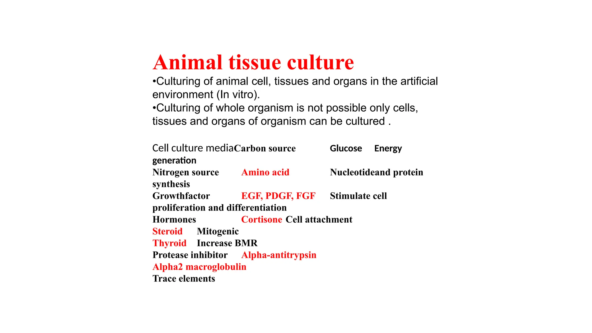
Animal tissue culture
•Culturingof animal cell, tissues and organs in the artificial
environment (In vitro).
•Culturing of whole organism is not possible only cells,
tissues and organs of organism can be cultured .
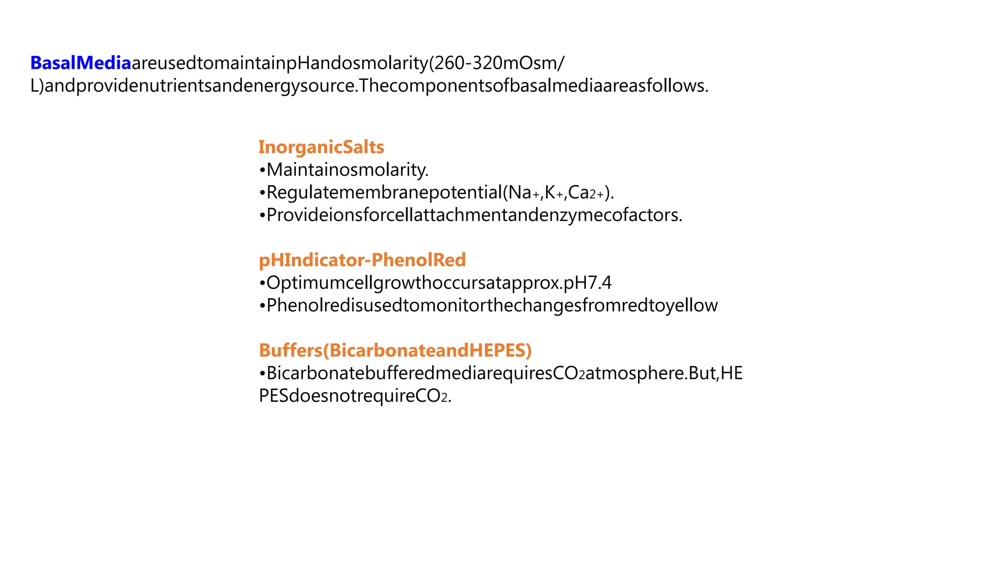
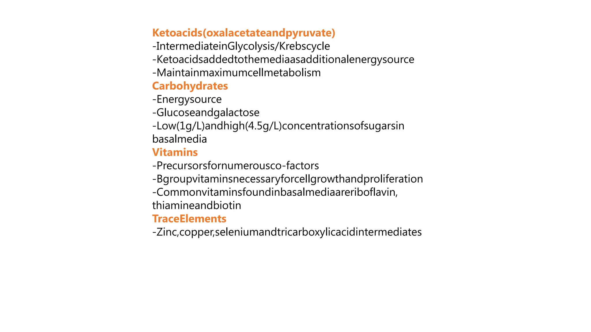
Cell culture mediaCarbon source Glucose Energy
generation
Nitrogen source Amino acid Nucleotideand protein
synthesis
Growthfactor EGF, PDGF, FGF Stimulate cell
proliferation and differentiation
Hormones Cortisone Cell attachment
Steroid Mitogenic
Thyroid Increase BMR
Protease inhibitor Alpha-antitrypsin
Alpha2 macroglobulin
Trace elements
29.
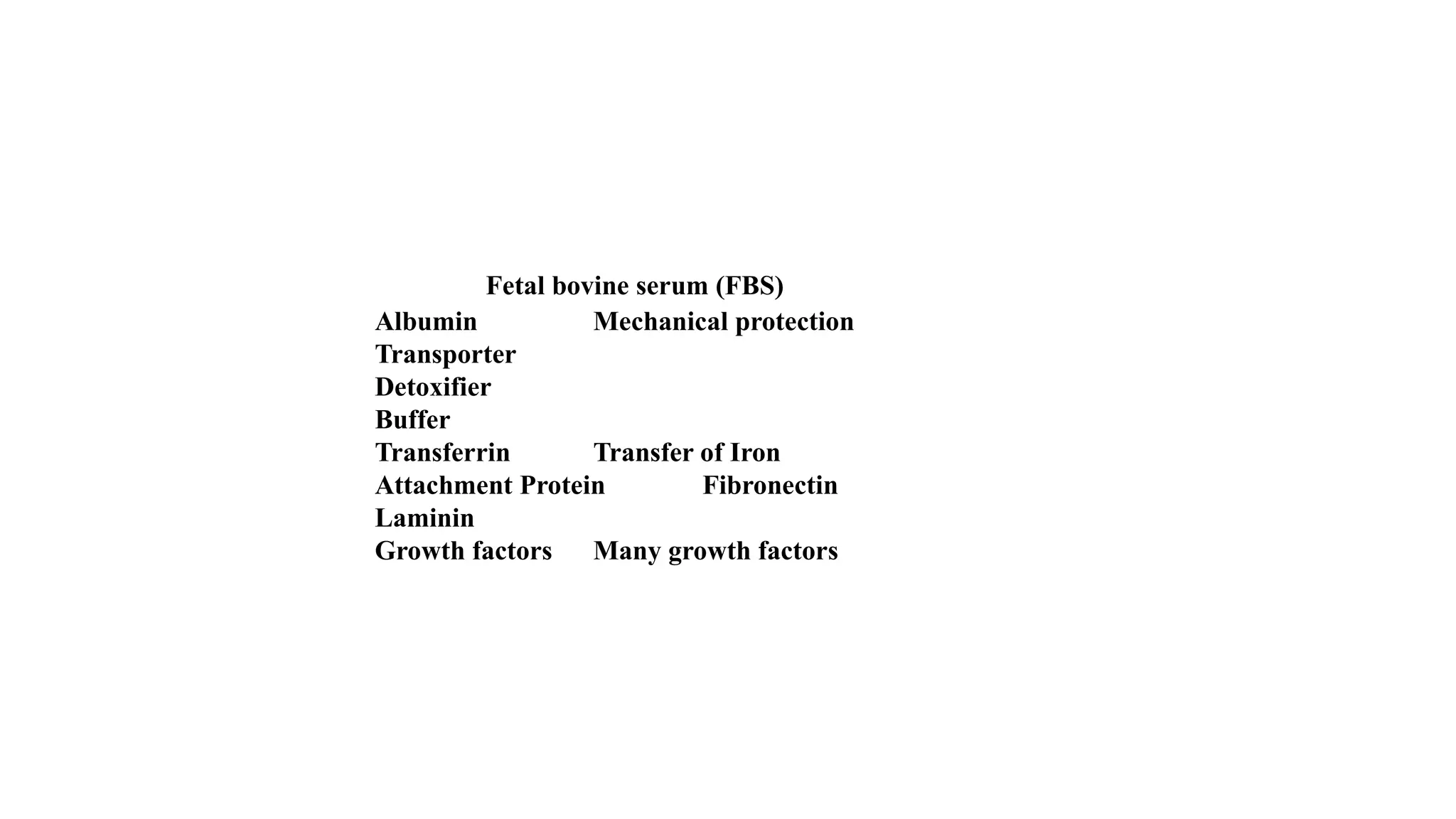
SerumFetal bovine serum(FBS)
Albumin Mechanical protection
Transporter
Detoxifier
Buffer
Transferrin Transfer of Iron
Attachment Protein Fibronectin
Laminin
Growth factors Many growth factors
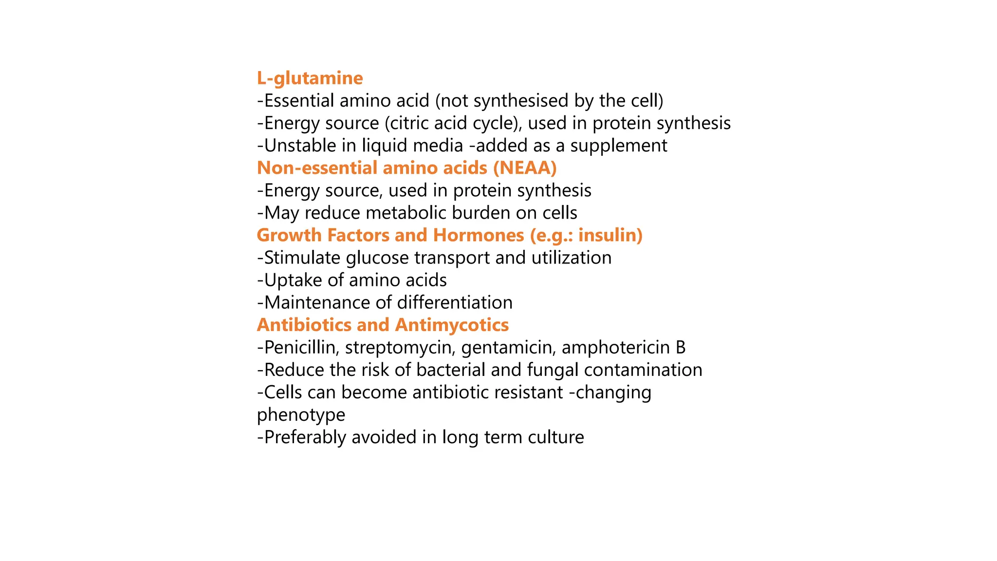
L-glutamine
-Essential amino acid(not synthesised by the cell)
-Energy source (citric acid cycle), used in protein synthesis
-Unstable in liquid media -added as a supplement
Non-essential amino acids (NEAA)
-Energy source, used in protein synthesis
-May reduce metabolic burden on cells
Growth Factors and Hormones (e.g.: insulin)
-Stimulate glucose transport and utilization
-Uptake of amino acids
-Maintenance of differentiation
Antibiotics and Antimycotics
-Penicillin, streptomycin, gentamicin, amphotericin B
-Reduce the risk of bacterial and fungal contamination
-Cells can become antibiotic resistant -changing
phenotype
-Preferably avoided in long term culture
34.
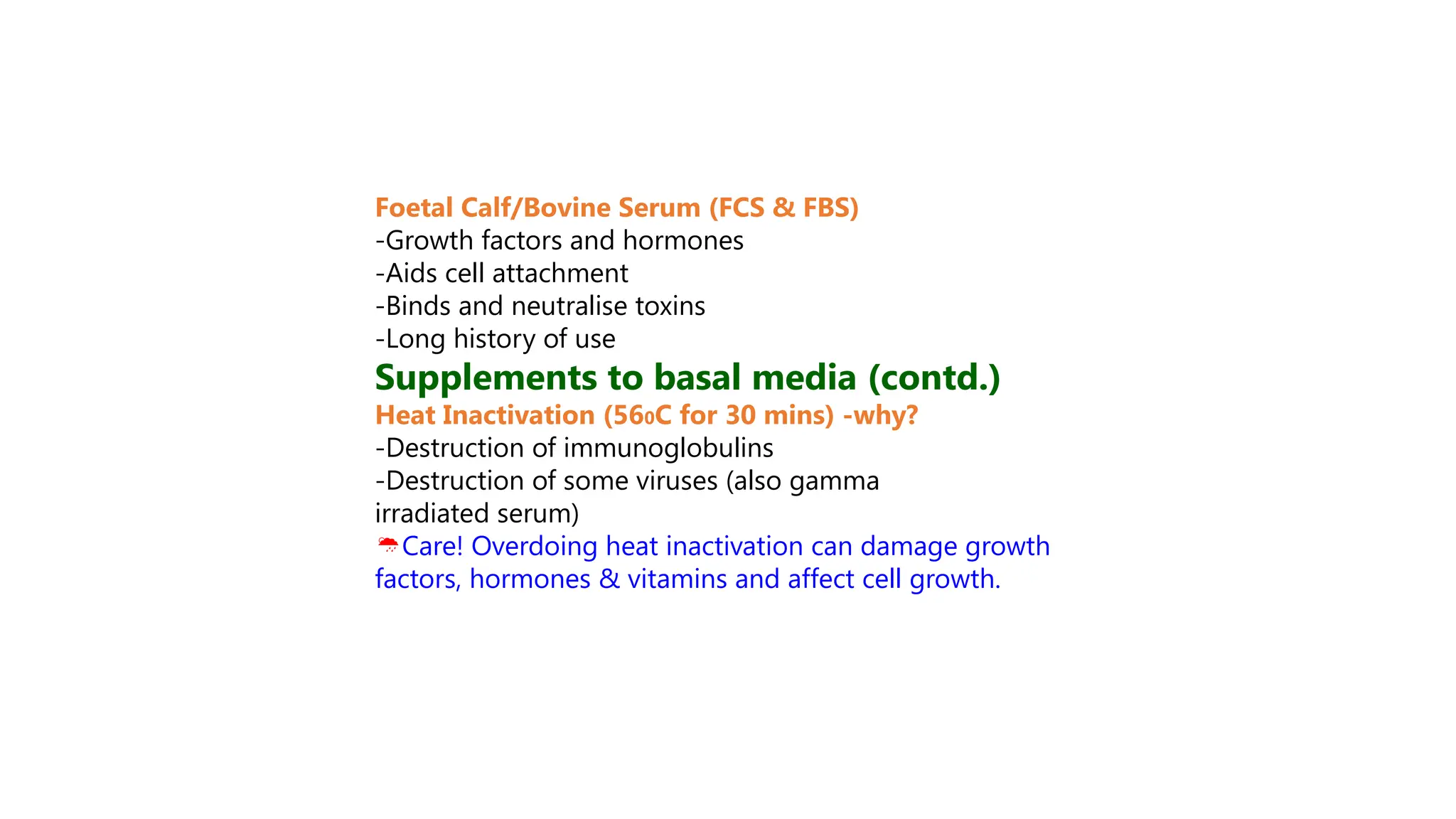
Foetal Calf/Bovine Serum(FCS & FBS)
-Growth factors and hormones
-Aids cell attachment
-Binds and neutralise toxins
-Long history of use
Supplements to basal media (contd.)
Heat Inactivation (560C for 30 mins) -why?
-Destruction of immunoglobulins
-Destruction of some viruses (also gamma
irradiated serum)
Care! Overdoing heat inactivation can damage growth
factors, hormones & vitamins and affect cell growth.
35.
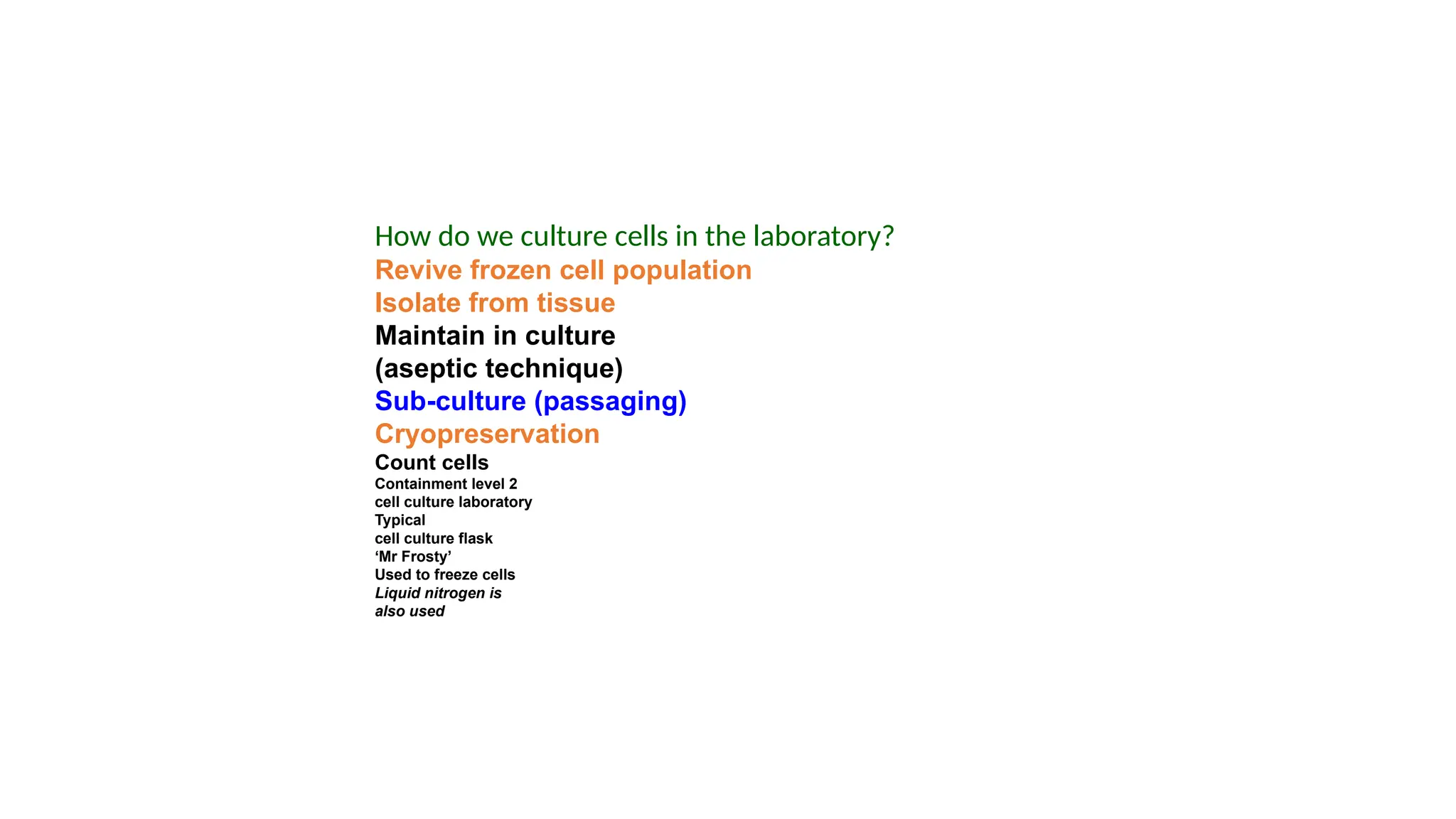
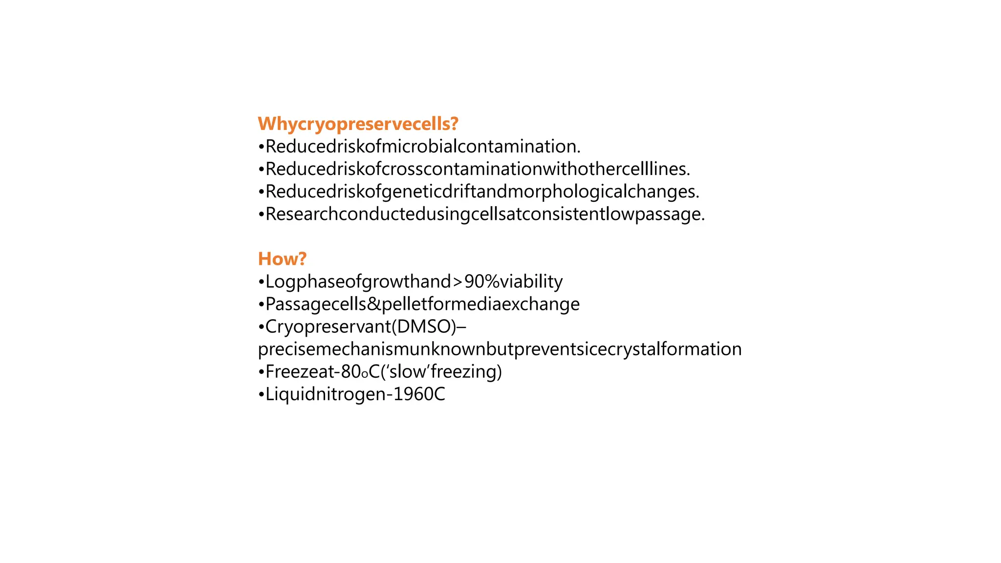
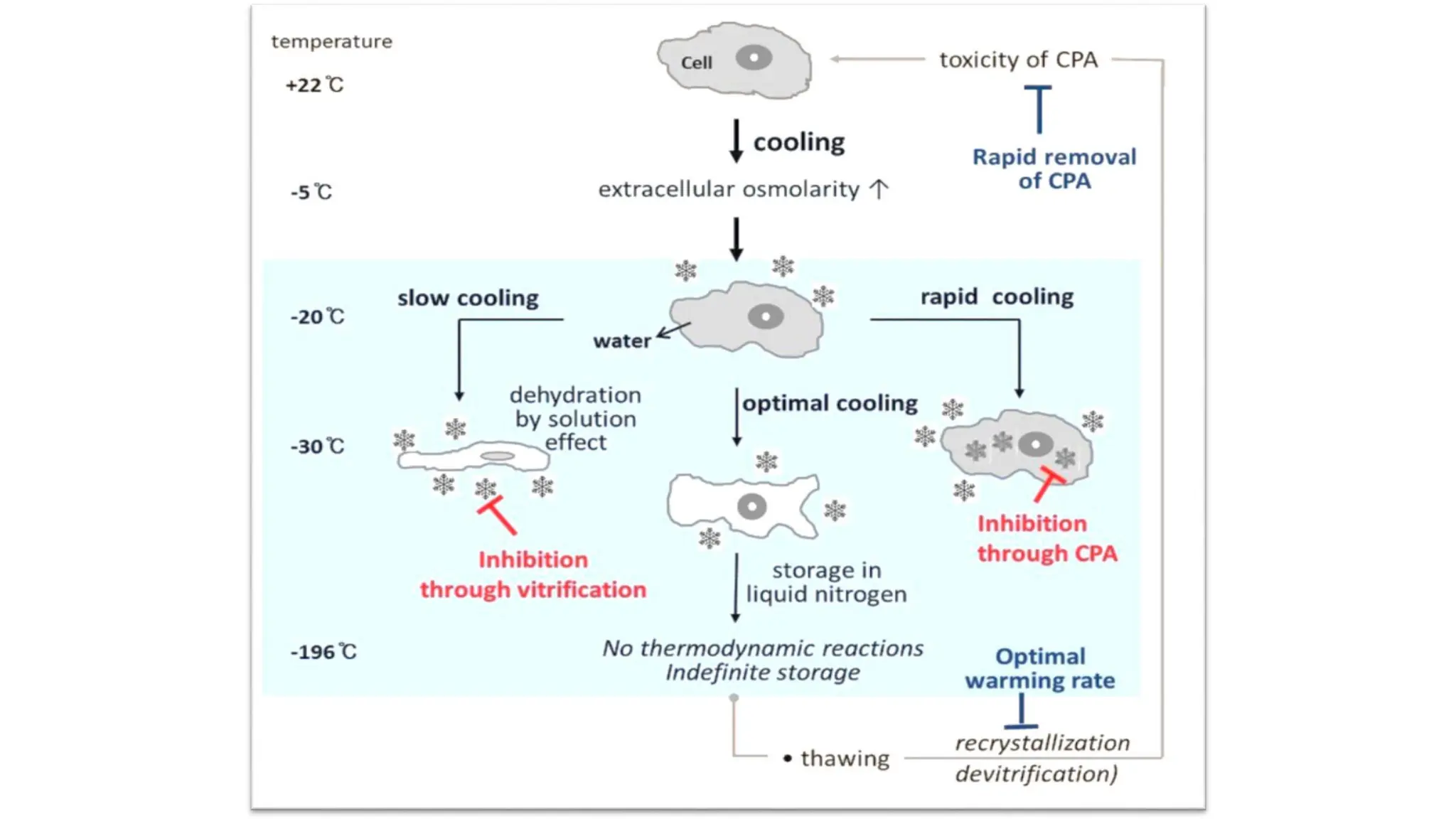
How do weculture cells in the laboratory?
Revive frozen cell population
Isolate from tissue
Maintain in culture
(aseptic technique)
Sub-culture (passaging)
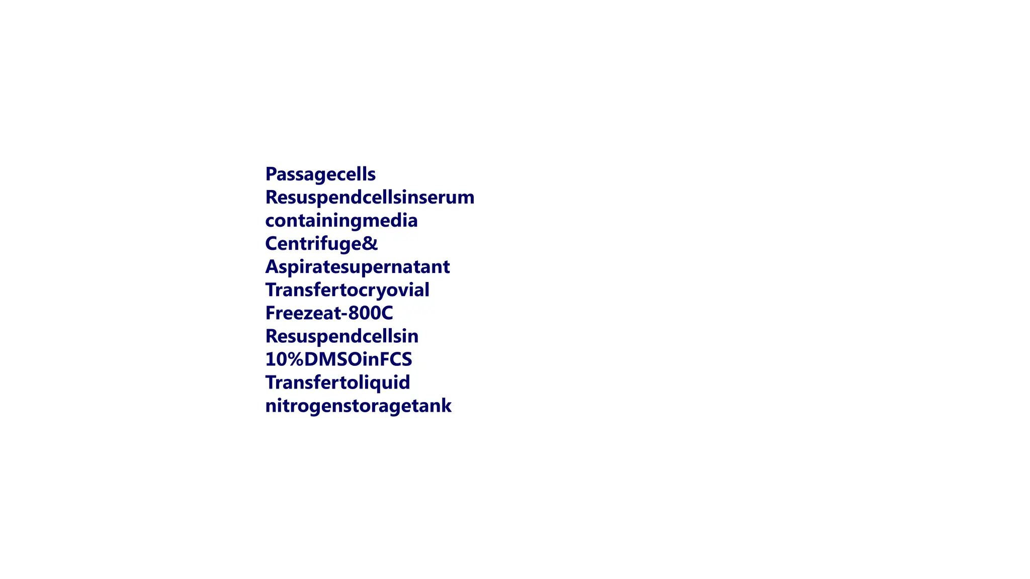
Cryopreservation
Count cells
Containment level 2
cell culture laboratory
Typical
cell culture flask
‘Mr Frosty’
Used to freeze cells
Liquid nitrogen is
also used
36.
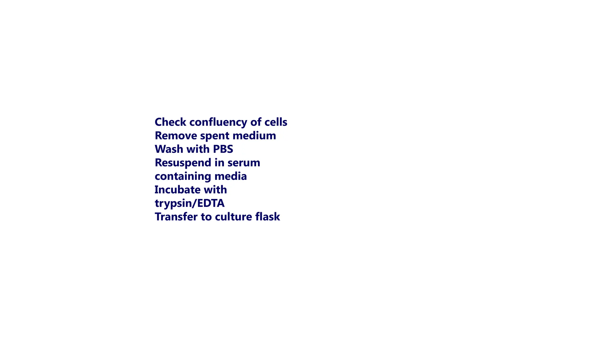
Check confluency ofcells
Remove spent medium
Wash with PBS
Resuspend in serum
containing media
Incubate with
trypsin/EDTA
Transfer to culture flask
37.
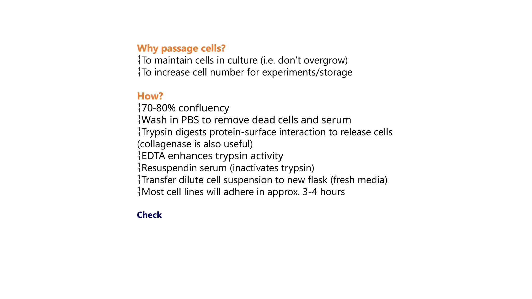
Why passage cells?
Tomaintain cells in culture (i.e. don’t overgrow)
To increase cell number for experiments/storage
How?
70-80% confluency
Wash in PBS to remove dead cells and serum
Trypsin digests protein-surface interaction to release cells
(collagenase is also useful)
EDTA enhances trypsin activity
Resuspendin serum (inactivates trypsin)
Transfer dilute cell suspension to new flask (fresh media)
Most cell lines will adhere in approx. 3-4 hours
Check