
Recommended
More Related Content
What's hot
What's hot (20)
Similar to Thyroid final [part 1]
Similar to Thyroid final [part 1] (20)
Recently uploaded
Recently uploaded (20)
Thyroid final [part 1]
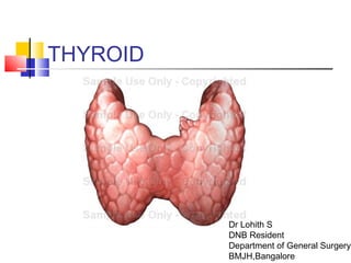
- 1. THYROID Dr Lohith S DNB Resident Department of General Surgery BMJH,Bangalore
- 2. HISTORY ● Term 'thyroid' was coined by Thomas Warton in 17th century ● Emil Theoder Kocher is considered as the Father of Modern Thyroid surgery ● First thyroidectomy is considered to be done more than 1000 years ago by Abu-al-Qasim ● The earliest account of thyroidectomy was probably given by Roger Frugardi, 1170
- 3. The thyroid glandThe thyroid gland ANATOMY AND EMBRYOLOGY Lobes Position Blood supply Development Parathyroid glands
- 4. The thyroid gland derives its name from the thyroid cartilage which resembles a shield (G. thyreos = shield)
- 5. Function The thyroid gland is an endocrine gland that is responsible for the secretion of thyroxin and thyrocalcitonin
- 6. Lobes The thyroid gland consists of two lobes united in front of the second, third and fourth tracheal rings by an isthmus of gland tissue. isthmusisthmus
- 7. Lobes Each lobe is pear- shaped consisting of a narrow upper pole and a broader lower pole upper poleupper pole lower polelower pole
- 8. Thyroid scan This nuclear scan uses an injectable radioactive compound. When injected into the bloodstream the compound will be concentrated in the thyroid gland resulting in an image of the gland The test can be useful in diagnosis of thyroid tumor
- 9. Position It lies under cover of sternothyroid and sternohyoid muscles on the side of the larynx and trachea sternothyroid sternohyoid
- 10. Position The upper pole of the thyroid cannot normally rise above the level of the oblique lineoblique line of the thyroid cartilage Thyroid, upper pole sternothyroid thyrohyoid cricothyroid
- 11. The thyroid gland is caught in the pocket of sternothyroid thyroid cricoidthyroidcartilage sternothyroidsternothyroid thyrohyoid cricothyroid Position
- 12. The lower pole of the thyroid gland extends along the side of the trachea as low as the sixth tracheal ring 1 2 3 4 5 6 Position
- 13. Because of the proximity of the thyroid gland to the trachea and esophagus, goiter causes compression of the trachea and esophagus resulting in dyspnea and dysphagia respectively esophagusesophagus
- 14. Retro-sternal goitre with tracheal deviationRetro-sternal goitre with tracheal deviation
- 15. Retro-sternal goitreRetro-sternal goitre with esophagealwith esophageal deviationdeviation
- 16. Pyramidal lobe In about 40% of people, there is a small upwards extension of the isthmus called the pyramidal lobe.
- 17. Levator glandulae thyroidae The pyramidal lobe may be attached to the hyoid bone by fibrous or muscular tissue (levator glandulae thyroidae).
- 18. Variations Bifurcation of the lower end of the pyramidal process, one part going to each lateral lobe
- 19. Variations Pyramidal process attached to the left lobe of the gland, isthmus absent.
- 20. Variations Both pyramidal process and isthmus are absent.
- 21. Pre-tracheal fascia The thyroid gland is surrounded by a fibrous capsule and is enclosed in the pre- tracheal fascia
- 22. Pre-tracheal fascia The pre-tracheal fascia attaches the thyroid gland to the trachea and larynx thus the thyroid moves upwards on swallowing, an important diagnostic feature for lumps in the neck thyroid larynx
- 23. Blood supply The thyroid gland is very vascular The vessels lie between the capsule and the pre-tracheal fascia. In some pathological conditions such as thyrotoxicosis, owing to its high vascularity, the blood flow can be heard with a stethoscope as a bruit
- 24. Thyroid arteries The main arteries are the superior and inferior thyroid arteries. superiorsuperior thyroid a.thyroid a. inferiorinferior thyroid a.thyroid a.
- 25. Superiorthyroidartery Arises from the anterior surface of the external carotid immediately distal to the carotid bifurcation. externalexternal carotid a.carotid a. carotidcarotid bifurcationbifurcation
- 26. Superior thyroid artery Arches downwards, giving a sternomastoid branch and a superior laryngeal branch that enters the larynx with the nerve of the same name superior laryngeal a. & n.
- 28. Superior thyroid artery enters deep to sternothyroid sternothyroid Superior thyroid vessels
- 29. Superior thyroid artery before reaching the upper pole of the gland, and within the pre-tracheal fascia, it divides into two main branches one for either surface of the gland anterior posterior
- 30. Superior thyroid artery the posterior branch anastomoses with the inferior thyroid artery posterior br. of superior thyroid a. inferior thyroid a.
- 31. Inferior thyroid artery Is a branch of the thyrocervical trunk from the subclavian artery . subclavian a.subclavian a. thyrocervicalthyrocervical trunktrunk inferiorinferior thyroid a.thyroid a.
- 32. Inferior thyroid artery Ascends and turns medially at the level of the cricoid cartilage to enter the back of the gland some distance above the lower pole.
- 33. Inferior thyroid artery The tortuous course of the inferior thyroid artery is due to the fact that in every swallow the thyroid gland ascends a few centimeters and must naturally drag its blood supply with it. If this artery has no capability to elongate, it would be traumatized
- 34. Inferior thyroid artery Divides outside the pre-tracheal fascia into four or five branches that pierce the fascia separately to reach the lower pole of the gland. Remember that the superior thyroidRemember that the superior thyroid artery divides within the pretrachealartery divides within the pretracheal fasciafascia
- 35. The recurrent laryngeal nerve lies normally behind the branches of the inferior thyroid artery
- 36. The recurrent laryngeal nerve lies normally behind the branches of the inferior thyroid artery but it is common for the nerve to pass between the artery branches before they pass through the fascia.
- 37. The recurrent laryngeal nerve always lies behind the pre- tracheal fascia and if this structure remains intact during thyroidectomy the nerve will not have been divided recurrent laryngeal n. inferior thyroid a.
- 38. Both thyroid arteries are related to nerves which must be avoided when tying the arteries.
- 39. A little distance behind the superior thyroid artery is the external laryngeal nerve. superior thyroid a. external laryngeal n. external laryngeal n. internal laryngeal n. superior laryngeal n.
- 40. Superior laryngeal nerve variations vagusvagus internalinternal externalexternal
- 41. To avoid injury to the external laryngeal nerve, the superior thyroid artery is ligated and sectioned near the superior pole of the thyroid gland where it is notnot so closely related to the nerve as it is at its origin.
- 42. Section of the external laryngeal nerve produces weakness of voice, since the vocal fold cannot be tensed. The cricothyroid muscle is paralyzed Cricothyroid tenses the vocal cordCricothyroid tenses the vocal cord
- 43. The recurrent laryngeal nerve has a variable relationship to the inferior thyroid artery because of its proximity to the inferior thyroid artery and the pre-tracheal fascia it may be injured while ligating the artery during thyroidectomy
- 44. hence the advisability of ligating the inferior thyroid artery well lateral to the gland before it begins to divide into its terminal branches. the inferior thyroid artery gives off esophageal and inferior laryngeal branches before its terminal distribution into the thyroid gland site of inferior thyroid a. ligation site of superior thyroid a. ligation
- 45. The variable relationship of the inferior thyroid artery to the recurrent laryngeal nerve makes thyroid surgery a potential risk to normal speech The recurrent laryngeal nerve supplies all the intrinsic muscles of the larynx
- 46. it is advisable that a surgeon about to perform a thyroidectomy examines the vocal cords prior to operation, so that if there is any problem postoperatively one knows at least the origin of the lesion.
- 47. Recurrent laryngeal nerve damage Is a complication of thyroid surgery that causes paralysis of the vocal cords When bilateral the voice is almost absent as the two vocal folds cannot be adducted.
- 48. Recurrent laryngeal nerve damage A unilateral recurrent laryngeal nerve injury may not be noticed in normal speech but would be very detrimental to a singers career.
- 49. The thyroid arteries anastomose freely with each other and with tracheal and esophageal arteries.
- 50. In operations of partial or sub-total thyroidectomy, all four arteries are tied
- 51. In operations of partial or sub- total thyroidectomy, all but the posterior part of the gland excised remaining thyroid tissue
- 52. the dangerous anatomy lies in the posterior lateral lobes (recurrent laryngeal nerve and the parathyroid glands) Recurrent laryngeal n. parathyroid gland
- 53. The remains of the gland are located alongside the trachea and contain the parathyroid glands, the whole being supplied with blood by the anastomosis
- 54. Thyroidae ima artery In about 10% of individuals, an unpaired artery, the thyroidae ima (L. ima = lowest) is a small occasional artery from the brachiocephalic trunk, or left common carotid artery, or direct from the arch of the aorta
- 55. Thyroidae ima artery Ascends anterior to trachea and supplies the isthmus of the thyroid gland.
- 56. Thyroidae ima artery The possible presence of the thyroid ima artery must be remembered when incising the trachea inferior to the isthmus. As the thyroidae ima runs anterior to the trachea, it is a potential source of serious bleeding
- 57. Thyroid veins The veins are three in number on each side the superior thyroid vein from the upper pole follows the artery and enters the internal jugular vein or the common facial vein Superior thyroid v. Internal jugular v.
- 58. The middle thyroid vein is short and wide, it enters the internal jugular vein Thyroid veins middle thyroid v. Internal jugular v.
- 59. From the isthmus and lower pole of the gland the inferior thyroid veins form a plexus within the pre-tracheal fascia that descends in front of the trachea to reach the left brachiocephalic vein Thyroid veins inferior thyroid vv. brachiocephalic v.
- 60. As the inferior thyroid veins cover the anterior surface of the trachea inferior to isthmus, they are potential sources of bleeding during tracheotomy (also remember the situation of the thyroidae ima artery). Inferior thyroid veins
- 61. Development of the thyroid gland The gland begins as a diverticulum from the floor of the embryonic pharynx
- 62. Development of the thyroid gland The diverticulum grows caudally superficial to the hyoid before dividing into two lobes The stem of the diverticulum, the thyroglossal duct, normally disappears hyoid Thyroglossal duct
- 63. Development of the thyroid gland After the tongue has developed, it can be seen that the point of outgrowth of the thyroglossal duct is the foramen cecum (of Morgagni) [Morgagni, Giovanni Battista, 1682-1771, a Padua anatomist and pathologist, also known for hydatid of Morgagni (appendix testis) and anal columns (of Morgagni)].
- 65. Thyroglossal cyst cysts derived from the duct may also appear anywhere between the foramen cecum and the normal position in the midline of the neck 1. Beneath foramen cecum 2. Floor of the mouth 3. Suprahyoid 4. Subhyoid 5. On thyroid cartilage 6. At level of cricoid cartilage
- 66. Thyroglossal cyst Can be diagnosed because characteristically it moves upwards as the patient puts his tongue out.
- 67. Infection of a thyroglossal cyst may spread to a persistent thyroglossal duct which must be then excised
- 68. Although the duct lies ventral to the hyoid bone, it passes up for a short distance behind the body, which therefore has to be excised with the duct
- 69. Accessory thyroid gland Aberrant thyroid tissue may appear between the foramen cecum and the normal position
- 70. Lingual thyroid Rarely the thyroid fails to descend during development resulting in the development of a lingual thyroid
- 71. Ectopic thyroid Failure of descent mar result in a superior cervical thyroid in the region of the hyoid bone the thyroid may sometimes descended too far and be found in the superior mediastinum
- 72. Parathyroid glands Two on each side They are yellow-brown endocrine glands, about the size of a small pea (about 0.5x0.8 cm ovoids) They are important because of their role in calcium metabolism. They secrete parathormone that mobilizes bone calcium and increases gut and kidney calcium absorption
- 73. Parathyroid glands Are located posterior to the thyroid gland between its capsule and fascial sheath
- 74. Superior parathyroid glands more constant in position embedded in the posterior surface of the thyroid gland, a short distance above the entry of inferior thyroid artery (and the level of the cricoid cartilage).
- 75. Inferior parathyroid glands variable in position usually embedded behind the lower pole but is often found elsewhere (they may even present in the superior mediastinum).
- 76. Parathyroid development The parathyroids develop from the endoderm of the third (inferior gland) and fourth (superior gland) pharyngeal pouches
- 77. The thymusthymus also develops from the third pouch and may therefore carry the inferior parathyroidparathyroid with it when it descends into the thorax. Parathyroid development
- 78. Parathyroid glands, blood supply The glands are usually supplied by the inferior thyroid arteries but may also be supplied by both superior and inferior thyroid arteries posterior br. of superior thyroid a. inferiorinferior thyroid a.thyroid a.
- 79. Parathyroid glands Awareness of the close relationship between the parathyroid glands and the thyroid gland is essential to prevent removal or damage of the parathyroid glands during thyroidectomy.
- 80. The parathyroid glands are usually safe during subtotal thyroidectomy because the posterior part of the thyroid gland is preserved
- 81. The variability in position of the parathyroid glands may create a problem during total thyroidectomy; in this case the parathyroid glands are saved by following their small vessels which are kept intact before the thyroid is removed.
- 82. LYMPHATICS ● Lymphatic drainage of thyroid gland has been proposed by Taylor. His studies shows clinically relevant lymphatic spread in thyroid malignancy ● Central compartment of neck - – Tracheal LN – Chain of LN which lie in tracheo-oesophageal groove – One or more LN lying above isthmus – 'delphian nodes' ● B/L central LN dissection (level 6 dissection) – Clears all LN from carotid artery to other and down into superior mediastinum.
- 83. ● Lateral compartment of neck ● A constant group of LN lies along IJV on each side (level 2,3,4). LN in supraclavicular fossa or more laterally level 5 LN may also be involved in thyroid malignancy ● Thoracic duct on left side of neck arches up out of mediastinum and passes forwards and laterally to drain into left subclavian vein / IJV ● Lateral LN dissection – ● removal of level 2, 3, 4 and 5 LN. Vagus N, symphatheticc ganglia, phrenic N, brachial plexus and spinal accessory N are preserved
- 85. Investigations
- 86. USG in Thyroid -gland enlarged or not -nodular/diffuse -single/multiple -lymphnode assessment -guide to FNAC -benign or malignant depending on vascularity Peripheral-benign Central -malignant
- 87. Disadvntage of USG in Thyroid No information about retrosternal thyroid staging.
- 88. Radioisotope Usually was preferred earlier But now avoid as much as possible 3 indications: - Toxicity associated with nodularity - To locate ectopic thyroid - To locate metastatic I123 should be avoided because of long t1/2. Tc99 should be used.
- 90. Important points Most common cause of nodularity in Thyroid –colloid>follicular adenoma 80% of thyroid nodule are benign, 20% are malignant. Chance of malignancy -Euthyroid > hypothyroid > hyperthyroid(<1%) -Cold > warm> hot So to summarize cold euthyroid is a deadly combination.
- 91. Important points IOC for systemic spread of carcinoma Thyroid –PET scan FNNAC(Fine Needle Non Aspiration Cytology) in this morphology is better accepted then FNAC. Therefore in thyroid gland FNNAC is preferred more than FNAC.
- 92. Thyroidectomy ● INDICATIONS ● As therapy for patients with thyrotoxicosis ● To treat benign and malignant thyroid tumours ● To alleviate pressure symptoms (respiratory distress, dysphagia) with benign/ malignant process ● Cosmetic purpose ● To establish a definitive diasgnosis of a mass within thyroid gland, especialy when cytological analysis is either non diagnostic or indeterminate ● Suspicion of malignancy in benign nodule like, hard nodule, sudden increase in size, involvement of adjacent structures, enlarged lymphnode and recurrent cyst.
- 93. TYPES ● Thyroid lobectomy / Hemithyroidectomy ● Subtotal thyroidectomy ● Near total thyroidectomy ● Total thyroidectomy ● Completion thyroidectomy
- 94. Types: Sub-total: about 8gms , or a tissue, size of pulp of finger is retained on lower pole on both sides and rest is removed. Commonly done in toxic thyroid, MNG. Total: entire gland is removed. Done in malignancy.
- 95. Near-total: both lobes except the lower pole which is very close to recurrent laryngeal nerve and parathyroid is removed. Here <2gm of tissue is left behind. Hemi: along with removal of one lobe, entire isthmus is removed. Done in benign disease of only one lobe, thyroid cyst, solitary nodule.
- 96. PRE OPERATIVE EVALUATION ● Ultrasonography ● Fine needle aspiration cytology – FNAC ● Thyroid function tests – TFT ● CT scan ● Thyroid uptake scan ● Laryngoscopy ● Serum Calcium, Parathormone (PTH)
- 97. PRE OPERATIVE PREPARATION Thyrotoxic patient are rendered euthyroid; Carbimazole 10-15mg 8hourly, when patient become euthyroid(in about 4weeks) they are maintained on 5-10mg Propranolol 80mg 6hourly 4-7days before operation. Symptoms and signs are usually controlled within 24hours. Continued 8- 10days post op Lugol’s iodine; 2weks pre-operatively to reduce the vascularity of the gland
- 98. PRE OPERATIVE CONSENT ● Scar ● Airway obstruction ● Voice changes ● Hypoparathyroidism ● Hypothyroidism
- 99. ANAESTHESIA Anaesthesia is general with cuffed endotracheal tube POSITION patient is placed in a supine position initially with the neck extended by placing a ring beneath the head and a sandbag roll beneath the shoulder. The table is tilted 20–30 degrees “head up” to aid in emptying the neck veins.11/26/16 99
- 100. The skin is prepped from the chin to the upper thorax Drapes are applied; head scarf, sides of the neck, chest-abd, large covering the legs. The are secured with clips Surgeon and assistant scrub and gown, the stands on the opposite side to be operated upon(usually the larger gland first)
- 101. Incision Site of incision is indented with suture A transverse skin crease incision is placed 2-3cm above the sternal notch about 8cm long extending to the lateral borders of sternocleidomastoid. The scapel (with size 15 blade) is slanted to divide the skin and platysma at different level to give a neater scar Hemostasis is controlled with electrocautary or prior infiltration with lidocaine and adrenaline11/26/16 101
- 102. 11/26/16 102
- 103. PROCEDURE Elevate the flap of skin with the platysma (the assistant lifts the skin and the platysma upward with double skin hooks to allow for the creation of a subplatysmal flap). Superiorly to the thyroid cartilage Inferiorly to the suprasternal notche Place Joll’s retractor to retract the skin flaps This procedure should be blood free, because the superficial veins lie beneath the cervical fascia. Divide the deep cervical fascia longitudinally in the midline, between the anterior jugular veins. At the lower part there is usually a transverse cervical vein that needs to be clamped, divided, and ligated with 3-0 silk sutures 11/26/16 103
- 104. 11/26/16 104
- 105. 11/26/16 105
- 106. The strap muscles (sternohyoid, and deeper sternothyroid) are carefully separated to allow their retraction laterally. Assess goiter; The loose areolar tissue(capsule) overlying the thyroid gland is divided with electrocautery. After the anterior surface of the thyroid has been thoroughly exposed, the entire gland is carefully explored and palpated. The strap muscles are firmly retracted with a small loop retractor while the thyroid gland is drawn medially Ligate and divide in continuity Middle thyroid vein Superior thyroid vessels close to the gland(to avoid injury to the external laryngeal nerve) between two proximal and one distal ligature. The recurrent laryngeal nerve and the parathyroids are identified and preserved then the terminal branches inferior thyroid artery are ligated and divided close to the capsule. Or the inferior thyroid artery is identified far away from the gland ligated in continuity to avoid injury to the recurrent laryngeal nerve. 11/26/16 106
- 107. The thyroid is then mobilized and removed; Divide isthmus and place hemostats around margin of resection (run with interlocking 3-0 absorbable suture) leaving about 4g of thyroid from each lobe for subtotal If a total thyroidectomy is being performed, the remaining lobe is removed in a similar fashion, with division of the middle thyroid vein, identification of the recurrent laryngeal nerve and parathyroid glands, and ligation and division of the superior pole and branches of the inferior thyroid vessels.11/26/16 107
- 108. CLOSURE Absolute haemostasis Suction drain to thyroid bed(beneath the strap muscles) Close loosely in layers with absorbable sutures Close the skin with sutures or clips Check vocal cords on extubation by direct laryngoscopy 11/26/16 bbinyunus2002@gmail.com 108
- 109. OPERATIVE STEPS ● Anaesthesia, Positioning & Draping
- 110. ● Skin incision and creation of flaps
- 112. Exposure of thyroid gland
- 113. Mobilization and dissection of upper pole
- 117. Dissection of ITA and removal of gland
- 119. Closure
- 120. POST OPERATIVE MGT Half-hourly observation until conscious At the bed side Michel clip remover in case of respiratory distress due to hematoma 10ml of 10% calcium gluconate in case of acute hypocalcamia Keep semi-recumbent Review indirect laryngoscopy(especially if there is cord impairment on extubation) Serum calcium regularly in the postoperative period Thyroid function tests at 6weeks postoperatively Remove Drain when dry, 24-48hours postoperatively Sutures/clips, 2-3days postoperatively 11/26/16 120
- 121. COMPLICATIONS EARLY Haemorrhage Tetany In first 3 days from corrected thyrotoxicosis After 1 week with hypoparathyroidism Recurrent laryngeal nerve palsy 95% neurapraxia and resolves If bilateral, cord adduct to midline so needs immediate reintubation Thyroid crisis, if throtoxic patient is inadequately prepared rare with modern technique Wound infection LATE Keloid Hypothroidism- 20% Recurrent thyrotoxicosis- <5% of patients undergoing thyroidectomy for grave disease 11/26/16 121
- 122. RECENT ADVANCES ● Minimally invasive thyroidectomy ● Robotic transaxillary thyroid surgery ● Transoral thyroidectomy
- 123. QUESTIONS 1. What is the blood supply to the thyroid gland 2. What are the preoperative measures prior to thyroidectomy for thyrotoxicosis 3. What are the types of thyroidectomy 4. Outline the steps of thyroidectomy 5. What are the complications of thyroidectomy 6. What does the recurrent laryngeal nerve supply and what is the consequence of it division 7. What does external laryngeal nerve supplies and what is the consequences of it division 8. What is the Simon’s triangle 11/26/16 123
Editor's Notes
- Strap muscles may need to be transected to gain better access to the thyroid gland; when necessary this should be done at the level of the thyroid cartilage to preserve their innervation from the ansa hypoglossi nerve. If there is local invasion by a thyroid neoplasm, the thyroid lobe is resected en bloc with its overlying strap muscles.
- The recurrent laryngeal nerve is usually found in Simon’s triangle, which is formed by the inferior thyroid artery superiorly, the common carotid artery laterally, and the esophagus medially