Objectives
1- Describe thedifferent parts of urinary system.
2- Describe the structure of kidney.
3- Define nephron, and list its histological parts.
a- Define renal corpuscle, describe its structure, and name
its parts.
b- Define Podocyte, and relate between its histological
structure and process of filtration.
c- Define filtration slit, describe its structure, and list its
components.
4- Recognize some clinical problems related to filtration.
5- Describe Mesangial cell, and identfy some of its functions.
3.
Urinary system
• Consistsof two kidneys, ureters, and one bladder and ureter.
• Function:
• 1- Maintenance of homeostasis by production of urine
through filtration, absorption, and secretion.
• 2- Regulation of fluid and electrolytes balance of the body.
• 3- Production of Renin (important in regulation of blood
pressure).
• 4- Production of Erythropoietin (stimulates the production of
erythrocytes).
4.
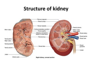
Kidney
Each kidney issurrounded by a connective tissue
capsule that is surrounded by a mass of peri
renal adipose tissue.
The kidney is divided into an outer cortex and an
inner medulla. Renal medulla consists of 10-18
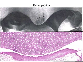
conical or pyramidal structures; the medullary
pyramids. The tips of pyramids are called renal
papillae, where the collecting ducts open into.
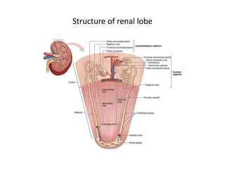
Kidney lobe consists of medullary pyramid and
associated cortical tissue at its base and sides.
Medical appliation
Polycystic kidneydisease is an inherited disorder
in which
normal cortical organization of both kidneys is
lost due to the formation of multiple, large,
fluid-filled cysts. The cysts may arise from any
epithelial cells of the nephron and can lead to
gross kidney enlargement and loss of renal
function.
Nephron
Is the functionalunit of the kidney. It consists of:
• Renal corpuscle, an initial dilated portion in the cortex
• Proximal convoluted tubule, located primarily in the cortex
• Thin and thick limbs of the nephron loop (loop of Henle),
which descend into the medulla, then ascend back to the
cortex
• Distal convoluted tubule
• Collecting tubule.
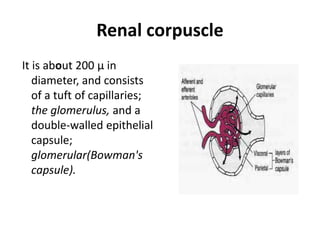
Renal corpuscle
It isabout 200 µ in
diameter, and consists
of a tuft of capillaries;
the glomerulus, and a
double-walled epithelial
capsule;
glomerular(Bowman's
capsule).
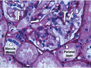
Bowman's capsule
Composed oftwo layers;
1- Parietal layer consists
of simple sequamus
epithelium supported
by a basal lamina and a
thin reticular fibers.
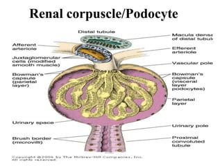
Podocyte
The cell bodygives rise to several
primary processes from which
arise numerous secondary
processes that called pedicels.
These pedicels embrace the
capillary of glomerulus and
come in contact with the basal
lamina at a periodic distance
of 25nm. The pedicels
interdigitate with each other
defining a space about 25nm
wide; the filtration slit which is
bridged by a diaphragm of
6nm thick.
A thick basement
membrane(0.1µm) is
formed by fusion of the
basal laminae of both
capillary and podocytes,
forming a filtration
barrier that separates
urinary space and
capillary blood.
21.
Filtration apparatus
• Consistsof:
• 1- Endothelium of glomerular capillaries: they are
fenestrated, with no diaphragm, and posse a large
number of aquaporin-1(AQP-1) water channels that
allow fast movement of water through epithelium.
• 2- Glomerular basement membrane.
• 3- Podocytes.
22.
Medical applications
• Indiseases such as diabetes mellitus and
glomerulonephritis, the glomerular filter is altered
and becomes much more permeable to proteins,
with the subsequent release of protein into the
urine (proteinuria).
23.
Mesangial cell
This cellis found adherent
to the wall of
glomerular capillaries
where the basal lamina
is shared by two or
more capillaries.
It has cytoplasmic
processes that extend
to the endothelial cells.
Functions of mesangialcell
• Physical support and contraction—the mesangium provides
internal structural support to the glomerulus and like
pericytes, its cells respond to vasoactivesubstances to help
maintain hydrostatic pressure for the optimal rate of filtration.
• Phagocytosis—mesangial cells phagocytose protein
aggregates that adhere to the glomerular filter, including
antibody-antigen complexes abundant in many pathological
conditions.
• Secretion—the cells synthesize and secrete several cytokines,
prostaglandins, and other factors important for immune
defense and repair in the glomerulus.