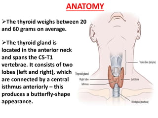
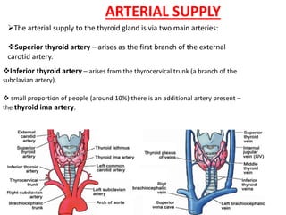
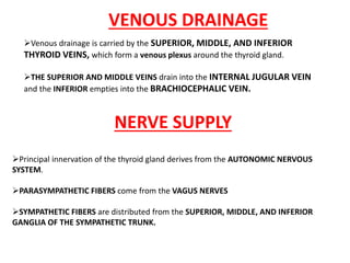
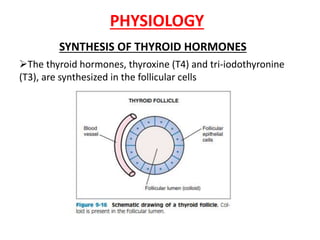
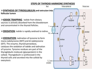
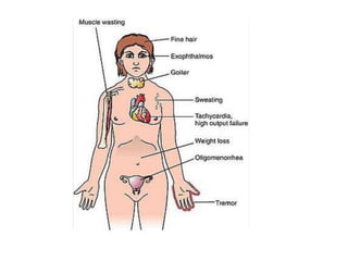
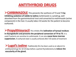
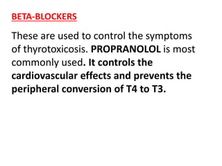
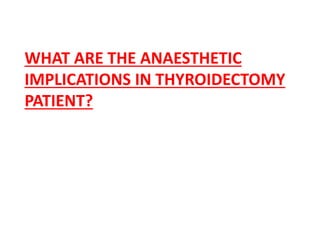
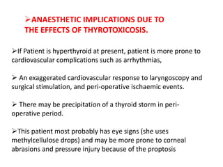
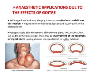
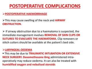
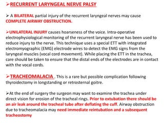
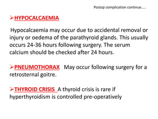
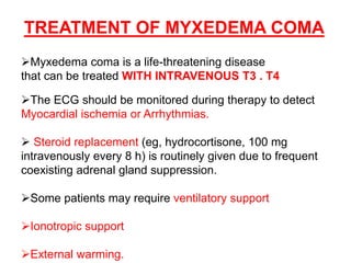
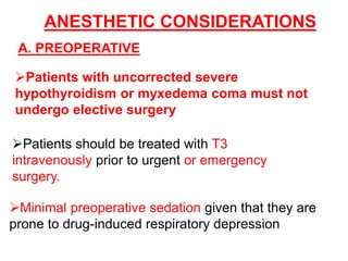
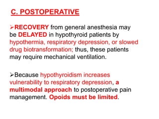
THE THYROID
DR RIZWAN ANSARI
FCPS
ASSISTANT PROFESSOR
BILAWAL MEDICAL COLLEGE LUMHS
The thyroid weighs between 20
and 60 grams on average.
The thyroid gland is
located in the anterior neck
and spans the C5-T1
vertebrae. It consists of two
lobes (left and right), which
are connected by a central
isthmus anteriorly – this
produces a butterfly-shape
appearance.
ANATOMY
The arterial supply to the thyroid gland is via two main arteries:
Superior thyroid artery – arises as the first branch of the external
carotid artery.
Inferior thyroid artery – arises from the thyrocervical trunk (a branch of the
subclavian artery).
 small proportion of people (around 10%) there is an additional artery present –
the thyroid ima artery.
ARTERIAL SUPPLY
Venous drainage is carried by the SUPERIOR, MIDDLE, AND INFERIOR
THYROID VEINS, which form a venous plexus around the thyroid gland.
THE SUPERIOR AND MIDDLE VEINS drain into the INTERNAL JUGULAR VEIN
and the INFERIOR empties into the BRACHIOCEPHALIC VEIN.
Principal innervation of the thyroid gland derives from the AUTONOMIC NERVOUS
SYSTEM.
PARASYMPATHETIC FIBERS come from the VAGUS NERVES
SYMPATHETIC FIBERS are distributed from the SUPERIOR, MIDDLE, AND INFERIOR
GANGLIA OF THE SYMPATHETIC TRUNK.
VENOUS DRAINAGE
NERVE SUPPLY
PHYSIOLOGY
SYNTHESIS OF THYROID HORMONES
The thyroid hormones, thyroxine (T4) and tri-iodothyronine
(T3), are synthesized in the follicular cells
IODIDE TRAPPING. Iodide from dietary
sources is actively absorbed from the bloodstream
and concentrated in the thyroid follicles.
SYNTHESIS OF TYROGLUBULIN and extrusion into
follicular lumen
OXIDATION. Iodide is rapidly oxidised to iodine.
STEPS OF THYROID HARMONE SYNTHESIS
IODINATION. Iodination of tyrosine to form
mono-iodotyrosine (MIT) and di-iodotyrosine
(DIT). The enzyme, thyroid peroxidase,
catalyses the oxidation of iodide and iodination
of tyrosine. Tyrosine residues are part of the
thyroglobulin molecule (glycoprotein) in the
colloid. Thyroglobulin is synthesised in the
thyroid cells and secreted into the colloid by
exocytosis
COMBINATION. T3 is formed by combining
one MIT and one DIT molecule. T4 is formed
by combining two DIT molecules.
PROTEASES DIGEST IODINATED
THYROGLOBULIN, releasing the hormones, T4
and T3, the biologically active agents central to
metabolic regulation
REGULATION OF THYROID HORMONE
SECRETION
The production of T4 and
T3 is regulated by TSH,
released by the anterior
pituitary, which in turn is
controlled by thyrotropin
releasing hormone released
by the hypothalamus. When
the T4 levels are high, TSH
production is suppressed
forming a negative feedback
loop
WHAT ARE THE NORMAL RANGE OF THYROID TSH T3 AND T4?
T4 = 77–155 nmol/l
T3 = 1.2–2.8 nmol/L
TSH to be 0.3–4 mU/l
The diagnosis of hyperthyroidism is confirmed by
abnormal thyroid function tests, which may include an
elevation in serum T4 and serum T3 and a reduced
TSH level.
 The free fraction of the hormone is
active.
 The half-life of T4 is 5-7 days.
 the half-life of T3 is only 24 hours.
 T3 is four times more active than the
more abundant T4.
HYPERTHYROIDISM
CLINICAL MANIFESTATIONS
CAUSES
 Graves’ disease (autoimmune).
 Toxic multinodular goitre.
Solitary toxic nodule/adenoma.
 Acute thyroiditis: viral, autoimmune and post-irradiation.
 Neonatal thyrotoxicosis (maternal thyroid antibodies).
Exogenous iodine.
Drugs (e.g. amiodarone)
WEIGHT LOSS
HEAT INTOLERANCE
MUSCLE WEAKNESS
DIARRHEA
HYPERACTIVE REFLEXES, AND NERVOUSNESS.
A FINE TREMOR, EXOPHTHALMOS, OR GOITER MAY BE NOTED,
PARTICULARLY WHEN THE CAUSE IS GRAVES DISEASE.
NEW ONSET OF ATRIAL FIBRILLATION IS A CLASSIC PRESENTATION OF
HYPERTHYROIDISM, BUT CARDIAC SIGNS MAY ALSO INCLUDE SINUS
TACHYCARDIA AND CONGESTIVE HEART FAILURE.
CLINICAL MANIFESTATIONS
ANTITHYROID DRUGS
CARBIMAZOLE. This prevents the synthesis of T3 and T4 by
inhibiting oxidation of iodide to iodine.Carbimazole is a prodrug, well
absorbed from the gastrointestinal tract and converted to methimazole (active
component) in the liver. It usually takes 4-8 weeks for the patient to become
euthyroid;
Propylthiouracil. This inhibits the iodination of tyrosyl residues
in thyroglobulin and prevents the peripheral conversion of T4 to T3. It is
used if patients are sensitive to carbimazole. It can also cause bone marrow
suppression. A euthyroid state is usually achieved after 4-8 weeks of treatment
Lugol’s iodine. Traditionally this has been used as an adjunct to
antithyroid drugs for 10-14 days before a partial thyroidectomy to reduce the
vascularity of the gland.
BETA-BLOCKERS
These are used to control the symptoms
of thyrotoxicosis. PROPRANOLOL is most
commonly used. It controls the
cardiovascular effects and prevents the
peripheral conversion of T4 to T3.
WHAT ARE THE ANAESTHETIC
IMPLICATIONS IN THYROIDECTOMY
PATIENT?
ANAESTHETIC IMPLICATIONS DUE TO THE
EFFECTS OF THYROTOXICOSIS.
ANAESTHETIC IMPLICATIONS DUE TO THE
EFFECTS OF GOITRE
.
If Patient is hyperthyroid at present, patient is more prone to
cardiovascular complications such as arrhythmias,
 An exaggerated cardiovascular response to laryngoscopy and
surgical stimulation, and peri-operative ischaemic events.
 There may be precipitation of a thyroid storm in peri-
operative period.
This patient most probably has eye signs (she uses
methylcellulose drops) and may be more prone to corneal
abrasions and pressure injury because of the proptosis
ANAESTHETIC IMPLICATIONS DUE TO
THE EFFECTS OF THYROTOXICOSIS.
 With regard to the airway, a large goitre may cause tracheal deviation or
obstruction. It may be worse in the supine position and usually eases in the
lateral position.
Postoperatively, after the removal of the thyroid gland, TRACHEOMALACIA
can lead to airway obstruction. There may be involvement of the recurrent
laryngeal nerve causing a hoarse voice (unilateral) or stridor (bilateral)
ANAESTHETIC IMPLICATIONS DUE TO
THE EFFECTS OF GOITRE
The following options for airway management are available when a
difficulty is anticipated:
 INHALATIONAL INDUCTION WITH SEVOFLURANE. The aim is to
maintain spontaneous ventilation until the airway is secured. Premedication with an anti-
sialogogue should be considered to dry the secretions in patients with a non-toxic goitre. A
nasopharyngeal or oral airway should be immediately available to overcome the airway
obstruction at the level of the oropharynx or nasopharynx. In the presence of airway
obstruction, inhalation induction may be prolonged. Airway collapse leading to complete
airway obstruction may occur as the patient is induced.
 AWAKE FIBREOPTIC INTUBATION. This is the technique of choice when the
airway anatomy is distorted. If the patient is in stridor, due to narrowing of the airway,
however, passage of the fibreoptic scope may result in complete airway obstruction
The presence of noisy breathing, dyspnoea and position-related
breathing difficulty indicates airway obstruction
HOW Would you anaesthetise
this patient?
Anesthetic Considerations continue…..
A. PREOPERATIVE
All elective surgical procedures, including subtotal thyroidectomy, should
be postponed until the patient is RENDERED CLINICALLY AND CHEMICALLY
EUTHYROID with medical treatment.
ANTITHYROID MEDICATIONS AND Β ADRENERGIC ANTAGONISTS ARE CONTINUED
THROUGH THE MORNING OF SURGERY. Administration of propylthiouracil and
methimazole is particularly important because of their relatively short half-
lives
Patients with larger goiters or thyroid masses will often HAVE PREOPERATIVE CT
OR MR IMAGING STUDIES TO RULE OUT EXTENSION INTO THE MEDIASTINUM. Such
extension might mandate sternotomy for complete resection.
MONITORING DURING ANAESTHESIA includes peripheral oxygen saturation
using pulse oximetry, continuous ECG and non-invasive blood pressure measurement.
THE CARDIOVASCULAR RESPONSE TO LARYNGOSCOPY can be minimised by using
OPIOIDS AT INDUCTION. LOCAL ANAESTHETIC SPRAY TO THE VOCAL CORDS AND TRACHEA
reduces the pressor response to tracheal manipulation during surgery.
 If there are no anticipated airway problems, PRE-OXYGENATION AND
INTRAVENOUS INDUCTION CAN BE USED.
Prior to administration of the muscle relaxant the ability to ventilate the lungs
should be checked with gentle ventilation through a face mask.
An ARMOURED ETT is usually chosen to secure the airway.
THE EYES SHOULD BE TAPED AND PADDED to protect the exophthalmic eye. If
intubation and/or ventilation should prove difficult, manually lifting forward a large
goitre may relieve airway obstruction.
INTRA-OPERATIVE
The trachea should be extubated once the patient is FULLY AWAKE.
While the patient is still ‘deep’ the ETT can be replaced by an LMA. This
ENABLES VISUALISATION OF THE VOCAL CORD MOVEMENT VIA THE LMA
USING A FIBREOPTIC SCOPE, after which the patient is allowed to wake up.
The patient should be monitored in the immediate postoperative period for
AIRWAY OBSTRUCTION, THYROID STORM, AND HYPOCALCAEMIA.
POSTOPERATIVE ANALGESIA should be provided with regular paracetamol
and NSAIDs such as ibuprofen or diclofenac sodium. Opioids such as codeine
phosphate and morphine can be administered to manage breakthrough pain
when required.
Supplementary oxygen and intravenous fluids should be continued in the
postoperative period to prevent dehydration and hypoxia.
 Sitting the patient up reduces oedema and venous engorgement.
RECOVERY AND POSTOPERATIVE PERIOD
WHAT ARE THE POSSIBLE
POSTOPERATIVE COMPLICATIONS
THAT CAN OCCUR FOLLOWING
THYROIDECTOMY?
POSTOPERATIVE HAEMORRHAGE
This may cause swelling of the neck and AIRWAY
OBSTRUCTION.
 If airway obstruction due to a haematoma is suspected, the
immediate management involves REMOVAL OF SKIN CLIPS OR
SUTURES TO EVACUATE THE HAEMATOMA. Clip removers or
stitch cutters should be available at the patient’s bed side.
LARYNGEAL OEDEMA
 This may be due to TRAUMATIC INTUBATION OR EXTENSIVE
NECK SURGERY. Dexamethasone 8mg administered intra-
operatively may reduce oedema. It can also be treated with
humidified oxygen and nebulised steroids
POSTOPERATIVE COMPLICATIONS
RECURRENT LARYNGEAL NERVE PALSY
 A BILATERAL partial injury of the recurrent laryngeal nerves may cause
COMPLETE AIRWAY OBSTRUCTION.
UNILATERAL INJURY causes hoarseness of the voice. Intra-operative
electrophysiological monitoring of the recurrent laryngeal nerve has been used to
reduce injury to the nerve. This technique uses a special ETT with integrated
electromyographic (EMG) electrode wires to detect the EMG signs from the
laryngeal muscles (vocal cord movement). While placing the ETT in the trachea,
care should be taken to ensure that the distal ends of the electrodes are in contact
with the vocal cords.
TRACHEOMALACIA . This is a rare but possible complication following
thyroidectomy in longstanding or retrosternal goitre.
At the end of surgery the surgeon may want to examine the trachea under
direct vision for erosion of the tracheal rings. Prior to extubation there should be
an air leak around the tracheal tube after deflating the cuff. Airway obstruction
due to tracheomalacia may need immediate reintubation and a subsequent
tracheostomy
HYPOCALCAEMIA
Hypocalcaemia may occur due to accidental removal or
injury or oedema of the parathyroid glands. This usually
occurs 24-36 hours following surgery. The serum
calcium should be checked after 24 hours.
PNEUMOTHORAX May occur following surgery for a
retrosternal goitre.
THYROID CRISIS A thyroid crisis is rare if
hyperthyroidism is controlled pre-operatively
Postop complication continue…..
The most serious threat to a hyperthyroid patient undergoing
surgery is thyroid storm, which is characterized by
HYPERPYREXIA, TACHYCARDIA, ALTERED CONSCIOUSNESS (Eg,
AGITATION, DELIRIUM, COMA), AND HYPOTENSION.
The onset is usually 6 to 24 h after surgery but can occur
intraoperatively, mimicking malignant hyperthermia. Unlike
malignant hyperthermia, thyroid storm is not associated with
muscle rigidity, elevated creatine kinase, or a marked degree
of metabolic (lactic) and respiratory acidosis.
THYROID STORM
SUPPORTIVE MEASURES
 Ensure and establish an adequate airway, breathing,
circulation and administer 100% oxygen.
 Secure venous access and administer cold
intravenous fluids (a litre of crystalloid infused rapidly
and further IV fluids should be given as required).
 Paracetamol 1g IV or orally should be given 6-hourly
to control the temperature.
TREATMENT
SPECIFIC DRUG THERAPY
 Propylthiouracil 600-1200mg given orally or via a nasogastric tube.
 Lugol’s iodine (potassium iodide) given orally, or sodium
iodide 0.25g IV. It should not be given until an hour after the
administration of antithyroid drugs. It acts immediately and prevents the
further release of thyroid hormones.
 Esmolol 0.5mg/kg as a bolus dose over one minute, followed by 50-
200mg/kg/minute as an infusion, or propranolol 1-5mg IV up to 10mg should
be used to control the sympathetic effects.
Hydrocortisone 100mg IV should be administered every 6-hourly.
 Plasma exchange may be needed.
Ionotropes and vasopressors may rarely be required
HYPOTHYROIDISM
CAUSES
AUTOIMMUNE DISEASE (EG, HASHIMOTO
THYROIDITIS)
THYROIDECTOMY
RADIOACTIVE IODINE, ANTITHYROID
MEDICATIONS,
IODINE DEFICIENCY
FAILURE OF THE HYPOTHALAMIC–PITUITARY
AXIS (SECONDARY HYPOTHYROIDISM).
CLINICAL MENIFESTATION
INFERTILITY, WEIGHT GAIN, COLD INTOLERANCE,
MUSCLE FATIGUE, LETHARGY, CONSTIPATION,
HYPOACTIVE REFLEXES, DULL FACIAL
EXPRESSION, AND DEPRESSION.
IN ADVANCED CASES HEART RATE, MYOCARDIAL
CONTRACTILITY, STROKE VOLUME, AND CARDIAC
OUTPUT ARE ALL DECREASED
 EXTREMITIES ARE COOL AND MOTTLED
BECAUSE OF PERIPHERAL VASOCONSTRICTION.
PLEURAL, ABDOMINAL, AND PERICARDIAL
EFFUSIONS ARE COMMON
Hypothyroidism may be diagnosed by
an ELEVATED TSH concentration, or a
REDUCED FREE (OR TOTAL) T3
LEVEL, OR BOTH.
Primary hypothyroidism, the more
common condition, is differentiated from
secondary disease by an ELEVATION
IN TSH IN SECONDARY CAUSE.
DIAGNOSIS
The treatment of hypothyroidism
consists of oral replacement
therapy with a thyroid hormone
preparation, which takes several
days to produce a physiological
effect and several weeks to evoke
clear-cut clinical improvement.
TREATMENT
MYXEDEMA COMA
Myxedema coma results from extreme
hypothyroidism and is characterized by coma,
hypoventilation, hypothermia, hyponatremia (from
inappropriate antidiuretic hormone secretion), and
congestive heart failure
It is more common in elderly patients and may
be precipitated BY INFECTION, SURGERY, OR
TRAUMA.
Myxedema coma is a life-threatening disease
that can be treated WITH INTRAVENOUS T3 . T4
The ECG should be monitored during therapy to detect
Myocardial ischemia or Arrhythmias.
 Steroid replacement (eg, hydrocortisone, 100 mg
intravenously every 8 h) is routinely given due to frequent
coexisting adrenal gland suppression.
Some patients may require ventilatory support
Ionotropic support
External warming.
TREATMENT OF MYXEDEMA COMA
ANESTHETIC CONSIDERATIONS
A. PREOPERATIVE
Patients with uncorrected severe
hypothyroidism or myxedema coma must not
undergo elective surgery
Patients should be treated with T3
intravenously prior to urgent or emergency
surgery.
Minimal preoperative sedation given that they are
prone to drug-induced respiratory depression
B. INTRAOPERATIVE
Clinically hypothyroid patients are more susceptible
to the hypotensive effect of anesthetic agents
because of diminished cardiac output, blunted
baroreceptor reflexes, and decreased intravascular
volume.
In this circumstance, ketamine or etomidate can be
recommended for induction of anesthesia.
Other potential coexisting conditions include
hypoglycemia, anemia, hyponatremia, difficulty during
intubation because of a large tongue, and hypothermia
from a low basal metabolic rate.
C. POSTOPERATIVE
RECOVERY from general anesthesia may
be DELAYED in hypothyroid patients by
hypothermia, respiratory depression, or slowed
drug biotransformation; thus, these patients
may require mechanical ventilation.
Because hypothyroidism increases
vulnerability to respiratory depression, a
multimodal approach to postoperative pain
management. Opoids must be limited.
THANK YOU

THE THYROID.ppt

  • 1.
    THE THYROID DR RIZWANANSARI FCPS ASSISTANT PROFESSOR BILAWAL MEDICAL COLLEGE LUMHS
  • 2.
    The thyroid weighsbetween 20 and 60 grams on average. The thyroid gland is located in the anterior neck and spans the C5-T1 vertebrae. It consists of two lobes (left and right), which are connected by a central isthmus anteriorly – this produces a butterfly-shape appearance. ANATOMY
  • 3.
    The arterial supplyto the thyroid gland is via two main arteries: Superior thyroid artery – arises as the first branch of the external carotid artery. Inferior thyroid artery – arises from the thyrocervical trunk (a branch of the subclavian artery).  small proportion of people (around 10%) there is an additional artery present – the thyroid ima artery. ARTERIAL SUPPLY
  • 4.
    Venous drainage iscarried by the SUPERIOR, MIDDLE, AND INFERIOR THYROID VEINS, which form a venous plexus around the thyroid gland. THE SUPERIOR AND MIDDLE VEINS drain into the INTERNAL JUGULAR VEIN and the INFERIOR empties into the BRACHIOCEPHALIC VEIN. Principal innervation of the thyroid gland derives from the AUTONOMIC NERVOUS SYSTEM. PARASYMPATHETIC FIBERS come from the VAGUS NERVES SYMPATHETIC FIBERS are distributed from the SUPERIOR, MIDDLE, AND INFERIOR GANGLIA OF THE SYMPATHETIC TRUNK. VENOUS DRAINAGE NERVE SUPPLY
  • 5.
    PHYSIOLOGY SYNTHESIS OF THYROIDHORMONES The thyroid hormones, thyroxine (T4) and tri-iodothyronine (T3), are synthesized in the follicular cells
  • 6.
    IODIDE TRAPPING. Iodidefrom dietary sources is actively absorbed from the bloodstream and concentrated in the thyroid follicles. SYNTHESIS OF TYROGLUBULIN and extrusion into follicular lumen OXIDATION. Iodide is rapidly oxidised to iodine. STEPS OF THYROID HARMONE SYNTHESIS IODINATION. Iodination of tyrosine to form mono-iodotyrosine (MIT) and di-iodotyrosine (DIT). The enzyme, thyroid peroxidase, catalyses the oxidation of iodide and iodination of tyrosine. Tyrosine residues are part of the thyroglobulin molecule (glycoprotein) in the colloid. Thyroglobulin is synthesised in the thyroid cells and secreted into the colloid by exocytosis
  • 7.
    COMBINATION. T3 isformed by combining one MIT and one DIT molecule. T4 is formed by combining two DIT molecules. PROTEASES DIGEST IODINATED THYROGLOBULIN, releasing the hormones, T4 and T3, the biologically active agents central to metabolic regulation
  • 8.
    REGULATION OF THYROIDHORMONE SECRETION The production of T4 and T3 is regulated by TSH, released by the anterior pituitary, which in turn is controlled by thyrotropin releasing hormone released by the hypothalamus. When the T4 levels are high, TSH production is suppressed forming a negative feedback loop
  • 9.
    WHAT ARE THENORMAL RANGE OF THYROID TSH T3 AND T4?
  • 10.
    T4 = 77–155nmol/l T3 = 1.2–2.8 nmol/L TSH to be 0.3–4 mU/l The diagnosis of hyperthyroidism is confirmed by abnormal thyroid function tests, which may include an elevation in serum T4 and serum T3 and a reduced TSH level.
  • 11.
     The freefraction of the hormone is active.  The half-life of T4 is 5-7 days.  the half-life of T3 is only 24 hours.  T3 is four times more active than the more abundant T4.
  • 13.
    HYPERTHYROIDISM CLINICAL MANIFESTATIONS CAUSES  Graves’disease (autoimmune).  Toxic multinodular goitre. Solitary toxic nodule/adenoma.  Acute thyroiditis: viral, autoimmune and post-irradiation.  Neonatal thyrotoxicosis (maternal thyroid antibodies). Exogenous iodine. Drugs (e.g. amiodarone)
  • 14.
    WEIGHT LOSS HEAT INTOLERANCE MUSCLEWEAKNESS DIARRHEA HYPERACTIVE REFLEXES, AND NERVOUSNESS. A FINE TREMOR, EXOPHTHALMOS, OR GOITER MAY BE NOTED, PARTICULARLY WHEN THE CAUSE IS GRAVES DISEASE. NEW ONSET OF ATRIAL FIBRILLATION IS A CLASSIC PRESENTATION OF HYPERTHYROIDISM, BUT CARDIAC SIGNS MAY ALSO INCLUDE SINUS TACHYCARDIA AND CONGESTIVE HEART FAILURE. CLINICAL MANIFESTATIONS
  • 16.
    ANTITHYROID DRUGS CARBIMAZOLE. Thisprevents the synthesis of T3 and T4 by inhibiting oxidation of iodide to iodine.Carbimazole is a prodrug, well absorbed from the gastrointestinal tract and converted to methimazole (active component) in the liver. It usually takes 4-8 weeks for the patient to become euthyroid; Propylthiouracil. This inhibits the iodination of tyrosyl residues in thyroglobulin and prevents the peripheral conversion of T4 to T3. It is used if patients are sensitive to carbimazole. It can also cause bone marrow suppression. A euthyroid state is usually achieved after 4-8 weeks of treatment Lugol’s iodine. Traditionally this has been used as an adjunct to antithyroid drugs for 10-14 days before a partial thyroidectomy to reduce the vascularity of the gland.
  • 17.
    BETA-BLOCKERS These are usedto control the symptoms of thyrotoxicosis. PROPRANOLOL is most commonly used. It controls the cardiovascular effects and prevents the peripheral conversion of T4 to T3.
  • 18.
    WHAT ARE THEANAESTHETIC IMPLICATIONS IN THYROIDECTOMY PATIENT?
  • 19.
    ANAESTHETIC IMPLICATIONS DUETO THE EFFECTS OF THYROTOXICOSIS. ANAESTHETIC IMPLICATIONS DUE TO THE EFFECTS OF GOITRE .
  • 20.
    If Patient ishyperthyroid at present, patient is more prone to cardiovascular complications such as arrhythmias,  An exaggerated cardiovascular response to laryngoscopy and surgical stimulation, and peri-operative ischaemic events.  There may be precipitation of a thyroid storm in peri- operative period. This patient most probably has eye signs (she uses methylcellulose drops) and may be more prone to corneal abrasions and pressure injury because of the proptosis ANAESTHETIC IMPLICATIONS DUE TO THE EFFECTS OF THYROTOXICOSIS.
  • 21.
     With regardto the airway, a large goitre may cause tracheal deviation or obstruction. It may be worse in the supine position and usually eases in the lateral position. Postoperatively, after the removal of the thyroid gland, TRACHEOMALACIA can lead to airway obstruction. There may be involvement of the recurrent laryngeal nerve causing a hoarse voice (unilateral) or stridor (bilateral) ANAESTHETIC IMPLICATIONS DUE TO THE EFFECTS OF GOITRE
  • 22.
    The following optionsfor airway management are available when a difficulty is anticipated:  INHALATIONAL INDUCTION WITH SEVOFLURANE. The aim is to maintain spontaneous ventilation until the airway is secured. Premedication with an anti- sialogogue should be considered to dry the secretions in patients with a non-toxic goitre. A nasopharyngeal or oral airway should be immediately available to overcome the airway obstruction at the level of the oropharynx or nasopharynx. In the presence of airway obstruction, inhalation induction may be prolonged. Airway collapse leading to complete airway obstruction may occur as the patient is induced.  AWAKE FIBREOPTIC INTUBATION. This is the technique of choice when the airway anatomy is distorted. If the patient is in stridor, due to narrowing of the airway, however, passage of the fibreoptic scope may result in complete airway obstruction The presence of noisy breathing, dyspnoea and position-related breathing difficulty indicates airway obstruction
  • 23.
    HOW Would youanaesthetise this patient?
  • 24.
    Anesthetic Considerations continue….. A.PREOPERATIVE All elective surgical procedures, including subtotal thyroidectomy, should be postponed until the patient is RENDERED CLINICALLY AND CHEMICALLY EUTHYROID with medical treatment. ANTITHYROID MEDICATIONS AND Β ADRENERGIC ANTAGONISTS ARE CONTINUED THROUGH THE MORNING OF SURGERY. Administration of propylthiouracil and methimazole is particularly important because of their relatively short half- lives Patients with larger goiters or thyroid masses will often HAVE PREOPERATIVE CT OR MR IMAGING STUDIES TO RULE OUT EXTENSION INTO THE MEDIASTINUM. Such extension might mandate sternotomy for complete resection.
  • 25.
    MONITORING DURING ANAESTHESIAincludes peripheral oxygen saturation using pulse oximetry, continuous ECG and non-invasive blood pressure measurement. THE CARDIOVASCULAR RESPONSE TO LARYNGOSCOPY can be minimised by using OPIOIDS AT INDUCTION. LOCAL ANAESTHETIC SPRAY TO THE VOCAL CORDS AND TRACHEA reduces the pressor response to tracheal manipulation during surgery.  If there are no anticipated airway problems, PRE-OXYGENATION AND INTRAVENOUS INDUCTION CAN BE USED. Prior to administration of the muscle relaxant the ability to ventilate the lungs should be checked with gentle ventilation through a face mask. An ARMOURED ETT is usually chosen to secure the airway. THE EYES SHOULD BE TAPED AND PADDED to protect the exophthalmic eye. If intubation and/or ventilation should prove difficult, manually lifting forward a large goitre may relieve airway obstruction. INTRA-OPERATIVE
  • 26.
    The trachea shouldbe extubated once the patient is FULLY AWAKE. While the patient is still ‘deep’ the ETT can be replaced by an LMA. This ENABLES VISUALISATION OF THE VOCAL CORD MOVEMENT VIA THE LMA USING A FIBREOPTIC SCOPE, after which the patient is allowed to wake up. The patient should be monitored in the immediate postoperative period for AIRWAY OBSTRUCTION, THYROID STORM, AND HYPOCALCAEMIA. POSTOPERATIVE ANALGESIA should be provided with regular paracetamol and NSAIDs such as ibuprofen or diclofenac sodium. Opioids such as codeine phosphate and morphine can be administered to manage breakthrough pain when required. Supplementary oxygen and intravenous fluids should be continued in the postoperative period to prevent dehydration and hypoxia.  Sitting the patient up reduces oedema and venous engorgement. RECOVERY AND POSTOPERATIVE PERIOD
  • 27.
    WHAT ARE THEPOSSIBLE POSTOPERATIVE COMPLICATIONS THAT CAN OCCUR FOLLOWING THYROIDECTOMY?
  • 28.
    POSTOPERATIVE HAEMORRHAGE This maycause swelling of the neck and AIRWAY OBSTRUCTION.  If airway obstruction due to a haematoma is suspected, the immediate management involves REMOVAL OF SKIN CLIPS OR SUTURES TO EVACUATE THE HAEMATOMA. Clip removers or stitch cutters should be available at the patient’s bed side. LARYNGEAL OEDEMA  This may be due to TRAUMATIC INTUBATION OR EXTENSIVE NECK SURGERY. Dexamethasone 8mg administered intra- operatively may reduce oedema. It can also be treated with humidified oxygen and nebulised steroids POSTOPERATIVE COMPLICATIONS
  • 29.
    RECURRENT LARYNGEAL NERVEPALSY  A BILATERAL partial injury of the recurrent laryngeal nerves may cause COMPLETE AIRWAY OBSTRUCTION. UNILATERAL INJURY causes hoarseness of the voice. Intra-operative electrophysiological monitoring of the recurrent laryngeal nerve has been used to reduce injury to the nerve. This technique uses a special ETT with integrated electromyographic (EMG) electrode wires to detect the EMG signs from the laryngeal muscles (vocal cord movement). While placing the ETT in the trachea, care should be taken to ensure that the distal ends of the electrodes are in contact with the vocal cords. TRACHEOMALACIA . This is a rare but possible complication following thyroidectomy in longstanding or retrosternal goitre. At the end of surgery the surgeon may want to examine the trachea under direct vision for erosion of the tracheal rings. Prior to extubation there should be an air leak around the tracheal tube after deflating the cuff. Airway obstruction due to tracheomalacia may need immediate reintubation and a subsequent tracheostomy
  • 30.
    HYPOCALCAEMIA Hypocalcaemia may occurdue to accidental removal or injury or oedema of the parathyroid glands. This usually occurs 24-36 hours following surgery. The serum calcium should be checked after 24 hours. PNEUMOTHORAX May occur following surgery for a retrosternal goitre. THYROID CRISIS A thyroid crisis is rare if hyperthyroidism is controlled pre-operatively Postop complication continue…..
  • 31.
    The most seriousthreat to a hyperthyroid patient undergoing surgery is thyroid storm, which is characterized by HYPERPYREXIA, TACHYCARDIA, ALTERED CONSCIOUSNESS (Eg, AGITATION, DELIRIUM, COMA), AND HYPOTENSION. The onset is usually 6 to 24 h after surgery but can occur intraoperatively, mimicking malignant hyperthermia. Unlike malignant hyperthermia, thyroid storm is not associated with muscle rigidity, elevated creatine kinase, or a marked degree of metabolic (lactic) and respiratory acidosis. THYROID STORM
  • 32.
    SUPPORTIVE MEASURES  Ensureand establish an adequate airway, breathing, circulation and administer 100% oxygen.  Secure venous access and administer cold intravenous fluids (a litre of crystalloid infused rapidly and further IV fluids should be given as required).  Paracetamol 1g IV or orally should be given 6-hourly to control the temperature. TREATMENT
  • 33.
    SPECIFIC DRUG THERAPY Propylthiouracil 600-1200mg given orally or via a nasogastric tube.  Lugol’s iodine (potassium iodide) given orally, or sodium iodide 0.25g IV. It should not be given until an hour after the administration of antithyroid drugs. It acts immediately and prevents the further release of thyroid hormones.  Esmolol 0.5mg/kg as a bolus dose over one minute, followed by 50- 200mg/kg/minute as an infusion, or propranolol 1-5mg IV up to 10mg should be used to control the sympathetic effects. Hydrocortisone 100mg IV should be administered every 6-hourly.  Plasma exchange may be needed. Ionotropes and vasopressors may rarely be required
  • 34.
    HYPOTHYROIDISM CAUSES AUTOIMMUNE DISEASE (EG,HASHIMOTO THYROIDITIS) THYROIDECTOMY RADIOACTIVE IODINE, ANTITHYROID MEDICATIONS, IODINE DEFICIENCY FAILURE OF THE HYPOTHALAMIC–PITUITARY AXIS (SECONDARY HYPOTHYROIDISM).
  • 35.
    CLINICAL MENIFESTATION INFERTILITY, WEIGHTGAIN, COLD INTOLERANCE, MUSCLE FATIGUE, LETHARGY, CONSTIPATION, HYPOACTIVE REFLEXES, DULL FACIAL EXPRESSION, AND DEPRESSION. IN ADVANCED CASES HEART RATE, MYOCARDIAL CONTRACTILITY, STROKE VOLUME, AND CARDIAC OUTPUT ARE ALL DECREASED  EXTREMITIES ARE COOL AND MOTTLED BECAUSE OF PERIPHERAL VASOCONSTRICTION. PLEURAL, ABDOMINAL, AND PERICARDIAL EFFUSIONS ARE COMMON
  • 36.
    Hypothyroidism may bediagnosed by an ELEVATED TSH concentration, or a REDUCED FREE (OR TOTAL) T3 LEVEL, OR BOTH. Primary hypothyroidism, the more common condition, is differentiated from secondary disease by an ELEVATION IN TSH IN SECONDARY CAUSE. DIAGNOSIS
  • 37.
    The treatment ofhypothyroidism consists of oral replacement therapy with a thyroid hormone preparation, which takes several days to produce a physiological effect and several weeks to evoke clear-cut clinical improvement. TREATMENT
  • 38.
    MYXEDEMA COMA Myxedema comaresults from extreme hypothyroidism and is characterized by coma, hypoventilation, hypothermia, hyponatremia (from inappropriate antidiuretic hormone secretion), and congestive heart failure It is more common in elderly patients and may be precipitated BY INFECTION, SURGERY, OR TRAUMA.
  • 40.
    Myxedema coma isa life-threatening disease that can be treated WITH INTRAVENOUS T3 . T4 The ECG should be monitored during therapy to detect Myocardial ischemia or Arrhythmias.  Steroid replacement (eg, hydrocortisone, 100 mg intravenously every 8 h) is routinely given due to frequent coexisting adrenal gland suppression. Some patients may require ventilatory support Ionotropic support External warming. TREATMENT OF MYXEDEMA COMA
  • 41.
    ANESTHETIC CONSIDERATIONS A. PREOPERATIVE Patientswith uncorrected severe hypothyroidism or myxedema coma must not undergo elective surgery Patients should be treated with T3 intravenously prior to urgent or emergency surgery. Minimal preoperative sedation given that they are prone to drug-induced respiratory depression
  • 42.
    B. INTRAOPERATIVE Clinically hypothyroidpatients are more susceptible to the hypotensive effect of anesthetic agents because of diminished cardiac output, blunted baroreceptor reflexes, and decreased intravascular volume. In this circumstance, ketamine or etomidate can be recommended for induction of anesthesia. Other potential coexisting conditions include hypoglycemia, anemia, hyponatremia, difficulty during intubation because of a large tongue, and hypothermia from a low basal metabolic rate.
  • 43.
    C. POSTOPERATIVE RECOVERY fromgeneral anesthesia may be DELAYED in hypothyroid patients by hypothermia, respiratory depression, or slowed drug biotransformation; thus, these patients may require mechanical ventilation. Because hypothyroidism increases vulnerability to respiratory depression, a multimodal approach to postoperative pain management. Opoids must be limited.
  • 44.