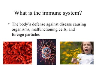
What is theimmune system?
• The body’s defense against disease causing
organisms, malfunctioning cells, and
foreign particles
3.
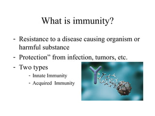
What is immunity?
-Resistance to a disease causing organism or
harmful substance
- Protection” from infection, tumors, etc.
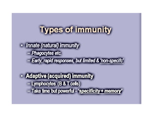
- Two types
- Innate Immunity
- Acquired Immunity
4.
What is immunity?
•“Innate immunity is always available
• Adaptive immunity distinguishes “self”
from “non-self”.
5.
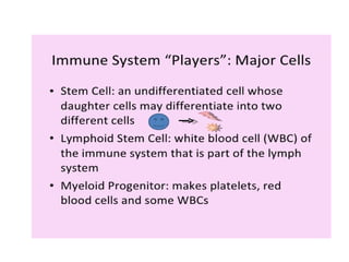
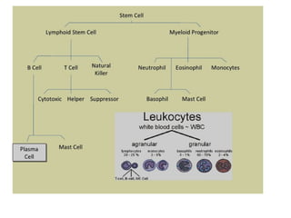
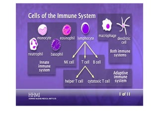
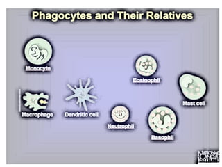
White Blood Cells
•Phagocytes - Neutrophils
- Macrophages
• Lymphocytes
Cells of the Immune System
• Innate immunity(not antigen-specific)
– Barriers
– Phagocytosis
– Complement system
– Additional components
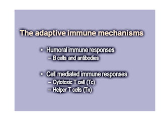
• Adaptive immunity (antigen-specific)
– Humoral
– Cellular
Two types of immunity
11.
• Stratified andcornified epithelium provides
a mechanical barrier
• Acid pH inhibits growth of disease
producing bacteria
• Bactericidal long chain fatty acids in
sebaceous gland secretions
Barriers-Skin
12.
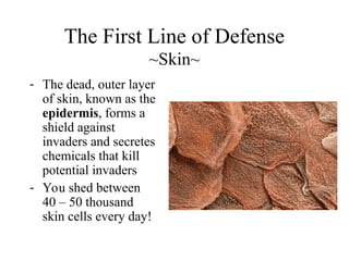
The First Lineof Defense
~Skin~
- The dead, outer layer
of skin, known as the
epidermis, forms a
shield against
invaders and secretes
chemicals that kill
potential invaders
- You shed between
40 – 50 thousand
skin cells every day!
13.
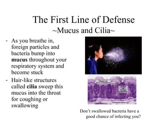
- As youbreathe in,
foreign particles and
bacteria bump into
mucus throughout your
respiratory system and
become stuck
- Hair-like structures
called cilia sweep this
mucus into the throat
for coughing or
swallowing
The First Line of Defense
~Mucus and Cilia~
Don’t swallowed bacteria have a
good chance of infecting you?
14.
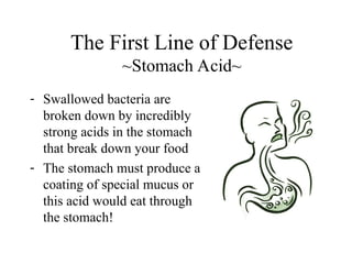
- Swallowed bacteriaare
broken down by incredibly
strong acids in the stomach
that break down your food
- The stomach must produce a
coating of special mucus or
this acid would eat through
the stomach!
The First Line of Defense
~Stomach Acid~
15.
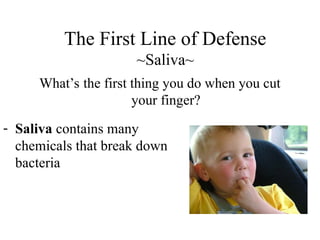
The First Lineof Defense
~Saliva~
What’s the first thing you do when you cut
your finger?
- Saliva contains many
chemicals that break down
bacteria
16.
• Flushing actionof tears which drain
through the lacrimal duct and deposit
bacteria in nasopharynx
• Tears contain a high concentration of
lysozyme (effective against gram
positive microorganisms)
Eye
17.
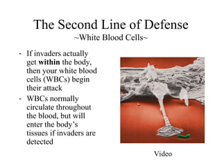
The Second Lineof Defense
~White Blood Cells~
- If invaders actually
get within the body,
then your white blood
cells (WBCs) begin
their attack
- WBCs normally
circulate throughout
the blood, but will
enter the body’s
tissues if invaders are
detected
Video
18.
• Produced throughoutlife by the bone
marrow.
• Scavengers – remove dead cells and
microorganisms.
Phagocytes
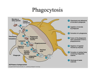
20.
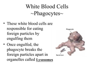
• These whiteblood cells are
responsible for eating
foreign particles by
engulfing them
• Once engulfed, the
phagocyte breaks the
foreign particles apart in
organelles called ________
White Blood Cells
~Phagocytes~
Lysosomes
21.
• 60% ofWBCs
• ‘Patrol tissues’ as they squeeze out of the
capillaries.
• Large numbers are released during infections
• Short lived – die after digesting bacteria
• Dead neutrophils make up a large proportion
of puss.
Neutrophils
22.
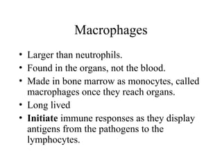
• Larger thanneutrophils.
• Found in the organs, not the blood.
• Made in bone marrow as monocytes, called
macrophages once they reach organs.
• Long lived
• Initiate immune responses as they display
antigens from the pathogens to the
lymphocytes.
Macrophages
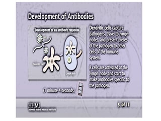
Antigen presenting cells(APC’s) are special cells that are capable
of phagocytosis.
These cells ingest invaders, break them up, and display parts of them on
their surface on special receptors. This presentation activates the specific
immune response. When specific immune cells recognize the presented
antigen, they become activated, produce an army of clones, and “seek
out” other invaders like the one that was presented.
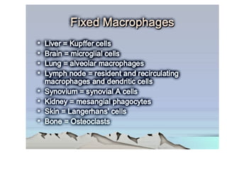
Cells that are classified as APC’s include dentritic cells, which are
specialized macrophage that are stationary in the connective tissue and
have lots and lots of processes, Langerhans cells, specialized macrophage
that are found in the epidermis, free macrophage, and B lymphocytes.
Some APC’s are found throughout the body, while others are localized.
For example, B lymphocytes and dendritic cells are found in the
germinal cells of lymph nodes.
29.
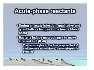
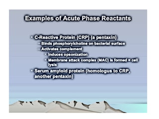
Complement system
• Thecomplement system is a part of the immune system that helps
or complements the ability of antibodies and phagocytic cells to
clear pathogens from an organism.
• The complement system consists of a number of small proteins found
in the blood, in general synthesized by the liver, and normally
circulating as inactive precursors (pro-proteins). When stimulated by
one of several triggers, proteases in the system cleave specific proteins
to release cytokines and initiate an amplifying cascade of further
cleavages. The end-result of this activation cascade is
massive amplification of the response and activation of the cell-
killing membrane attack complex. Over 30 proteins and protein
fragments make up the complement system.
• Three biochemical pathways activate the complement system:
the classical complement pathway, the alternative complement
pathway, and the lectin pathway.
30.
Membrane attack complex(MAC), consists of
C5b, C6, C7, C8, and polymeric C9. MAC is the
cytolytic end-product of the complement cascade;
it forms a transmembrane channel, which
causes osmotic lysis of the target cell. Kupffer
cells and other macrophage cell types help clear
complement-coated pathogens.
31.
Functions
The following arethe basic functions of complement:
1. Opsonization – enhancing phagocytosis of antigens
2. Chemotaxis – attracting macrophages and neutrophils
3. Cell Lysis – rupturing membranes of foreign cells
4. Agglutination – clustering and binding of pathogens
together (sticking)
33.
Opsonisation (“to maketasty” - Greek)
Opsonins are molecules, which enhance the
efficiency of the phagocytic process by
coating the microbe and effectively marking
them for their destruction. Important opsonins
are the complement component C3b and
antibodies.
34.
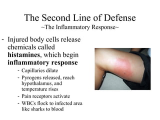
- Injured bodycells release
chemicals called
histamines, which begin
inflammatory response
- Capillaries dilate
- Pyrogens released, reach
hypothalamus, and
temperature rises
- Pain receptors activate
- WBCs flock to infected area
like sharks to blood
The Second Line of Defense
~The Inflammatory Response~
37.
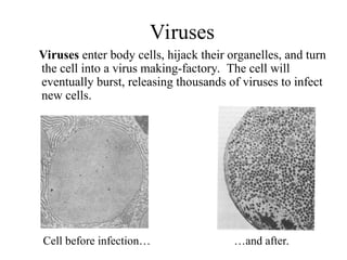
Viruses
Viruses enter bodycells, hijack their organelles, and turn
the cell into a virus making-factory. The cell will
eventually burst, releasing thousands of viruses to infect
new cells.
Cell before infection… …and after.
38.
- Virus-infected body
cellsrelease
interferon when an
invasion occurs
- Interferon – chemical
that interferes with the
ability to viruses to
attack other body cells
The Second Line of Defense
~Interferon~
39.
Lysozyme is anti-bacterial
Interferoneis anti-viral
Defensins peptides: appear to act by binding to outer
membrane of bacteria, resulting in increased
membrane permeability.
May also play a role in inflammation and wound
repair.
Antimicrobial components
40.
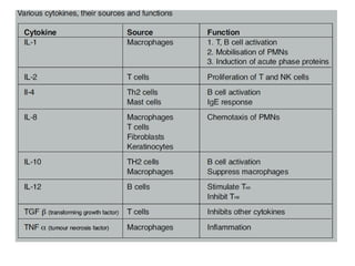
Cytokines
Cytokines (also termedinterleukins [IL] meaning
“between white blood cells”) are small molecules
that act as a signal between cells and have a variety
of roles including chemotaxis, cellular growth and
cytotoxicity. Owing to their ability to control
immune activity, they have been described as the
“hormones” of the immune system.
42.
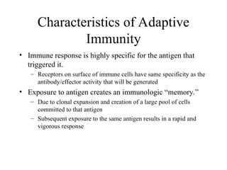
• Immune responseis highly specific for the antigen that
triggered it.
– Receptors on surface of immune cells have same specificity as the
antibody/effector activity that will be generated
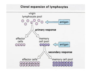
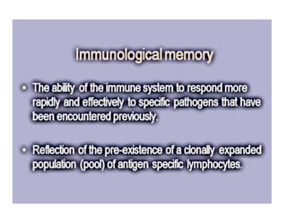
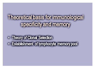
• Exposure to antigen creates an immunologic “memory.”
– Due to clonal expansion and creation of a large pool of cells
committed to that antigen
– Subsequent exposure to the same antigen results in a rapid and
vigorous response
Characteristics of Adaptive
Immunity
44.
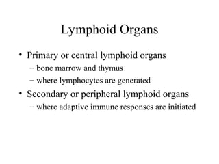
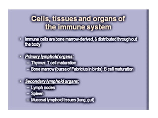
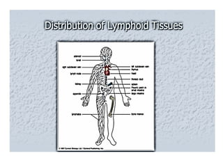
• Primary orcentral lymphoid organs
– bone marrow and thymus
– where lymphocytes are generated
• Secondary or peripheral lymphoid organs
– where adaptive immune responses are initiated
Lymphoid Organs
47.
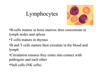
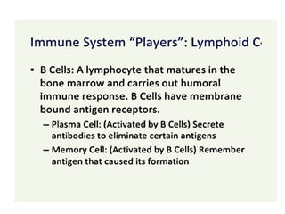
•B-cells mature inbone marrow then concentrate in
lymph nodes and spleen
•T-cells mature in thymus
•B and T cells mature then circulate in the blood and
lymph
•Circulation ensures they come into contact with
pathogens and each other
•Null cells (NK cells)
Lymphocytes
49.
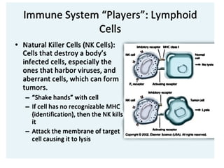
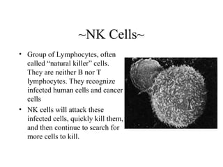
~NK Cells~
• Groupof Lymphocytes, often
called “natural killer” cells.
They are neither B nor T
lymphocytes. They recognize
infected human cells and cancer
cells
• NK cells will attack these
infected cells, quickly kill them,
and then continue to search for
more cells to kill.
51.
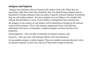
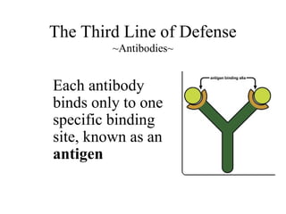
antigens and haptens
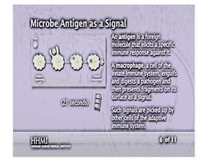
Antigensare substances that are found on the surface of all cells. When they are
somewhere other than where they should be, they are called foreign antigens and are
described as foreign substances that can induce a specific immune response. Essentially,
they are cell surface markers. You have antigens on your kidneys, for example, that
indicate that the kidney is yours. If your kidney is transplanted into someone else,
the antigens on the surface of your kidney will be identified as foreign by the immune
system of the recipient. This is why immune suppressants have to be taken by
individuals who receive a transplant. Complete antigens are those that have two
properties;
immunogenicity = they are able to stimulate an immune response, and
reactivity = they can react with immune effector cells and substances.
An incomplete antigen is called a hapten. These are smaller molecules that don’t elicit
an immune response on their own, but can if they bind to larger molecules.
52.
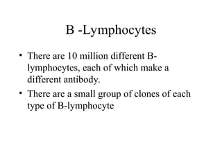
• There are10 million different B-
lymphocytes, each of which make a
different antibody.
• There are a small group of clones of each
type of B-lymphocyte
B -Lymphocytes
55.
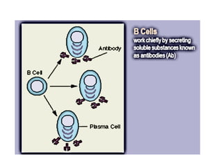
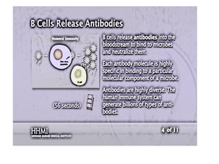
• Some activatedB cells PLASMA CELLS
these produce lots of antibodies, < 1000/sec
• The antibodies travel to the blood, lymph,
lining of gut and lungs.
• The number of plasma cells goes down after a
few weeks
• Antibodies stay in the blood longer but
eventually their numbers go down too.
B -Lymphocytes
56.
• Some activatedB cells MEMORY
CELLS.
• Memory cells divide rapidly as soon as the
antigen is reintroduced.
• When the pathogen/infection infects again it
is destroyed before any symptoms show.
B -Lymphocytes
57.
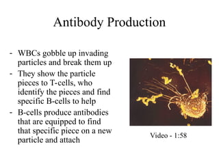
Antibody Production
- WBCsgobble up invading
particles and break them up
- They show the particle
pieces to T-cells, who
identify the pieces and find
specific B-cells to help
- B-cells produce antibodies
that are equipped to find
that specific piece on a new
particle and attach
Video - 1:58
58.
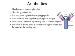
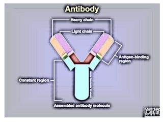
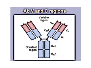
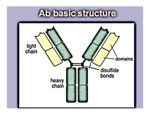
• Also knownas immunoglobulins
• Globular glycoproteins
• The heavy and light chains are polypeptides
• The chains are held together by disulphide bridges
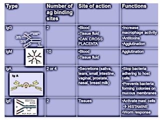
• Each ab has 2 identical ag binding sites – variable regions.
• The order of amino acids in the variable region determines
the shape of the binding site
Antibodies
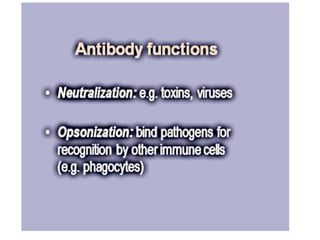
60.
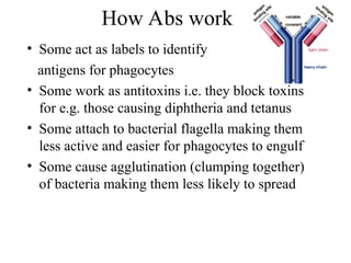
• Some actas labels to identify
antigens for phagocytes
• Some work as antitoxins i.e. they block toxins
for e.g. those causing diphtheria and tetanus
• Some attach to bacterial flagella making them
less active and easier for phagocytes to engulf
• Some cause agglutination (clumping together)
of bacteria making them less likely to spread
How Abs work
• Almost allof biology occurs because
recognition
– Enzymatic action
– Interactions between cells
(cooperation/activation)
– Communication between cells
• Innate and adaptive immunity requires it
Receptors
72.
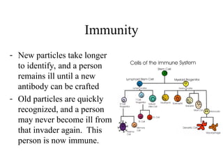
Immunity
- New particlestake longer
to identify, and a person
remains ill until a new
antibody can be crafted
- Old particles are quickly
recognized, and a person
may never become ill from
that invader again. This
person is now immune.
73.
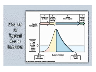
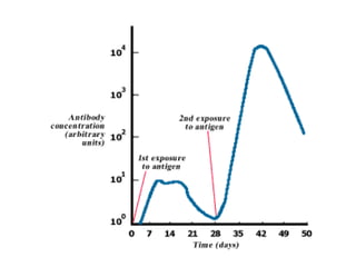
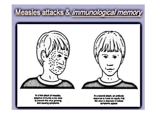
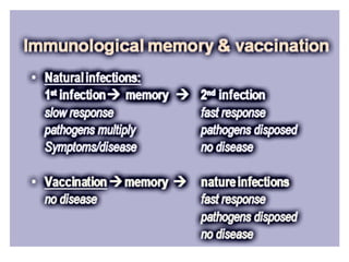
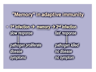
• Primary versusSecondary Immune Response
• The primary immune response occurs the first time that the
immune system comes in contact with the antigen. During this
time the immune system has to learn to recognize antigen and how
to make antibody against it and eventually gain immunological
memory. This primary response takes time (about two weeks) and
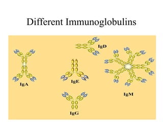
during this time the person experiences signs of illness. IgM
antibodies are the hallmark of a new infection because they are the
first antibodies made when a person is exposed to an antigen for
the first time. After the body learns to make IgM antibodies, it will
start making IgG antibodies to the antigen.
74.
• The secondaryimmune response occurs the second time
(3rd
, 4th
, etc.) the person is exposed to the same antigen. At
this point immunological memory has been established and
the immune system can start making antibodies
immediately. The antigen usually is killed within minutes
and the person is not aware that he/she was attacked. The
antibodies in this response are IgG and IgA or (in the case
of allergy IgE).
75.
• and IgAor (in the case of allergy IgE).
• Titers of antibody refer to the amount of antibody
you find in the blood. If the titer is high it means that
you have recently been exposed to the antigen. If you
have no antibody titer, it means that you have never
been exposed or have been exposed so recently that
the immune system hasn’t started to make antibodies.
You also can analyze the type of antibodies involved.
If the antibodies are IgM, it mean a new infection.
•
78.
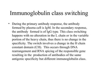
Immunoglobulin class switching
•During the primary antibody response, the antibody
formed by plasma cell is IgM. In the secondary response,
the antibody formed is of IgG type. This class switching
happens with no alteration in the L chain or in the variable
portion of the heavy chain, thus there is no change in the
specificity. The switch involves a change in the H chain
constant domain (CH). This occurs through DNA
rearrangement and RNA spicing of the responsible gene
resulting in the production of antibodies of the same
antigenic specificity but different immunoglobulin class.
80.
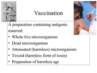
A preparation containingantigenic
material:
• Whole live microorganism
• Dead microorganism
• Attenuated (harmless) microorganism
• Toxoid (harmless form of toxin)
• Preparation of harmless ags
Vaccination
81.
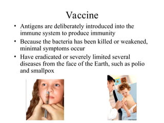
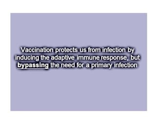
Vaccine
• Antigens aredeliberately introduced into the
immune system to produce immunity
• Because the bacteria has been killed or weakened,
minimal symptoms occur
• Have eradicated or severely limited several
diseases from the face of the Earth, such as polio
and smallpox
89.
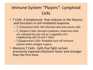
T lymphocytes
T lymphocytesare involved in cellular immunity. These are cells that
are activated only by presented antigen and are part of the specific immune response.
There are three populations of T lymphocytes; helper T cells = which increase the
immune response of both B and T lymphocytes,
killer T cells = cells which directly attach to and destroy specific cells, and
suppressor T cells = cells which wind down the immune response when it is
complete.
The helper T cells are the major target cell for HIV. When HIV invades these cells
and becomes active, it destroys the helper T lymphocytes, severely debilitating the
specific immune response. Affected individuals become very ill because their
immune system is severely
compromised.
90.
Aquired Immune DeficiencySyndrome
• Caused by the Human
Immunodeficiency Virus
• Discovered in 1983
• Specifically targets and kills
T-cells
91.
• Mature T-cellshave T cell receptors which
have a very similar structure to antibodies
and are specific to 1 antigen.
T-Lymphocytes
92.
• After activationthe cell divides to form:
• T-helper cells – secrete CYTOKINES
help B cells divide
stimulate macrophages
• Cytotoxic T cells (killer T cells)
Kill body cells displaying antigen
• Memory T cells
remain in body
T-Lymphocytes
93.
Active and PassiveImmunity
Passive immunity
B and T cells are not activated and plasma
cells have not produced antibodies.
The antigen doesn’t have to be encountered
for the body to make the antibodies.
Antibodies appear immediately in blood but
protection is only temporary.
94.
Active and PassiveImmunity
Natural passive immunity
A mother’s antibodies pass across the
placenta to the foetus and remain for several
months.
Colostrum (the first breast milk) contains lots
of IgA which remain on surface of the
baby’s gut wall and pass into blood
95.
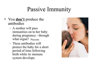
Passive Immunity
• Youdon’t produce the
antibodies
– A mother will pass
immunities on to her baby
during pregnancy - through
what organ?
– These antibodies will
protect the baby for a short
period of time following
birth while its immune
system develops.
Placenta
96.
A Artificial passiveimmunity
Used when a very rapid immune response is needed
e.g. after infection with tetanus.
Human antibodies are injected. In the case of tetanus
these are antitoxin antibodies.
Antibodies come from blood donors who have
recently had the tetanus vaccination.
Only provides short term protection as abs destroyed
by phagocytes in spleen and liver
97.
Active immunity
Lymphocytes areactivated by antigens on the
surface of pathogens
Natural active immunity - acquired due to
infection
Artificial active immunity – vaccination
Takes time for enough B and T cells to be
produced to mount an effective response.
Active and Passive Immunity
98.
Active Immunity
- Youproduce the antibodies
- Your body has been exposed to the antigen in
the past either through:
- Exposure to the actual disease causing antigen –
You fought it, you won, you remember it
- Planned exposure to a form of the antigen that has
been killed or weakened – You detected it,
eliminated it, and remember it
99.
How long doesactive immunity
last?
• It depends on the antigen
• Some disease-causing
bacteria multiply into new
forms that our body doesn’t
recognize, requiring annual
vaccinations, like the flu shot
• Booster shot - reminds the
immune system of the antigen
• Others last for a lifetime, such
as chicken pox
![Cytokines
Cytokines (also termed interleukins [IL] meaning
“between white blood cells”) are small molecules
that act as a signal between cells and have a variety
of roles including chemotaxis, cellular growth and
cytotoxicity. Owing to their ability to control
immune activity, they have been described as the
“hormones” of the immune system.](https://image.slidesharecdn.com/thehumanimmunesystem-250809181650-dd4139fe/85/The-Human-Immune-System-ppt-40-320.jpg)