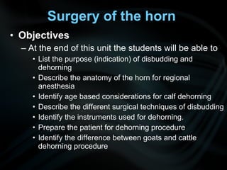
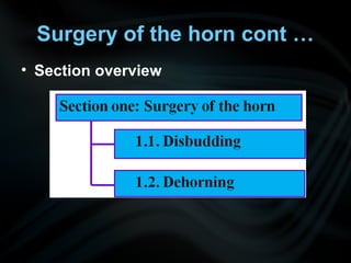
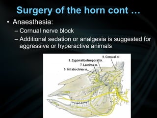
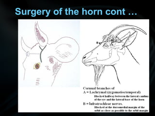
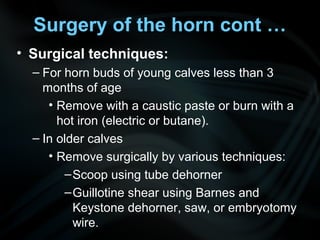
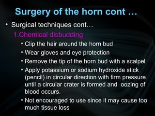
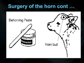
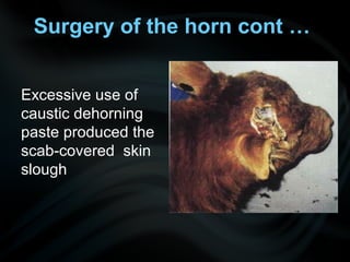
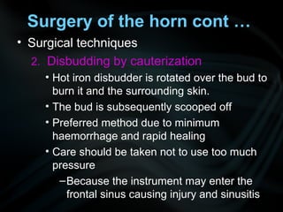
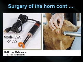
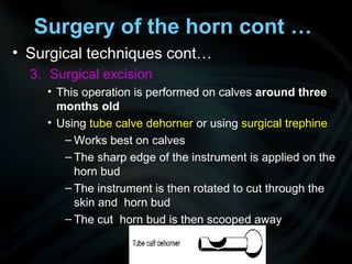
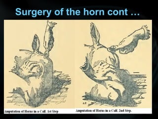
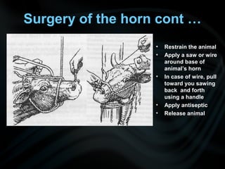
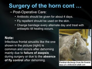
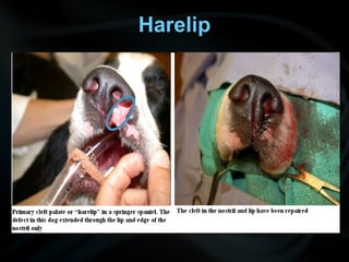
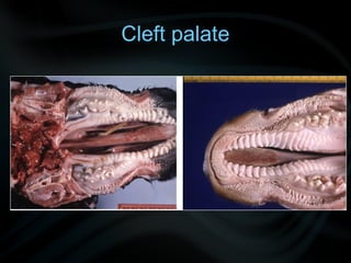
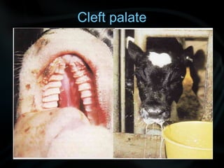
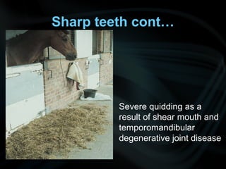
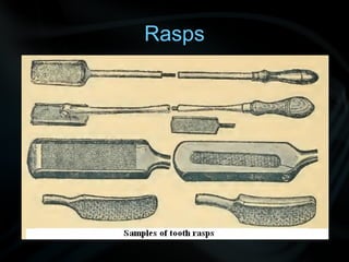
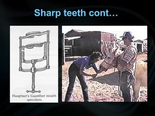
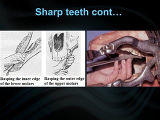
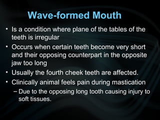
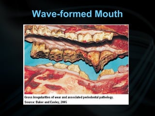
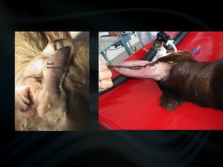
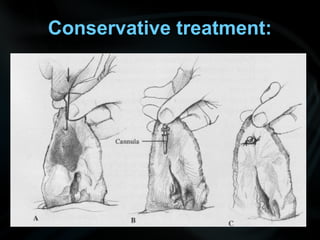
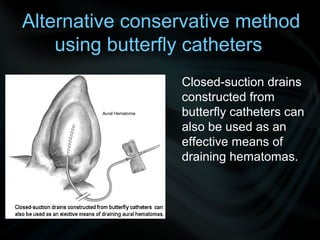
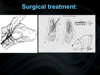
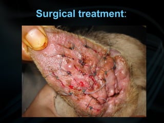
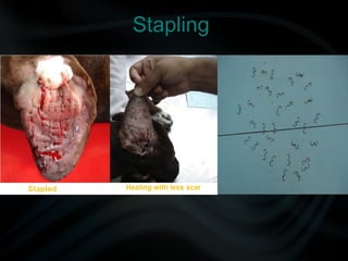
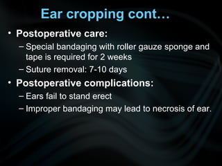
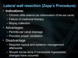
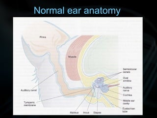
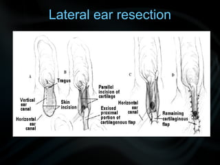
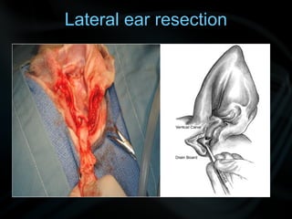
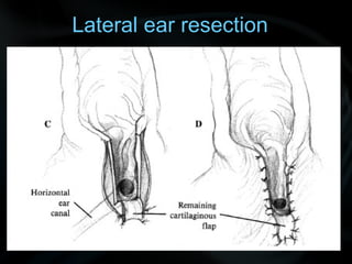
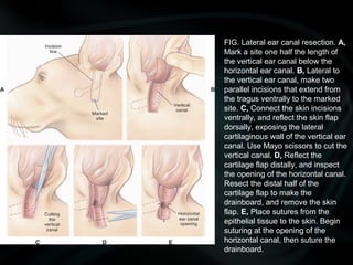
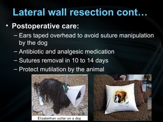
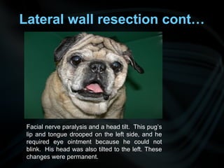
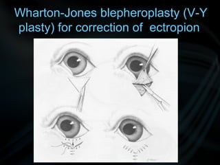
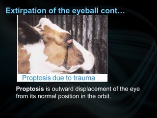
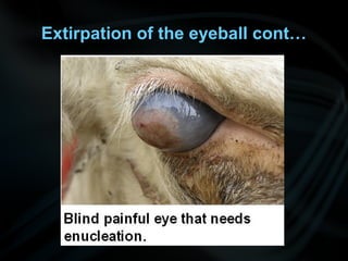
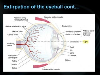
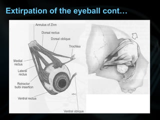
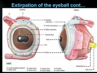
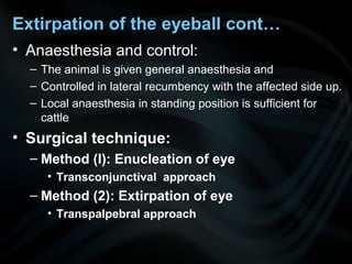
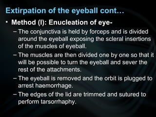
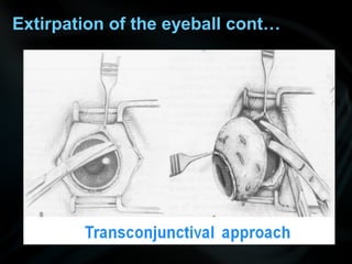
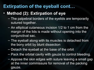
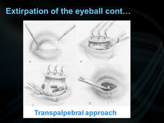
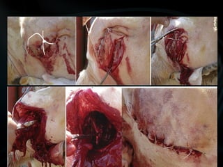
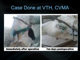
Vet. Operative Surgery
Unit One:
Surgery of head and neck
Section one: Surgery of the horn
• Objectives
– At the end of this unit the students will be able to
• List the purpose (indication) of disbudding and
dehorning
• Describe the anatomy of the horn for regional
anesthesia
• Identify age based considerations for calf dehorning
• Describe the different surgical techniques of disbudding
• Identify the instruments used for dehorning.
• Prepare the patient for dehorning procedure
• Identify the difference between goats and cattle
dehorning procedure
Surgery of the horn
• Section overview
Surgery of the horn cont …
• Anaesthesia:
– Cornual nerve block
– Additional sedation or analgesia is suggested for
aggressive or hyperactive animals
Surgery of the horn cont …
• Disbudding:
– Disbudding involves the removal of the horn bud
in exotic cow calves, lambs and kids.
– Indication
• To reduce the risk of injury to farm personnel and other
animals.
• For easy and safe restraint of the animals
– Cattle with horns are harder to handle in chutes and
require three times more space at feed bunkers and
during transport
• For aesthetic purpose in show animals.
• The most suitable age for disbudding is when the
animals get 5-15 days old. At this age, the horn buds
are button like and easy to remove.
Surgery of the horn cont …
• Surgical techniques:
– For horn buds of young calves less than 3
months of age
• Remove with a caustic paste or burn with a
hot iron (electric or butane).
– In older calves
• Remove surgically by various techniques:
–Scoop using tube dehorner
–Guillotine shear using Barnes and
Keystone dehorner, saw, or embryotomy
wire.
Surgery of the horn cont …
• Surgical techniques cont…
1.Chemical disbudding
• Clip the hair around the horn bud
• Wear gloves and eye protection
• Remove the tip of the horn bud with a scalpel
• Apply potassium or sodium hydroxide stick
(pencil) in circular direction with firm pressure
until a circular crater is formed and oozing of
blood occurs.
• Not encouraged to use since it may cause too
much tissue loss
Surgery of the horn cont …
Excessive use of
caustic dehorning
paste produced the
scab-covered skin
slough
Surgery of the horn cont …
• Surgical techniques
2. Disbudding by cauterization
• Hot iron disbudder is rotated over the bud to
burn it and the surrounding skin.
• The bud is subsequently scooped off
• Preferred method due to minimum
haemorrhage and rapid healing
• Care should be taken not to use too much
pressure
–Because the instrument may enter the
frontal sinus causing injury and sinusitis
Surgery of the horn cont …
• Surgical techniques cont…
3. Surgical excision
• This operation is performed on calves around three
months old
• Using tube calve dehorner or using surgical trephine
– Works best on calves
– The sharp edge of the instrument is applied on the
horn bud
– The instrument is then rotated to cut through the
skin and horn bud
– The cut horn bud is then scooped away
Surgery of the horn cont …
• Using Barns Dehorner
– Best for
1. Smaller size is for six to eight month old calves
2. Larger size is for dehorning yearling or older cattle.
– Restrain animal
– Place dehorner over horn and down against skull of
animal.
– To remove horn, correctly place and spread handles
apart quickly.
– Close the bleeding artery
– Treat the wound with antiseptic spray or solution
– Repeat w/ other horn
– Release animal
Surgery of the horn cont …
Instruments for dehorning
Surgery of the horn cont …
• Using Keystone dehorner
– Used primarily for older calves (8 month and
older) because of its ease of cutting.
– Apply local anaesthetic and restrain the animal
– Apply clippers over the horn & clamp down
– Stop bleeding & apply antiseptic
– Repeat with the other horn & release
Surgery of the horn cont …
• Dehorning
– Is amputation of the horn in adult animals
– Indication
• Irreparable injuries or horn cancer
• To make management and control easier
• Misdirected or overgrown horn to the extent of causing
cutaneous injuries due to pressure
Surgery of the horn cont …
– Surgical techniques of dehorning
• Elliptical incision is made around the base of the horn
• The skin on both sides of the incision is reflected to
form a flap.
• Cornual artery is ligated
• The exposed horn is then cut close to its base either
using a saw or dehorning gigli wire.
• KMNO4 crystals can be used to arrest haemorrhage.
• The entire skin flap is sutured by interrupted or
mattress sutures using non- absorbable suture
material
• A protective bandage is applied after covering the
wound with antiseptics
Surgery of the horn cont …
• Restrain the animal
• Apply a saw or wire
around base of
animal’s horn
• In case of wire, pull
toward you sawing
back and forth
using a handle
• Apply antiseptic
• Release animal
– Post-Operative Care:
• Antibiotic should be given for about 4 days.
• Fly repellent should be used on the skin.
• Change bandage every alternate day and treat with
antiseptic till healing occurs.
Surgery of the horn cont …
Note:
Infectious frontal sinusitis like the one
shown in the picture (right) is
common and occurs after dehorning
mainly due to failure of asepsis
during surgery or due to the absence
of fly control after dehorning.
Section Two: Congenital abnormalities
• Overview:
– Incidence in cattle is low ranging between 0.2–3%
– 40–50% are born dead.
– Congenital defects reduce the value of affected
calves and their normal relatives.
– A limited number of conditions can be corrected
surgically
• When it is likely that the condition is inherited, castration
or sterilization should be done to avoid breeding
• Not possible to deal with all the recorded congenital
defects here
– So discussion is limited to those defects that are thought to be
of general importance
• Section overview
Congenital defects
• Section objectives
– At the end of this unit, you are expected to
• List the common congenital abnormalities in organs of
the head region in domestic animals
• List the problems the congenital abnormalities create on
the animals survival and productivity
• Classify cleft palate
• Mention the common clinical presentation of an animal
with cleft palate
• Describe the treatment for the common congenital
abnormalities
Congenital defects
Congenital Defects
• Ankyloglossia (persistent lingual frenulum)
– Ankylose - meaning - to fuse together,
– The condition may be lethal at birth
– In surviving animals, may produce varying degrees
of difficulty in eating or drinking.
• Treatment
– Incising the overlong lingual frenulum so that a full
range of tongue motion is possible.
• A congenital condition of exotic
cattle
– The dorsal surface of the tongue
is smooth either due to
insignificant number or lack of
horny papillae.
– Calves with smooth tongue show
signs of
• Poor body condition and retarded
growth as soon as they are put to
graze due to poor prehension
• Treatment
– No treatment for this abnormality
– May be maintained on stall fed
with soft diet
Smooth Tongue
Hare - lip
Harelip
• Is congenital fissure of the upper lip
• It may be unilateral or bilateral and extended
for a variable distance into the lip
• It may exist alone or be accompanied by cleft
palate.
• Treatment:
– Remove a thin slice of tissue from each border of
the fissure and bring them into contact with deep
sutures, not going through the mucous
membrane.
– Apply suitable topical antiseptic on the wound.
Harelip
Cleft palate
Cleft palate
Cleft palate:
• It is congenital affection generally seen in
newborn animals.
• Occurs due to failure of fusion of lateral
palatine process during embryonic
development.
Cleft palate: cont…
• Classification of cleft palate
– Clefts of the prepalate (Primary palate)
• Cleft lip or harelip (Cheiloschisis)
–Read as (kĭ-lŏs′kĭ-sĭs)
• Cleft alveolar process (Alveoloschisis)
• Cleft prepalate (Cheilo+alveolo+schisis)
– Clefts of the palate (Secondary palate)
• Cleft soft palate (Staphyloschisis)
• Cleft hard palate (Uranoschisis)
• Cleft hard and soft palate (Urano+staphylo+schisis)
– Clefts of the prepalate and palate
(Gnatho+palato+schisis)
Cleft palate: cont…
• Clinical signs:
–Clinical signs depend upon site and
severity of cleft palate and includes:
• Regurgitation of ingested liquid or milk from
nostrils
• Frequent sneezing
• Malnutrition and aspiratory pneumonia leading
to death
Cleft palate: cont…
• Preoperative evaluation and care:
– For secondary palate
• Tube feeding and early repair (7-9 weeks) is
recommended.
– Check for other congenital abnormalities
– Thoracic radiography
– Bacterial culture and antibiotic therapy
– Rhinoscopy or endoscopy and flushing of nasal
cavity to remove foreign materials
Cleft palate: cont…
• Treatment
– Surgical correction of defect (palatoplasty).
– Main objective of surgery
• To establish normal separation between oral and nasal
cavities
– Anesthesia
• Done under general anesthesia and dorsal recumbency
– In dogs:
• Head is placed on a soft pad and
• Maxilla is immobilized with a tape placed over the
incisors or canine teeth and secured on the operating
table on either side to approach cleft palate.
Cleft palate: cont…
Cleft palate: cont…
• Treatment cont..
– In horses
• Mandibular symphysiotomy and transhyoid
pharyngostomy are done to approach cleft palate.
Cleft palate: cont…
• Surgical technique
• Unilateral (overlapping) flap technique
Cleft palate: cont…
• Bilateral flap technique:
– Hinged flaps are elevated bilaterally, rolled back
and sutured together over the middle of the
defect.
– For second later closure, the palatine mucosa is
incised just medial to the dental arcade, leaving
a flap attached rostrally and caudally.
– The flaps are advanced towards the midline and
sutured.
Cleft palate: cont…
Cleft palate: cont…
• Postoperative care:
– Liquid / milk diet
– Feeding with nasogastric intubation
– Feeding with oesophagostomy tube placement
– Avoid hay feeding for 2 weeks
– Antibiotic and analgesics
Cleft palate: cont…
Section Three:
Surgical conditions of the teeth
• Teeth abnormalities of wear
– Abnormal wear patterns develop 2o
to poor dental
occlusion or altered masticatory patterns.
– Cheek teeth abnormalities of wear that we will
discuss:
• Section overview
Teeth abnormalities of wear
• Section objectives
– At the end of this you will be able to
• Describe how shear mouth (sharp teeth is formed)
• List the clinical symptoms of common abnormal wear
conditions: shear mouth and wave mouth
• Describe the surgical treatment of shear mouth and
wave mouth
Sharp teeth
• Common in cattle and horses.
• The sharpness is develops due to insufficient
wear on
– The outer border of the upper molars and
– Inner border of lower molars.
• It occurs when the side-to-side movement of the
jaws becomes restricted due to some reason
• Weakness of masseter muscles,
• Painful lesions in the mouth
• The sharp borders cause injury to the cheek and
tongue
Sharp teeth
Sharp teeth cont…
• Symptoms are
– There is pain.
– Imperfect grinding of food.
– Quidding.
– Foaming saliva may be seen during chewing
– Wounds or ulcers on the tongue
– Loss of general condition of the animal
• Treatment
– Rasping of the sharp teeth is carried out with the
horse standing until the teeth is reduced to a
blunt roundness
Sharp teeth cont…
Severe quidding as a
result of shear mouth and
temporomandibular
degenerative joint disease
Rasps
Sharp teeth cont…
Sharp teeth cont…
Wave-formed Mouth
• Is a condition where plane of the tables of the
teeth is irregular
• Occurs when certain teeth become very short
and their opposing counterpart in the opposite
jaw too long
• Usually the fourth cheek teeth are affected.
• Clinically animal feels pain during mastication
– Due to the opposing long tooth causing injury to
soft tissues.
Wave-formed Mouth
• Treatment:
– Extraction of the tooth or removal of the sharp
points and edges of the long tooth with molar
cutter
Wave-formed Mouth
Section four:
Surgical affections of the ear
• Section overview
Surgical affections of the ear
• Section objectives
– At the end of this unit you should be able to
• List the common diseases of the ear that need
surgical intervention
• Mention the name of appropriate surgical procedure
for each of the common ear diseases
• Describe the indicated surgical procedure for each of
the common ear diseases
• Describe the proper postoperative care
Aural or ear haematoma
• Etiology:
– Scratching or head shaking by the animal
• Underlying causes:
– Inflammation, parasites, allergy or foreign bodies.
• Site:
– Usually on the concave surface of the ear.
• Treatment objective:
– To remove the hematoma, prevent recurrence, and retain
the natural appearance of the ears
• Conservative treatment:
• Needle aspiration of blood in fresh and small
haematoma cases and then doing pressure
bandaging.
• A teat canula can also be placed in fresh cases for
continued drainage for 2-3 weeks.
• Recurrence is common problem.
Aural or ear haematoma
Conservative treatment:
Alternative conservative method
using butterfly catheters
Closed-suction drains
constructed from
butterfly catheters can
also be used as an
effective means of
draining hematomas.
Aural or ear haematoma
• Surgical treatment:
• An incision is given on the concave aspect of the ear
(straight, cruciate or S shaped) over the swelling.
• Fibrin clots curetted and cavity irrigated with NSS and
2% povidone iodine solution.
• Loosely tied through and through mattress sutures are
applied using monofilament non-absorbable suture
material such as nylon.
• Sutures are applied parallel to the major vessels starting
from the periphery of ear towards the incision line.
• Incision is not sutured rather allowed to gape for
drainage.
Surgical treatment:
Surgical treatment:
Stapling
• Postoperative care:
– Light protective bandaging with ear against the
dorsum of head
• To protect the ear from contamination and self-inflicted
trauma
– Antiseptic dressing on every alternative days
– Suture removal from 7-10 days
Aural or ear haematoma
• Postoperative complications:
– If sutures are too tight can cause
• Edema, pain and surgical failure
• Wrinkling of ear
• Drooping of ear
• Fibrosis of ear
• Necrosis of the pinna and infection of the surgical site
Aural or ear haematoma
Ear cropping/ cosmetic
otoplasty/ ear trimming
• It is done on ears of certain breeds to meet their
breed specifications
• This operation is done for cosmetic appearance but
not for medical reason.
• Now it is banned in most of the countries.
– Allowed only when injured or traumatized pina
needs such kinds of surgery
• Prerequisites:
– General health of puppy should be good
– Puppy should be vaccinated
– Age 8-12 weeks
Ear cropping cont…
Breed Age Ear to be cropped
Boxer 9-10 weeks 2/3rd
– 3/4th
of ear left
Doberman 8-9 weeks 3/4th
of the ear left
Great Dane 9 weeks 3/4th
of the ear left
Ear cropping cont…
• Surgical technique:
– Operation is done under GA
– Animal is secured in sternal recumbency
– Aseptic preparation of ear
– First incision beneath the tragus and antitragus
cartilages
– With straight ruler the desired length of ear is
marked on the medial margin of ear.
– Both ear are extended fully and marked identical.
– With the help of Doyen’s intestinal forceps, the ear
is hold at the 2 marks and incised.
Doyen intestinal forceps
Doyen’s intestinal forceps
Ear cropping cont…
• Surgical technique:
– The distal third of incision is made with finely
serrated cartilage scissors
– Sutures materials and technique:
• Main incision on skin on the sides of cartilage
– Closed with monofilament non-absorbable suture
materials
– Using simple continuous suture pattern beginning at
ventral ear.
– Sutures are applied inside out
Ear cropping cont…
• Postoperative care:
– Special bandaging with roller gauze sponge and
tape is required for 2 weeks
– Suture removal: 7-10 days
• Postoperative complications:
– Ears fail to stand erect
– Improper bandaging may lead to necrosis of ear.
Ear cropping cont…
Lateral wall resection (Zepp’s Procedure):
• Indications:
– Chronic otitis externa (an inflammation of the ear canal)
– Failure of medicinal therapy
– Biopsy collection
• Advantages:
– Permits ear canal drainage
– Provides proper ventilation
• Disadvantage
– Requires topical and systemic management
afterwards
– Should not be done if irreversible hyperplastic
changes have occurred
Normal ear anatomy
Lateral wall resection
• Surgical technique:
– Ear canal irrigated
– Positioning: Lateral recumbency with Pinna overhead
– Incision given at rostral and caudal borders of vertical
ear canal extending from the tragus dorsally to 1-2 cm
below the horizontal canal.
– Dorsal and ventral ends of incision are joined and skin
removed.
– Lateral half of vertical canal is reflected and one third
is sutured below to skin margins to act as a drain
board (baffle plate) with monofilament non-
absorbable sutures
– Trauma to parotid gland should be avoided
Lateral ear resection
FIG. Lateral ear canal resection. A,
Mark a site one half the length of
the vertical ear canal below the
horizontal ear canal. B, Lateral to
the vertical ear canal, make two
parallel incisions that extend from
the tragus ventrally to the marked
site. C, Connect the skin incisions
ventrally, and reflect the skin flap
dorsally, exposing the lateral
cartilaginous wall of the vertical ear
canal. Use Mayo scissors to cut the
vertical canal. D, Reflect the
cartilage flap distally, and inspect
the opening of the horizontal canal.
Resect the distal half of the
cartilage flap to make the
drainboard, and remove the skin
flap. E, Place sutures from the
epithelial tissue to the skin. Begin
suturing at the opening of the
horizontal canal, then suture the
drainboard.
Lateral wall resection cont…
• Postoperative care:
– Ears taped overhead to avoid suture manipulation
by the dog
– Antibiotic and analgesic medication
– Sutures removal in 10 to 14 days
– Protect mutilation by the animal
• Post operative complications:
– Failure to drain the horizontal canal
– Stricture of horizontal canal
– Exudation for some days
– Partial wound dehiscence (go for secondary
healing)
– Facial nerve damage
Lateral wall resection cont…
Facial nerve paralysis and a head tilt. This pug’s
lip and tongue drooped on the left side, and he
required eye ointment because he could not
blink. His head was also tilted to the left. These
changes were permanent.
Section five:
Surgical affections of the eye
• Section overview
Surgical affections of the eye
• Section objectives
– At the end of this unit you are expected to
• Define entropion and ectropion
• List the clinical presentation of entropion and ectropion
• Mention causes of entropion and ectropion
• Describe the surgical procedures of entropion and
ectropion
• List the indications for extirpation of the eyeball
• Describe the different methods of eyeball extirpation
Surgical affections of the eye: cont…
• Entropion
– It is an inversion of the eyelid margin where the
eyelashes rub the cornea.
– Clinical Signs:
• Superficial irritation of conjunctiva and cornea
• Chronic ocular discharge
• Blepharospasm
– Etiology:
• Congenital Entropion: bilateral and common in young
dogs
• Spastic Entropion: unilateral can occur at any age
• Aquired Entropion: due to endopthalomos from loss of
orbital fat or temporal muscle atrophy
Surgical affections of the eye: cont…
Prior to definitive repair for younger animals (< 8 month
for small breeds, < 2 years for giant breeds
• Temporary tarsorraphy or tacking sutures are indicated
• Use vertical mattress suture or surgical staples to
temporarily correct entropion or
• Suture eyelids closed (temporary tarsorraphy) to
protect cornea until definitive surgery can be done at
the appropriate age
Surgical affections of the eye: cont…
• Entropion cont…
• Surgical correction
• Modified Holtz-Celsus procedure
– One of the most common eye surgical procedures
– It is reserved for fully grown animals
• Half-moon shaped flap of skin 2-3 mm from the lid margin is
removed.
• Incision length extended 1-2 mm beyond the affected portion
• The skin excision is 3-4 mm wider than the affected area of the
lid.
• The area of skin to be removed is clamped using Halsted or
Crile forceps and adjustments made, the forceps is kept there
for 30 seconds.
• The skin is closed with 5-0 or 6-0 simple interrupted silk sutures
placed 2 mm apart.
• Subcutaneous suturing is not done.
Modified Holtz-Celsus procedure
to correct entropion
Modified Holtz-Celsus procedure
Surgical affections of the eye: cont…
• Entropion cont…
– Post-operative Care:
• For first few days the lid seems to be overcorrected but
when the inflammatory swelling subsides the lids may
come in normal position.
• Protect self mutilation by the animal
• Suture removal at 10-14 days
Surgical affections of the eye: cont…
• Ectropion
– Eversion of the lid margin
– Common in Saint Bernard, Blood hound, American
cocker spaniel, basset hound and bull dog.
– Usually congenital or due to scarring
– Generally lower eyelid is involved
– If not causing any keratitis or conjunctivitis ,
surgery is not necessary.
Breeds of dogs susceptible to Ectropion
Surgical affections of the eye:
cont…
• Ectropion cont…
• Surgical Technique
– Wharton-Jones blepheroplasty (V-Y plasty)
– Triangle is outlined under the defect and incision
made
– Skin flap is elevated and dissected away
– Triangular defect sutured in Y’ shape using 6-0 or
4-0 slik.
Wharton-Jones blepheroplasty (V-Y
plasty) for correction of ectropion
Wharton-Jones blepheroplasty (V-Y
plasty) for correction of ectropion
Surgical affections of the eye: cont…
• Ectropion
• Post-operative care:
– Topical antibiotic and corticosteroid ointment applied to
eyelid tid for 5-7 days
– Prevent self mutilation
• Post-operative Complication:
– Under correction
– Overcorrection leading to entropion
Extirpation (removal) of the eyeball
• Indications:
– Irreparable injury
– Orbital abscesses
– Malignant disease of eye ball and adjacent tissue.
– Blind, painful glaucomatous eye
– Proptosis of the globe with severance of muscle
attachments, vascular supply and optic nerve
damage
SCC
Extirpation of the eyeball cont…
Proptosis is outward displacement of the eye
from its normal position in the orbit.
Extirpation of the eyeball cont…
• Anaesthesia and control:
– The animal is given general anaesthesia and
– Controlled in lateral recumbency with the affected side up.
– Local anaesthesia in standing position is sufficient for
cattle
• Surgical technique:
– Method (I): Enucleation of eye
• Transconjunctival approach
– Method (2): Extirpation of eye
• Transpalpebral approach
Extirpation of the eyeball cont…
• Method (I): Enucleation of eye-
– The conjunctiva is held by forceps and is divided
around the eyeball exposing the scleral insertions
of the muscles of eyeball.
– The muscles are then divided one by one so that it
will be possible to turn the eyeball and sever the
rest of the attachments.
– The eyeball is removed and the orbit is plugged to
arrest haemorrhage.
– The edges of the lid are trimmed and sutured to
perform tarsorrhaphy.
Extirpation of the eyeball cont…
• Method (2): Extirpation of eye
– The palpebral borders of the eyelids are temporarily
sutured together.
– An elliptical cutaneous incision 1/2 to 1 cm from the
margin of the lids is made without opening into the
conjunctival sac.
– The eyeball along with its muscles is detached from
the bony orbit by blunt dissection
– Detach the eyeball at the base of the orbit
– Pack the orbital cavity with gauze to control bleeding.
– Appose the skin edges with suture leaving a small gap
at the inner commissure for removal of the packing
gauze.
Extirpation of the eyeball cont…
Case Done at VTH, CVMA
• Transconjunctival approach
– Easy dissection through better anterior
visualization.
• Transpalpebral approach
– Improved exposure of the globe for posterior
dissection and
– To keep nearly sterile environment at operation
site.
• Therefore it is preferable when there is conjunctival or
intraocular infection.
Extirpation of the eyeball cont…
• Postoperative complication
– Orbital hemorrhage associated with swelling of
surgical site
– Serosanguinous discharge from the nostril may
occur for the first one to two days
– Orbital infection and autotrauma
Extirpation of the eyeball cont…
How would you treat this?
Section six:
Surgical affections on the neck
• Section overview
• Section objectives
– At the end of this unit the students are
expected to
• Define yoke gall and list its causes
• List predisposing factors to yoke gall
• List the fates of yoke gall
• Describe the treatments for yoke gall at different
progressive stages
Yoke Gall
Yoke Gall
• Is localized acute inflammation of the skin and
subcutis on the neck of cattle
– It is due to constant friction caused by the yoke.
– It is characterized by a swelling due to the
accumulation of inflammatory exudate collected
subcutaneously or between the layers of the skin
Yoke Gall cont…
• Predisposing factors:
– Moist skin due to rain or sweat
– Tender skin in young animals
– Prolonged work
– Yoke with an irregular surface
– Irregular pressure of the yoke
• Improper adjustment of weight
• Improper selection of the pair of animals
Yoke Gall cont…
• Clinical signs:
– Initial stages
• Acute painful swelling
– In later stages
• Yoke gall may become infected, leading to formation of
abscess.
– Progress of the yoke gall
• May resolve, exudate may get reabsorbed.
• Pyogenic organisms may enter leading to suppuration.
• In severe cases may predispose to gangrene and
sloughing
• Swelling may persist, become fibrous leading to formation
of fibrous tumour
Yoke Gall cont…
• Diagnosis:
– From clinical signs and exploratory puncture into
the swelling.
• Treatment
– Acute stages:
• Fomentations and application of freshly prepared acetic
acid chalk paste, Kaolin paste or Magnesium sulphate,
Glycerin paste may reduce the swelling.
• Rubbing with iodine ointment may facilitate reabsorption
of the exudate.
Yoke Gall cont…
• Treatment cont…
– Chronic abscess:
• Opening of the mature abscess, draining the pus and
flush the cavity with hypertonic saline (2-5%), followed
by iodine solution.
• Packing the cavity with moist gauze dipped in Tincture
iodine.
– Tumour neck swelling:
• Large and hard swelling developed from yoke gall in the
form of a fibrous mass (Fibrous tumor/ Fibroma).
– Treated by enucleation (extirpation) under local anaesthesia
– Surgical technique for tumour neck swelling:
• Local infiltration anaesthesia is used around the base
of the tumour.
• An elliptical skin incision is made across the swelling.
• The incision is extended at either end a little beyond
the swelling and should enclose necrosed areas of skin
that need to be removed.
• After cutting through the skin, dissect outwards
subcutaneously to reach the base of the fibrous mass
and enucleate it completely.
• Stop haemorrhage and suture the wound and allow it to
heal by first intention.
Yoke Gall cont…
• Post operative complications
– Failure or delay of healing may be seen due to:
• Infection
• Improper apposition of the cut ends
• Failure of arrest of haemorrhage
• Use of irritant antiseptics
• Excessive trauma during operation and/or
interference by the animal after operation.
Yoke Gall cont…
THANKS
Boxer
Doberman
Great Dane

Surgery of the head

  • 1.
  • 2.
    Unit One: Surgery ofhead and neck
  • 3.
  • 4.
    • Objectives – Atthe end of this unit the students will be able to • List the purpose (indication) of disbudding and dehorning • Describe the anatomy of the horn for regional anesthesia • Identify age based considerations for calf dehorning • Describe the different surgical techniques of disbudding • Identify the instruments used for dehorning. • Prepare the patient for dehorning procedure • Identify the difference between goats and cattle dehorning procedure Surgery of the horn
  • 5.
    • Section overview Surgeryof the horn cont …
  • 6.
    • Anaesthesia: – Cornualnerve block – Additional sedation or analgesia is suggested for aggressive or hyperactive animals Surgery of the horn cont …
  • 7.
    Surgery of thehorn cont …
  • 8.
    • Disbudding: – Disbuddinginvolves the removal of the horn bud in exotic cow calves, lambs and kids. – Indication • To reduce the risk of injury to farm personnel and other animals. • For easy and safe restraint of the animals – Cattle with horns are harder to handle in chutes and require three times more space at feed bunkers and during transport • For aesthetic purpose in show animals. • The most suitable age for disbudding is when the animals get 5-15 days old. At this age, the horn buds are button like and easy to remove. Surgery of the horn cont …
  • 9.
    • Surgical techniques: –For horn buds of young calves less than 3 months of age • Remove with a caustic paste or burn with a hot iron (electric or butane). – In older calves • Remove surgically by various techniques: –Scoop using tube dehorner –Guillotine shear using Barnes and Keystone dehorner, saw, or embryotomy wire. Surgery of the horn cont …
  • 10.
    • Surgical techniquescont… 1.Chemical disbudding • Clip the hair around the horn bud • Wear gloves and eye protection • Remove the tip of the horn bud with a scalpel • Apply potassium or sodium hydroxide stick (pencil) in circular direction with firm pressure until a circular crater is formed and oozing of blood occurs. • Not encouraged to use since it may cause too much tissue loss Surgery of the horn cont …
  • 11.
    Surgery of thehorn cont …
  • 12.
    Excessive use of causticdehorning paste produced the scab-covered skin slough Surgery of the horn cont …
  • 13.
    • Surgical techniques 2.Disbudding by cauterization • Hot iron disbudder is rotated over the bud to burn it and the surrounding skin. • The bud is subsequently scooped off • Preferred method due to minimum haemorrhage and rapid healing • Care should be taken not to use too much pressure –Because the instrument may enter the frontal sinus causing injury and sinusitis Surgery of the horn cont …
  • 14.
    Surgery of thehorn cont …
  • 15.
    • Surgical techniquescont… 3. Surgical excision • This operation is performed on calves around three months old • Using tube calve dehorner or using surgical trephine – Works best on calves – The sharp edge of the instrument is applied on the horn bud – The instrument is then rotated to cut through the skin and horn bud – The cut horn bud is then scooped away Surgery of the horn cont …
  • 16.
    Surgery of thehorn cont …
  • 17.
    • Using BarnsDehorner – Best for 1. Smaller size is for six to eight month old calves 2. Larger size is for dehorning yearling or older cattle. – Restrain animal – Place dehorner over horn and down against skull of animal. – To remove horn, correctly place and spread handles apart quickly. – Close the bleeding artery – Treat the wound with antiseptic spray or solution – Repeat w/ other horn – Release animal Surgery of the horn cont …
  • 18.
  • 19.
    Surgery of thehorn cont …
  • 20.
    • Using Keystonedehorner – Used primarily for older calves (8 month and older) because of its ease of cutting. – Apply local anaesthetic and restrain the animal – Apply clippers over the horn & clamp down – Stop bleeding & apply antiseptic – Repeat with the other horn & release Surgery of the horn cont …
  • 21.
    Surgery of thehorn cont …
  • 22.
    • Dehorning – Isamputation of the horn in adult animals – Indication • Irreparable injuries or horn cancer • To make management and control easier • Misdirected or overgrown horn to the extent of causing cutaneous injuries due to pressure Surgery of the horn cont …
  • 23.
    – Surgical techniquesof dehorning • Elliptical incision is made around the base of the horn • The skin on both sides of the incision is reflected to form a flap. • Cornual artery is ligated • The exposed horn is then cut close to its base either using a saw or dehorning gigli wire. • KMNO4 crystals can be used to arrest haemorrhage. • The entire skin flap is sutured by interrupted or mattress sutures using non- absorbable suture material • A protective bandage is applied after covering the wound with antiseptics Surgery of the horn cont …
  • 24.
    Surgery of thehorn cont …
  • 25.
    Surgery of thehorn cont … • Restrain the animal • Apply a saw or wire around base of animal’s horn • In case of wire, pull toward you sawing back and forth using a handle • Apply antiseptic • Release animal
  • 26.
    – Post-Operative Care: •Antibiotic should be given for about 4 days. • Fly repellent should be used on the skin. • Change bandage every alternate day and treat with antiseptic till healing occurs. Surgery of the horn cont … Note: Infectious frontal sinusitis like the one shown in the picture (right) is common and occurs after dehorning mainly due to failure of asepsis during surgery or due to the absence of fly control after dehorning.
  • 27.
    Section Two: Congenitalabnormalities • Overview: – Incidence in cattle is low ranging between 0.2–3% – 40–50% are born dead. – Congenital defects reduce the value of affected calves and their normal relatives. – A limited number of conditions can be corrected surgically • When it is likely that the condition is inherited, castration or sterilization should be done to avoid breeding • Not possible to deal with all the recorded congenital defects here – So discussion is limited to those defects that are thought to be of general importance
  • 28.
  • 29.
    • Section objectives –At the end of this unit, you are expected to • List the common congenital abnormalities in organs of the head region in domestic animals • List the problems the congenital abnormalities create on the animals survival and productivity • Classify cleft palate • Mention the common clinical presentation of an animal with cleft palate • Describe the treatment for the common congenital abnormalities Congenital defects
  • 30.
    Congenital Defects • Ankyloglossia(persistent lingual frenulum) – Ankylose - meaning - to fuse together, – The condition may be lethal at birth – In surviving animals, may produce varying degrees of difficulty in eating or drinking. • Treatment – Incising the overlong lingual frenulum so that a full range of tongue motion is possible.
  • 31.
    • A congenitalcondition of exotic cattle – The dorsal surface of the tongue is smooth either due to insignificant number or lack of horny papillae. – Calves with smooth tongue show signs of • Poor body condition and retarded growth as soon as they are put to graze due to poor prehension • Treatment – No treatment for this abnormality – May be maintained on stall fed with soft diet Smooth Tongue
  • 32.
  • 33.
    Harelip • Is congenitalfissure of the upper lip • It may be unilateral or bilateral and extended for a variable distance into the lip • It may exist alone or be accompanied by cleft palate. • Treatment: – Remove a thin slice of tissue from each border of the fissure and bring them into contact with deep sutures, not going through the mucous membrane. – Apply suitable topical antiseptic on the wound.
  • 34.
  • 35.
  • 36.
  • 37.
    Cleft palate: • Itis congenital affection generally seen in newborn animals. • Occurs due to failure of fusion of lateral palatine process during embryonic development.
  • 38.
    Cleft palate: cont… •Classification of cleft palate – Clefts of the prepalate (Primary palate) • Cleft lip or harelip (Cheiloschisis) –Read as (kĭ-lŏs′kĭ-sĭs) • Cleft alveolar process (Alveoloschisis) • Cleft prepalate (Cheilo+alveolo+schisis) – Clefts of the palate (Secondary palate) • Cleft soft palate (Staphyloschisis) • Cleft hard palate (Uranoschisis) • Cleft hard and soft palate (Urano+staphylo+schisis) – Clefts of the prepalate and palate (Gnatho+palato+schisis)
  • 39.
    Cleft palate: cont… •Clinical signs: –Clinical signs depend upon site and severity of cleft palate and includes: • Regurgitation of ingested liquid or milk from nostrils • Frequent sneezing • Malnutrition and aspiratory pneumonia leading to death
  • 40.
    Cleft palate: cont… •Preoperative evaluation and care: – For secondary palate • Tube feeding and early repair (7-9 weeks) is recommended. – Check for other congenital abnormalities – Thoracic radiography – Bacterial culture and antibiotic therapy – Rhinoscopy or endoscopy and flushing of nasal cavity to remove foreign materials
  • 41.
    Cleft palate: cont… •Treatment – Surgical correction of defect (palatoplasty). – Main objective of surgery • To establish normal separation between oral and nasal cavities – Anesthesia • Done under general anesthesia and dorsal recumbency – In dogs: • Head is placed on a soft pad and • Maxilla is immobilized with a tape placed over the incisors or canine teeth and secured on the operating table on either side to approach cleft palate.
  • 42.
  • 43.
    Cleft palate: cont… •Treatment cont.. – In horses • Mandibular symphysiotomy and transhyoid pharyngostomy are done to approach cleft palate.
  • 44.
    Cleft palate: cont… •Surgical technique • Unilateral (overlapping) flap technique
  • 45.
  • 46.
    • Bilateral flaptechnique: – Hinged flaps are elevated bilaterally, rolled back and sutured together over the middle of the defect. – For second later closure, the palatine mucosa is incised just medial to the dental arcade, leaving a flap attached rostrally and caudally. – The flaps are advanced towards the midline and sutured. Cleft palate: cont…
  • 47.
  • 48.
    • Postoperative care: –Liquid / milk diet – Feeding with nasogastric intubation – Feeding with oesophagostomy tube placement – Avoid hay feeding for 2 weeks – Antibiotic and analgesics Cleft palate: cont…
  • 49.
    Section Three: Surgical conditionsof the teeth • Teeth abnormalities of wear – Abnormal wear patterns develop 2o to poor dental occlusion or altered masticatory patterns. – Cheek teeth abnormalities of wear that we will discuss: • Section overview
  • 50.
    Teeth abnormalities ofwear • Section objectives – At the end of this you will be able to • Describe how shear mouth (sharp teeth is formed) • List the clinical symptoms of common abnormal wear conditions: shear mouth and wave mouth • Describe the surgical treatment of shear mouth and wave mouth
  • 51.
    Sharp teeth • Commonin cattle and horses. • The sharpness is develops due to insufficient wear on – The outer border of the upper molars and – Inner border of lower molars. • It occurs when the side-to-side movement of the jaws becomes restricted due to some reason • Weakness of masseter muscles, • Painful lesions in the mouth • The sharp borders cause injury to the cheek and tongue
  • 52.
  • 53.
    Sharp teeth cont… •Symptoms are – There is pain. – Imperfect grinding of food. – Quidding. – Foaming saliva may be seen during chewing – Wounds or ulcers on the tongue – Loss of general condition of the animal • Treatment – Rasping of the sharp teeth is carried out with the horse standing until the teeth is reduced to a blunt roundness
  • 54.
    Sharp teeth cont… Severequidding as a result of shear mouth and temporomandibular degenerative joint disease
  • 55.
  • 56.
  • 57.
  • 58.
    Wave-formed Mouth • Isa condition where plane of the tables of the teeth is irregular • Occurs when certain teeth become very short and their opposing counterpart in the opposite jaw too long • Usually the fourth cheek teeth are affected. • Clinically animal feels pain during mastication – Due to the opposing long tooth causing injury to soft tissues.
  • 59.
  • 60.
    • Treatment: – Extractionof the tooth or removal of the sharp points and edges of the long tooth with molar cutter Wave-formed Mouth
  • 61.
    Section four: Surgical affectionsof the ear • Section overview
  • 62.
    Surgical affections ofthe ear • Section objectives – At the end of this unit you should be able to • List the common diseases of the ear that need surgical intervention • Mention the name of appropriate surgical procedure for each of the common ear diseases • Describe the indicated surgical procedure for each of the common ear diseases • Describe the proper postoperative care
  • 63.
    Aural or earhaematoma • Etiology: – Scratching or head shaking by the animal • Underlying causes: – Inflammation, parasites, allergy or foreign bodies. • Site: – Usually on the concave surface of the ear. • Treatment objective: – To remove the hematoma, prevent recurrence, and retain the natural appearance of the ears
  • 65.
    • Conservative treatment: •Needle aspiration of blood in fresh and small haematoma cases and then doing pressure bandaging. • A teat canula can also be placed in fresh cases for continued drainage for 2-3 weeks. • Recurrence is common problem. Aural or ear haematoma
  • 66.
  • 67.
    Alternative conservative method usingbutterfly catheters Closed-suction drains constructed from butterfly catheters can also be used as an effective means of draining hematomas.
  • 68.
    Aural or earhaematoma • Surgical treatment: • An incision is given on the concave aspect of the ear (straight, cruciate or S shaped) over the swelling. • Fibrin clots curetted and cavity irrigated with NSS and 2% povidone iodine solution. • Loosely tied through and through mattress sutures are applied using monofilament non-absorbable suture material such as nylon. • Sutures are applied parallel to the major vessels starting from the periphery of ear towards the incision line. • Incision is not sutured rather allowed to gape for drainage.
  • 69.
  • 70.
  • 71.
  • 72.
    • Postoperative care: –Light protective bandaging with ear against the dorsum of head • To protect the ear from contamination and self-inflicted trauma – Antiseptic dressing on every alternative days – Suture removal from 7-10 days Aural or ear haematoma
  • 73.
    • Postoperative complications: –If sutures are too tight can cause • Edema, pain and surgical failure • Wrinkling of ear • Drooping of ear • Fibrosis of ear • Necrosis of the pinna and infection of the surgical site Aural or ear haematoma
  • 74.
    Ear cropping/ cosmetic otoplasty/ear trimming • It is done on ears of certain breeds to meet their breed specifications • This operation is done for cosmetic appearance but not for medical reason. • Now it is banned in most of the countries. – Allowed only when injured or traumatized pina needs such kinds of surgery • Prerequisites: – General health of puppy should be good – Puppy should be vaccinated – Age 8-12 weeks
  • 75.
    Ear cropping cont… BreedAge Ear to be cropped Boxer 9-10 weeks 2/3rd – 3/4th of ear left Doberman 8-9 weeks 3/4th of the ear left Great Dane 9 weeks 3/4th of the ear left
  • 76.
    Ear cropping cont… •Surgical technique: – Operation is done under GA – Animal is secured in sternal recumbency – Aseptic preparation of ear – First incision beneath the tragus and antitragus cartilages – With straight ruler the desired length of ear is marked on the medial margin of ear. – Both ear are extended fully and marked identical. – With the help of Doyen’s intestinal forceps, the ear is hold at the 2 marks and incised.
  • 77.
  • 78.
    Ear cropping cont… •Surgical technique: – The distal third of incision is made with finely serrated cartilage scissors – Sutures materials and technique: • Main incision on skin on the sides of cartilage – Closed with monofilament non-absorbable suture materials – Using simple continuous suture pattern beginning at ventral ear. – Sutures are applied inside out
  • 79.
    Ear cropping cont… •Postoperative care: – Special bandaging with roller gauze sponge and tape is required for 2 weeks – Suture removal: 7-10 days • Postoperative complications: – Ears fail to stand erect – Improper bandaging may lead to necrosis of ear.
  • 80.
  • 81.
    Lateral wall resection(Zepp’s Procedure): • Indications: – Chronic otitis externa (an inflammation of the ear canal) – Failure of medicinal therapy – Biopsy collection • Advantages: – Permits ear canal drainage – Provides proper ventilation • Disadvantage – Requires topical and systemic management afterwards – Should not be done if irreversible hyperplastic changes have occurred
  • 82.
  • 83.
    Lateral wall resection •Surgical technique: – Ear canal irrigated – Positioning: Lateral recumbency with Pinna overhead – Incision given at rostral and caudal borders of vertical ear canal extending from the tragus dorsally to 1-2 cm below the horizontal canal. – Dorsal and ventral ends of incision are joined and skin removed. – Lateral half of vertical canal is reflected and one third is sutured below to skin margins to act as a drain board (baffle plate) with monofilament non- absorbable sutures – Trauma to parotid gland should be avoided
  • 84.
  • 85.
  • 86.
  • 87.
    FIG. Lateral earcanal resection. A, Mark a site one half the length of the vertical ear canal below the horizontal ear canal. B, Lateral to the vertical ear canal, make two parallel incisions that extend from the tragus ventrally to the marked site. C, Connect the skin incisions ventrally, and reflect the skin flap dorsally, exposing the lateral cartilaginous wall of the vertical ear canal. Use Mayo scissors to cut the vertical canal. D, Reflect the cartilage flap distally, and inspect the opening of the horizontal canal. Resect the distal half of the cartilage flap to make the drainboard, and remove the skin flap. E, Place sutures from the epithelial tissue to the skin. Begin suturing at the opening of the horizontal canal, then suture the drainboard.
  • 88.
    Lateral wall resectioncont… • Postoperative care: – Ears taped overhead to avoid suture manipulation by the dog – Antibiotic and analgesic medication – Sutures removal in 10 to 14 days – Protect mutilation by the animal
  • 89.
    • Post operativecomplications: – Failure to drain the horizontal canal – Stricture of horizontal canal – Exudation for some days – Partial wound dehiscence (go for secondary healing) – Facial nerve damage Lateral wall resection cont…
  • 90.
    Lateral wall resectioncont… Facial nerve paralysis and a head tilt. This pug’s lip and tongue drooped on the left side, and he required eye ointment because he could not blink. His head was also tilted to the left. These changes were permanent.
  • 91.
    Section five: Surgical affectionsof the eye • Section overview
  • 92.
    Surgical affections ofthe eye • Section objectives – At the end of this unit you are expected to • Define entropion and ectropion • List the clinical presentation of entropion and ectropion • Mention causes of entropion and ectropion • Describe the surgical procedures of entropion and ectropion • List the indications for extirpation of the eyeball • Describe the different methods of eyeball extirpation
  • 93.
    Surgical affections ofthe eye: cont… • Entropion – It is an inversion of the eyelid margin where the eyelashes rub the cornea. – Clinical Signs: • Superficial irritation of conjunctiva and cornea • Chronic ocular discharge • Blepharospasm – Etiology: • Congenital Entropion: bilateral and common in young dogs • Spastic Entropion: unilateral can occur at any age • Aquired Entropion: due to endopthalomos from loss of orbital fat or temporal muscle atrophy
  • 94.
    Surgical affections ofthe eye: cont… Prior to definitive repair for younger animals (< 8 month for small breeds, < 2 years for giant breeds • Temporary tarsorraphy or tacking sutures are indicated • Use vertical mattress suture or surgical staples to temporarily correct entropion or • Suture eyelids closed (temporary tarsorraphy) to protect cornea until definitive surgery can be done at the appropriate age
  • 95.
    Surgical affections ofthe eye: cont… • Entropion cont… • Surgical correction • Modified Holtz-Celsus procedure – One of the most common eye surgical procedures – It is reserved for fully grown animals • Half-moon shaped flap of skin 2-3 mm from the lid margin is removed. • Incision length extended 1-2 mm beyond the affected portion • The skin excision is 3-4 mm wider than the affected area of the lid. • The area of skin to be removed is clamped using Halsted or Crile forceps and adjustments made, the forceps is kept there for 30 seconds. • The skin is closed with 5-0 or 6-0 simple interrupted silk sutures placed 2 mm apart. • Subcutaneous suturing is not done.
  • 96.
  • 97.
  • 98.
    Surgical affections ofthe eye: cont… • Entropion cont… – Post-operative Care: • For first few days the lid seems to be overcorrected but when the inflammatory swelling subsides the lids may come in normal position. • Protect self mutilation by the animal • Suture removal at 10-14 days
  • 99.
    Surgical affections ofthe eye: cont… • Ectropion – Eversion of the lid margin – Common in Saint Bernard, Blood hound, American cocker spaniel, basset hound and bull dog. – Usually congenital or due to scarring – Generally lower eyelid is involved – If not causing any keratitis or conjunctivitis , surgery is not necessary.
  • 100.
    Breeds of dogssusceptible to Ectropion
  • 101.
    Surgical affections ofthe eye: cont… • Ectropion cont… • Surgical Technique – Wharton-Jones blepheroplasty (V-Y plasty) – Triangle is outlined under the defect and incision made – Skin flap is elevated and dissected away – Triangular defect sutured in Y’ shape using 6-0 or 4-0 slik.
  • 102.
    Wharton-Jones blepheroplasty (V-Y plasty)for correction of ectropion
  • 103.
    Wharton-Jones blepheroplasty (V-Y plasty)for correction of ectropion
  • 104.
    Surgical affections ofthe eye: cont… • Ectropion • Post-operative care: – Topical antibiotic and corticosteroid ointment applied to eyelid tid for 5-7 days – Prevent self mutilation • Post-operative Complication: – Under correction – Overcorrection leading to entropion
  • 105.
    Extirpation (removal) ofthe eyeball • Indications: – Irreparable injury – Orbital abscesses – Malignant disease of eye ball and adjacent tissue. – Blind, painful glaucomatous eye – Proptosis of the globe with severance of muscle attachments, vascular supply and optic nerve damage
  • 106.
    SCC Extirpation of theeyeball cont…
  • 107.
    Extirpation of theeyeball cont… Proptosis is outward displacement of the eye from its normal position in the orbit.
  • 108.
    Extirpation of theeyeball cont…
  • 109.
    Extirpation of theeyeball cont…
  • 110.
    Extirpation of theeyeball cont…
  • 111.
    Extirpation of theeyeball cont…
  • 112.
    • Anaesthesia andcontrol: – The animal is given general anaesthesia and – Controlled in lateral recumbency with the affected side up. – Local anaesthesia in standing position is sufficient for cattle • Surgical technique: – Method (I): Enucleation of eye • Transconjunctival approach – Method (2): Extirpation of eye • Transpalpebral approach Extirpation of the eyeball cont…
  • 113.
    • Method (I):Enucleation of eye- – The conjunctiva is held by forceps and is divided around the eyeball exposing the scleral insertions of the muscles of eyeball. – The muscles are then divided one by one so that it will be possible to turn the eyeball and sever the rest of the attachments. – The eyeball is removed and the orbit is plugged to arrest haemorrhage. – The edges of the lid are trimmed and sutured to perform tarsorrhaphy. Extirpation of the eyeball cont…
  • 114.
    Extirpation of theeyeball cont…
  • 115.
    • Method (2):Extirpation of eye – The palpebral borders of the eyelids are temporarily sutured together. – An elliptical cutaneous incision 1/2 to 1 cm from the margin of the lids is made without opening into the conjunctival sac. – The eyeball along with its muscles is detached from the bony orbit by blunt dissection – Detach the eyeball at the base of the orbit – Pack the orbital cavity with gauze to control bleeding. – Appose the skin edges with suture leaving a small gap at the inner commissure for removal of the packing gauze. Extirpation of the eyeball cont…
  • 116.
    Extirpation of theeyeball cont…
  • 118.
    Case Done atVTH, CVMA
  • 119.
    • Transconjunctival approach –Easy dissection through better anterior visualization. • Transpalpebral approach – Improved exposure of the globe for posterior dissection and – To keep nearly sterile environment at operation site. • Therefore it is preferable when there is conjunctival or intraocular infection. Extirpation of the eyeball cont…
  • 120.
    • Postoperative complication –Orbital hemorrhage associated with swelling of surgical site – Serosanguinous discharge from the nostril may occur for the first one to two days – Orbital infection and autotrauma Extirpation of the eyeball cont…
  • 121.
    How would youtreat this?
  • 122.
    Section six: Surgical affectionson the neck • Section overview
  • 123.
    • Section objectives –At the end of this unit the students are expected to • Define yoke gall and list its causes • List predisposing factors to yoke gall • List the fates of yoke gall • Describe the treatments for yoke gall at different progressive stages Yoke Gall
  • 124.
    Yoke Gall • Islocalized acute inflammation of the skin and subcutis on the neck of cattle – It is due to constant friction caused by the yoke. – It is characterized by a swelling due to the accumulation of inflammatory exudate collected subcutaneously or between the layers of the skin
  • 125.
    Yoke Gall cont… •Predisposing factors: – Moist skin due to rain or sweat – Tender skin in young animals – Prolonged work – Yoke with an irregular surface – Irregular pressure of the yoke • Improper adjustment of weight • Improper selection of the pair of animals
  • 126.
    Yoke Gall cont… •Clinical signs: – Initial stages • Acute painful swelling – In later stages • Yoke gall may become infected, leading to formation of abscess. – Progress of the yoke gall • May resolve, exudate may get reabsorbed. • Pyogenic organisms may enter leading to suppuration. • In severe cases may predispose to gangrene and sloughing • Swelling may persist, become fibrous leading to formation of fibrous tumour
  • 127.
    Yoke Gall cont… •Diagnosis: – From clinical signs and exploratory puncture into the swelling. • Treatment – Acute stages: • Fomentations and application of freshly prepared acetic acid chalk paste, Kaolin paste or Magnesium sulphate, Glycerin paste may reduce the swelling. • Rubbing with iodine ointment may facilitate reabsorption of the exudate.
  • 128.
    Yoke Gall cont… •Treatment cont… – Chronic abscess: • Opening of the mature abscess, draining the pus and flush the cavity with hypertonic saline (2-5%), followed by iodine solution. • Packing the cavity with moist gauze dipped in Tincture iodine. – Tumour neck swelling: • Large and hard swelling developed from yoke gall in the form of a fibrous mass (Fibrous tumor/ Fibroma). – Treated by enucleation (extirpation) under local anaesthesia
  • 129.
    – Surgical techniquefor tumour neck swelling: • Local infiltration anaesthesia is used around the base of the tumour. • An elliptical skin incision is made across the swelling. • The incision is extended at either end a little beyond the swelling and should enclose necrosed areas of skin that need to be removed. • After cutting through the skin, dissect outwards subcutaneously to reach the base of the fibrous mass and enucleate it completely. • Stop haemorrhage and suture the wound and allow it to heal by first intention. Yoke Gall cont…
  • 130.
    • Post operativecomplications – Failure or delay of healing may be seen due to: • Infection • Improper apposition of the cut ends • Failure of arrest of haemorrhage • Use of irritant antiseptics • Excessive trauma during operation and/or interference by the animal after operation. Yoke Gall cont…
  • 131.
  • 132.
  • 133.
  • 134.