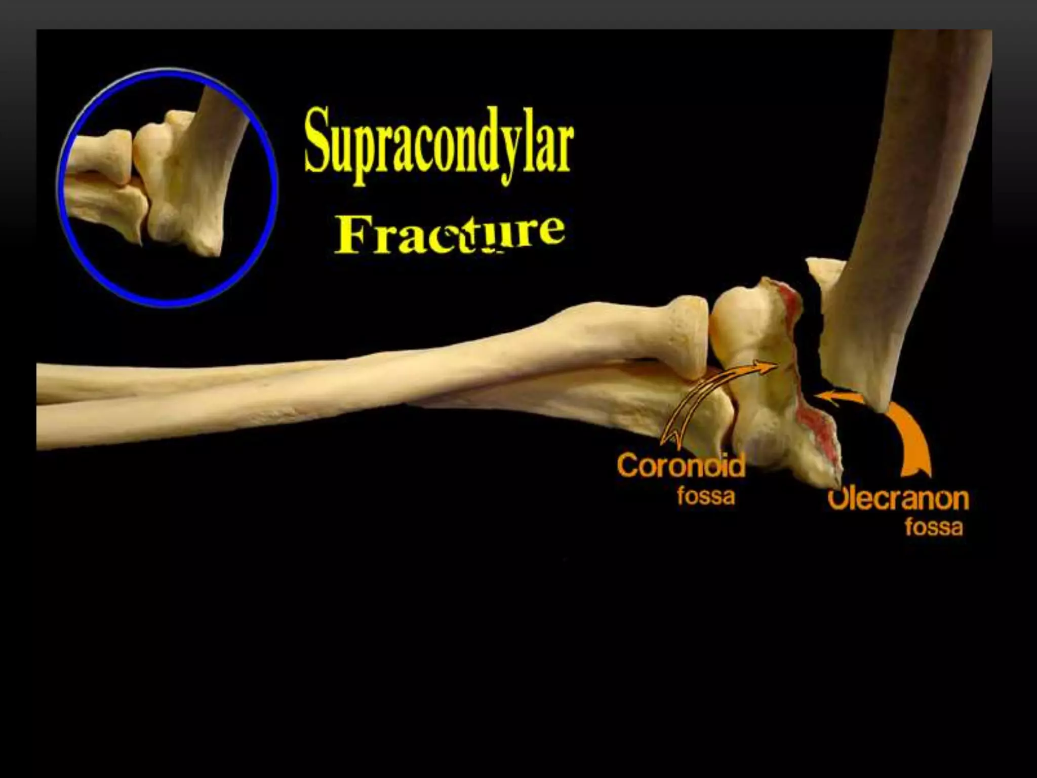
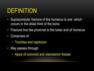
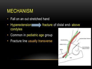
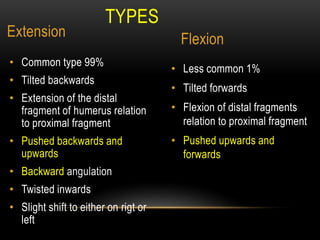
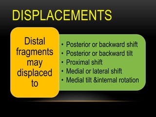
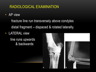
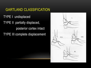
SUPRA CONDYLAR FRACTURE
DEFINITION
• Supracondylar fracture of the humerus is one which
occurs in the distal third of the bone
• Fracture line lies proximal to the lower end of humerus
• Comprises of
• Trochlea and capitulum
• May passes through
• Apice of coronoid and olecreanon fossae
MECHANISM
• Fall on an out stretched hand
• Hyperextension fracture of distal end- above
condyles
• Common in pediatric age group
• Fracture line usually transverse
TYPES
Extension
Flexion
• Less common 1%
• Tilted forwards
• Flexion of distal fragments
relation to proximal fragment
• Pushed upwards and
forwards
• Common type 99%
• Tilted backwards
• Extension of the distal
fragment of humerus relation
to proximal fragment
• Pushed backwards and
upwards
• Backward angulation
• Twisted inwards
• Slight shift to either on rigt or
left
TYPES
Extension Flexion
DISPLACEMENTS
• Posterior or backward shift
• Posterior or backward tilt
• Proximal shift
• Medial or lateral shift
• Medial tilt &internal rotation
Distal
fragments
may
displaced
to
PRESENTING COMPLAINTS
History of
Trauma
Pain
Swelling deformity inability to move
affected elbow
CLINICAL FEATURES
• Patient hold the elbow in a flexed position by holding
the forearm with other elbow
• Swelling
• Tense ,filling up the hollow around the elbow and
obscuring bony points
• Displaced backward upward medially
• Movements restricted and painful
EXAMINATION
• Posterior prominence of elbow joint
• Equilateral triangular relationship of
OLECRANON
MEDIAL LATERAL
• Careful palpation elicit tenderness –distal humerus
• Restricted movements
• RADIAL & ULNAR PULSE
• Median nerve
• Radial nerve
RADIOLOGICAL EXAMINATION
• AP view
fracture line run transversely above condyles
distal fragment – dispaced & rotated laterally.
• LATERAL view
line runs upwards
& backwards
GARTLAND CLASSIFICATION
TYPE I undisplaced
TYPE II partially displaced,
posterior cortex intact
TYPE III complete displacement
TREATMENT
• Undisplaced fracture
immobilisation + posterior elbow slab, elbow in 90o flexion
• Displaced fracture
1. closed reduction & percutaneous K wire fixation
2. continuous traction -SMITH’S, DUNLOP’S
3. open reduction & K wire.
BAUMANN’S ANGLE
 intersection of a line drawn perpendicular to the
humeral axis and line drawn along the growth plate of
the lateral condyle of the elbow.
COMPLICATIONS
IMMEDIATE
Injury to brachial artery, nerves.
EARLY
volkmann’s ischaemia
LATE
1. malunion- cubitus varus or gun stock deformity
2. myositis ossificans
3. volkmann’s ischaemic contracture.
THANK YOU

Supra condylar fracture.pptx

  • 1.
  • 2.
    DEFINITION • Supracondylar fractureof the humerus is one which occurs in the distal third of the bone • Fracture line lies proximal to the lower end of humerus • Comprises of • Trochlea and capitulum • May passes through • Apice of coronoid and olecreanon fossae
  • 3.
    MECHANISM • Fall onan out stretched hand • Hyperextension fracture of distal end- above condyles • Common in pediatric age group • Fracture line usually transverse
  • 4.
  • 6.
    • Less common1% • Tilted forwards • Flexion of distal fragments relation to proximal fragment • Pushed upwards and forwards • Common type 99% • Tilted backwards • Extension of the distal fragment of humerus relation to proximal fragment • Pushed backwards and upwards • Backward angulation • Twisted inwards • Slight shift to either on rigt or left TYPES Extension Flexion
  • 7.
    DISPLACEMENTS • Posterior orbackward shift • Posterior or backward tilt • Proximal shift • Medial or lateral shift • Medial tilt &internal rotation Distal fragments may displaced to
  • 8.
    PRESENTING COMPLAINTS History of Trauma Pain Swellingdeformity inability to move affected elbow
  • 9.
    CLINICAL FEATURES • Patienthold the elbow in a flexed position by holding the forearm with other elbow • Swelling • Tense ,filling up the hollow around the elbow and obscuring bony points • Displaced backward upward medially • Movements restricted and painful
  • 10.
    EXAMINATION • Posterior prominenceof elbow joint • Equilateral triangular relationship of OLECRANON MEDIAL LATERAL • Careful palpation elicit tenderness –distal humerus • Restricted movements
  • 11.
    • RADIAL &ULNAR PULSE • Median nerve • Radial nerve
  • 12.
    RADIOLOGICAL EXAMINATION • APview fracture line run transversely above condyles distal fragment – dispaced & rotated laterally. • LATERAL view line runs upwards & backwards
  • 13.
    GARTLAND CLASSIFICATION TYPE Iundisplaced TYPE II partially displaced, posterior cortex intact TYPE III complete displacement
  • 14.
    TREATMENT • Undisplaced fracture immobilisation+ posterior elbow slab, elbow in 90o flexion • Displaced fracture 1. closed reduction & percutaneous K wire fixation 2. continuous traction -SMITH’S, DUNLOP’S 3. open reduction & K wire.
  • 15.
    BAUMANN’S ANGLE  intersectionof a line drawn perpendicular to the humeral axis and line drawn along the growth plate of the lateral condyle of the elbow.
  • 16.
    COMPLICATIONS IMMEDIATE Injury to brachialartery, nerves. EARLY volkmann’s ischaemia LATE 1. malunion- cubitus varus or gun stock deformity 2. myositis ossificans 3. volkmann’s ischaemic contracture.
  • 17.