1. BIO 0304214
Comparative Vertebrate Anatomy
Lecture Notes 2 - Vertebrate Skeletal Systems
Bone:
• inorganic components of bone comprise 60% of the dry weight (largely calcium hydroxy-
appetite crystals) & provide the compressive strength of bone. The organic component is
primarily collagen, which gives bone great tensile strength.
• provides support and movement via attachments for soft tissue and muscle, protects vital
organs, is a major site for red marrow for production of blood cells, and plays a role in the
metabolism of minerals such as calcium and phosphorus.
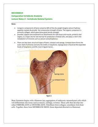
• There are two basic structural types of bone, compact and spongy. Compact bone forms the
outer shell of all bones and also the shafts in long bones. Spongy bone is found at the expanded
heads of long bones and fills most irregular bones. .
Figure 1
Bone formation begins with a blastema (any aggregation of embryonic mesenchymal cells which
will differentiate into tissue such as muscle, cartilage, or bone). These cells then develop into
either FIBROBLASTS or OSTEOBLASTS. Fibroblasts form collagen; osteoblasts form bone
cells. Together, these form MEMBRANE BONE (bone deposited directed in a blastema).
2. Intramembranous ossification is the process of membrane bone formation. This process give
rise to:
• bones of the lower jaw, skull, & pectoral girdle
• dentin & other bone that develops in the skin
• vertebrae in some vertebrates (teleosts, urodeles, & apodans)
Endochondral ossification is the process in which bone is deposited in pre-existing cartilage, &
such bone is called REPLACEMENT BONE.
Figure 2
Skeletal elements:
• Dermal skeleton
o skin of most living vertebrates has no hard skeletal parts but dermal bone elements are
usually present in the head region
o early vertebrates (ostracoderms) had so much dermal bone they were called 'armored
fishes'
3. Source: http://www.auburn.edu/academic/classes/zy/0301/Topic3b/Topic3b.html
o after ostracoderms, fish continued to develop much bone in skin but that bone has
become 'thinner' over time
• Endoskeleton
o Somatic - axial & appendicular skeletons
o Visceral - cartilage or bone associated with gills & skeletal elements (such as jaw
cartilages) derived from them
Dermal bone of fishes:
• Basic structure includes lamellar (compact) bone, spongy bone, dentin, &, often, a surface with a
layer of enamel-like material
• Evolutionary 'trend' = large bony plates giving way to smaller, thinner bony scales
o Ancient armor - not found on living fish
o Ganoid scales - found only on Latimeria (coelocanth) & sturgeons
4. o Placoid scales - elasmobranchs (diagram to the right; pulp cavity > dentin layer >
enamel)
o Ctenoid & Cycloid scales - modern bony fish
Also check: http://courses.washington.edu/vertebra/453/photos/skin_photos/special_integument1.htm
5. 1 = lamellar bone, 2 = spongy bone, 3 = dentin, 4 = enameloid, & 5 = fibrous plate (collagen)
Source: http://www.uta.edu/biology/restricted/3452int.htm
Tetrapods - retain dermal elements in the skull, jaws, & pectoral girdle
Somatic skeleton = axial skeleton (vertebral column, ribs, sternum, & skull) + appendicular
skeleton
Vertebral column:
Vertebrae - consist of a centrum (or body), 1 or 2 arches, plus various processes
• Amphicelous
o concave at both ends
o most fish, a few salamanders (Necturus), & caecilians
• Opisthocoelous
o convex in front & concave in back
o most salamanders
• Procelous
o concave in front & convex in back
o anurans & present-day reptiles
• Acelous
o flat-ended
o mammals
• Heterocelous
o saddle-shaped centrum at both ends
o birds
6. Vertebral arches:
• Neural arch - on top of centrum
• Hemal arch (also called chevrons) - beneath centrum in caudal vertebrae of fish, salamanders,
most reptiles, some birds, & many long-tailed mammals
Vertebral processes:
• projections from arches & centra
• some give rigidity to the column, articulate with ribs, or serve as sites of muscle attachment
7. Transverse processes - most common type of process; extend laterally from the base of a neural
arch or centrum & separate the epaxial & hypaxial muscles
Diapophyses & parapophyses - articulate with ribs
Prezygapophyses (cranial zygapophyses) & postzygapophyses (caudal zygapophyses) -
articulate with one another & limit flexion & torsion of the vertebral column
Vertebral columns:
• Cartilaginous fishes
o do not have typical fish vertebral columns
o vertebrae include neural arches (cartilaginous dorsal plates) & dorsal intercalary plates
are located between successive arches
8. • Teleosts
o well-ossified amphicelous vertebrae
o notochord persists within each centrum (but constricted)
o neural arch associated with each centrum & hemal arches in tail (caudal) vertebrae
• Chondrosteans (sturgeons & paddlefish) & modern lungfishes
o incomplete centra
o notochord is not constricted
o cartilage deposited in notochord sheath provides structural support
Diplospondyly = 2 centra and 2 sets of arches per body segment; occurs in some fish (including sharks)
Agnathans - only skeletal elements associated with the notochord are paired, lateral neural
cartilages
9. Vertebral columns of tetrapods
• Cervical region
o Amphibians - single cervical vertebra; allows little head movement
o Reptiles - increased numbers of cervical vertebrae (usually 7) & increased flexibility of
head
o Birds - variable number of cervical vertebrae (as many as 25 in swans)
o Mammals - usually 7 cervical vertebrae
o Reptiles, birds, & mammals - 1st two cervical vertebrae are modified & called the atlas &
axis
atlas - 1st cervical vertebra; ring-like (most of centrum gone); provides 'cradle' in
which skull can 'rock' (as when nodding 'yes')
axis - 2nd cervical vertebra
o
Transverse foramen (#6 in above caudal view of a cervical vertebra)
found in cervical vertebrae of birds & mammals
provides canal for vertebral artery & vein
• Dorsal region
o Dorsals - name given to vertebrae between cervicals & sacrals when all
articulate with similar ribs (e.g., fish, amphibians, & snakes)
o Crocodilians, lizards, birds, & mammals - ribs are confined to anterior region of trunk
thoracic - vertebrae with ribs
10. lumbar - vertebrae without ribs
• Sacrum & Synsacrum
o sacral vertebrae - have short transverse processes that brace the pelvic girdle &
hindlimbs against the vertebral column
Amphibians - 1 sacral vertebra
Living reptiles & most birds - 2 sacral vertebrae
Most mammals - 3 to 5 sacral vertebrae
o Sacrum - single bony complex consisting of fused sacral vertebrae; found when there is
more than 1 sacral vertebra (see examples below):
o Synsacrum
found in birds
produced by fusion of last thoracics, all lumbars, all sacrals, & first few caudals
fused with pelvic girdle
provides rigid support for bipedal locomotion
11. • Caudal region
o Primitive tetrapods - 50 or more caudal vertebrae
o Present-day tetrapods
number of caudal vertebrae is reduced
arches & processes get progressively shorter (the last few caudals typically
consist of just cylindrical centra as shown below)
o Anurans - unique terminal segment called the urostyle (section of unsegmented
vertebral column probably derived from separate caudals of early anurans)
o Birds - last 4 or 5 caudal vertebrae fused to form pygostyle (see drawing above)
o Apes & humans - last 3 to 5 caudal vertebrae fused to form coccygeal (or tail bone)
EVOLUTION OF VERTEBRAE
• Unlike most tetrapods today, vertebral column of earliest tetrapods did not consist of 1
bone/body segment.
• Crossopterygian vertebrae consisted of an hypocentrum (a large, wedge-shaped piece) plus 2
pleurocentra (smaller, intersegmental pieces). This type of vertebra is called a rachitomous
vertebra.
12. • The 'trend' in vertebra evolution has been for pleurocentra to increase in size (and, of course,
for the hypocentrum to decrease in size). This trend is apparent in this diagram:
Ribs - may be long or short, cartilaginous or bony; articulate medially with vertebrae & extend
into the body wall
• A few teleosts - have 2 pair of ribs for each centrum of trunk (dorsal rib separates epaxial &
hypaxial muscles)
• Most teleosts - ventral ribs only
• Sharks - dorsal ribs only
• Agnathans - no ribs
• Tetrapods - ribs usually articulate with vertebrae in moveable joints (see above drawing)
13. o Early tetrapods - ribs articulated with every vertebra from the atlas to the end of the
trunk
o Later tetrapods - long ribs limited to thoracic region
Thoracic ribs - most composed of a dorsal element (vertebral rib) & a ventral
element (sternal rib)
Sternal rib - may be ossified (birds) or remain cartilaginous (mammals); usually
articulate with sternum (except 'floating ribs')
Uncinate processes - found in birds; provides rib-cage with additional support
Sternum - strictly a tetrapod structure &, primarily, an amniote structure.
• Amphibians - no sternum in early amphibians &, among present-day amphibians, only anurans
have one
• Amniotes
o sternum is a plate of cartilage & replacement bone
o sternum articulates with the pectoral girdle anteriorly & with a variable number of ribs