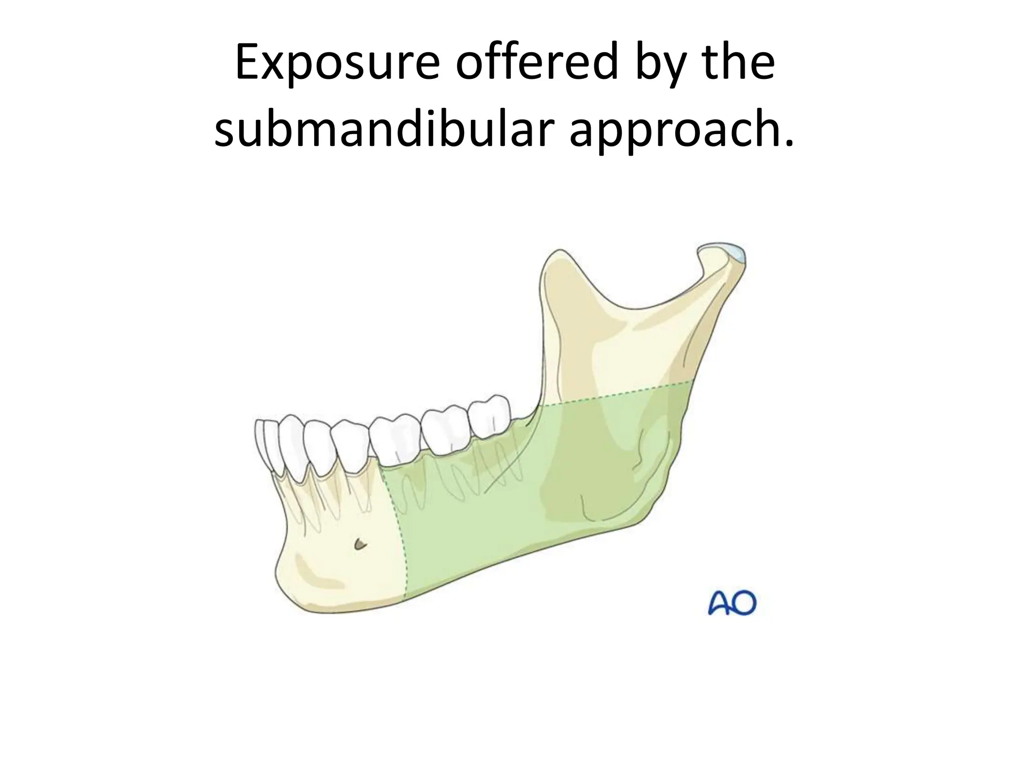
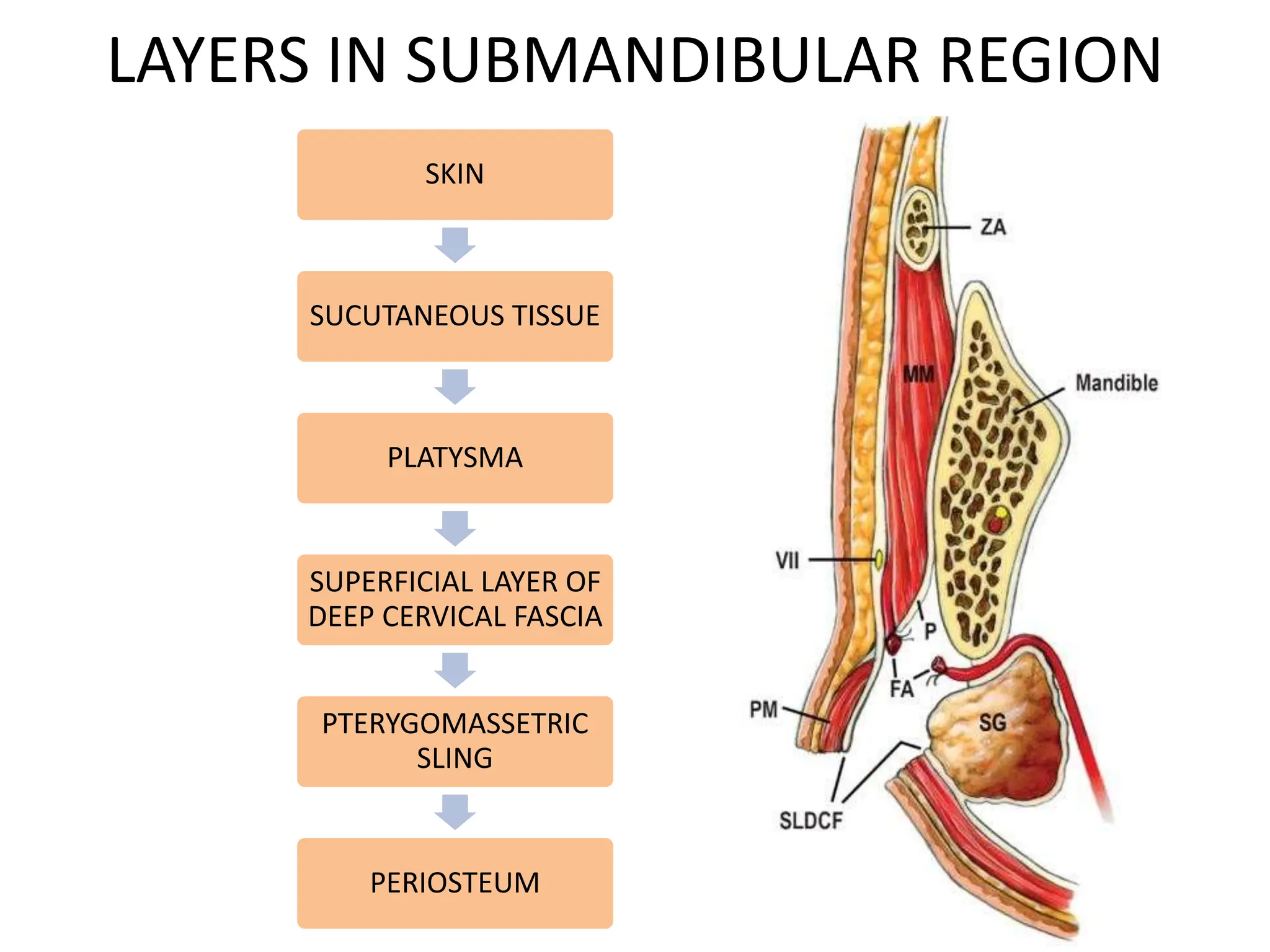
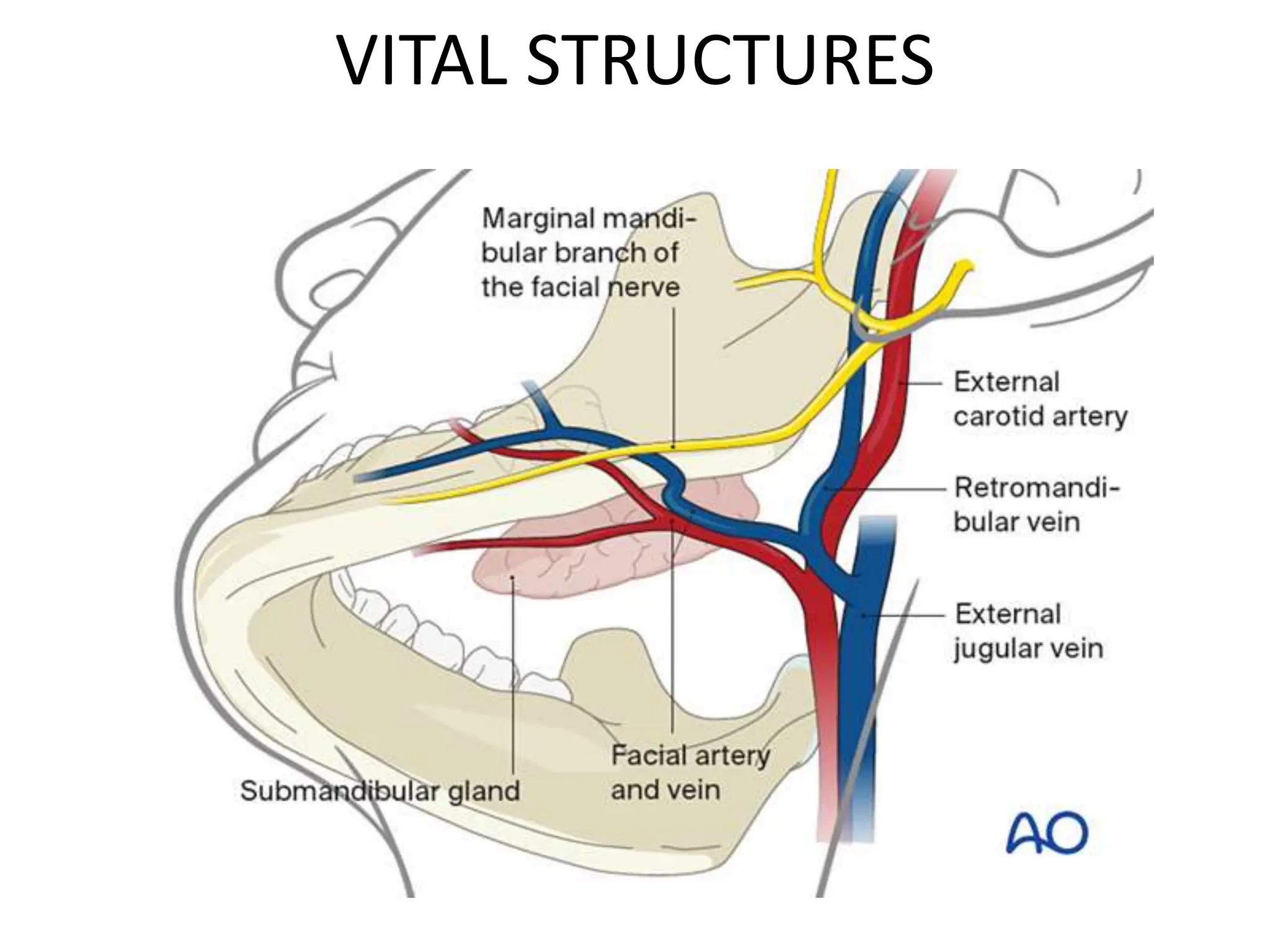
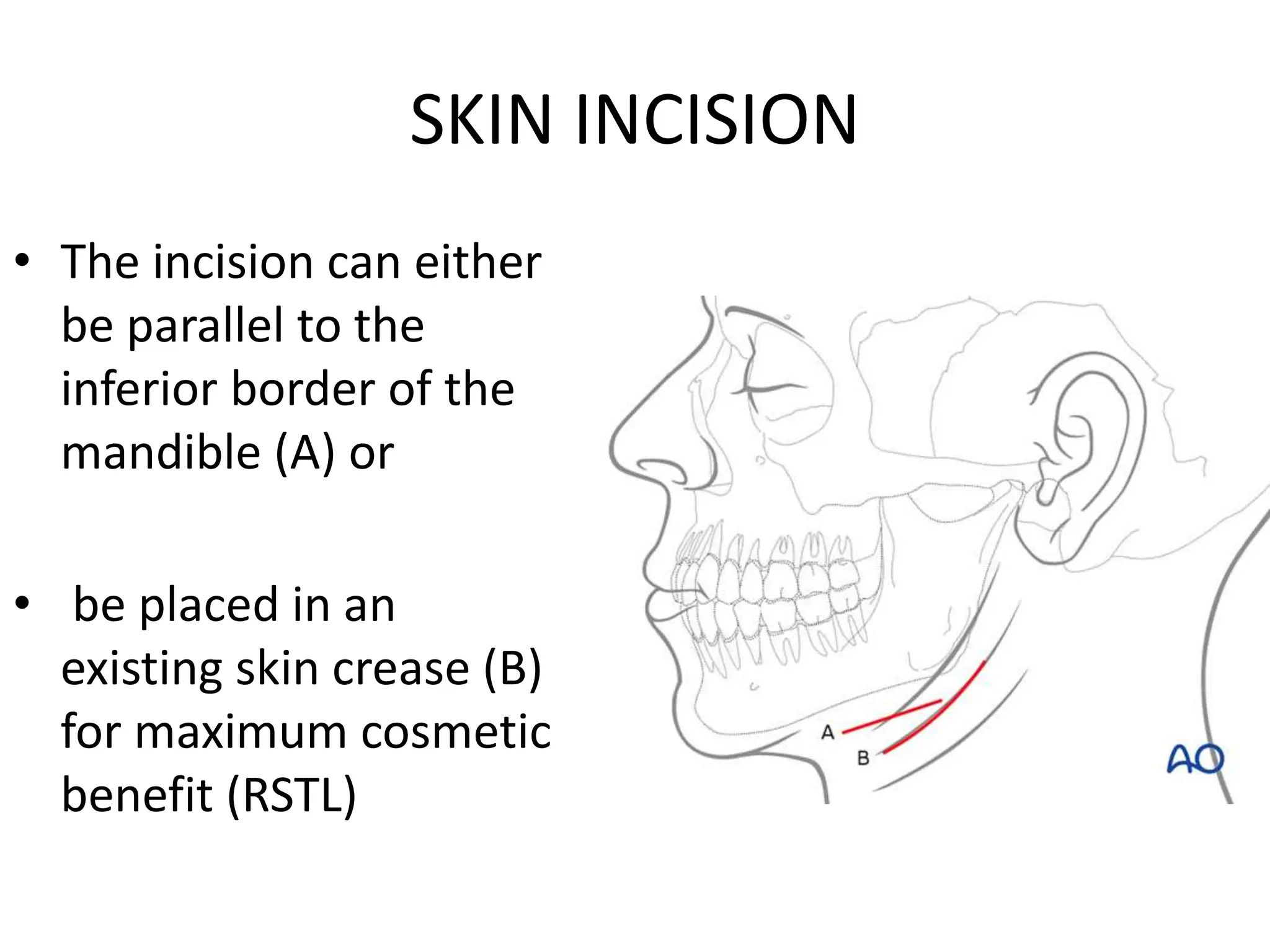
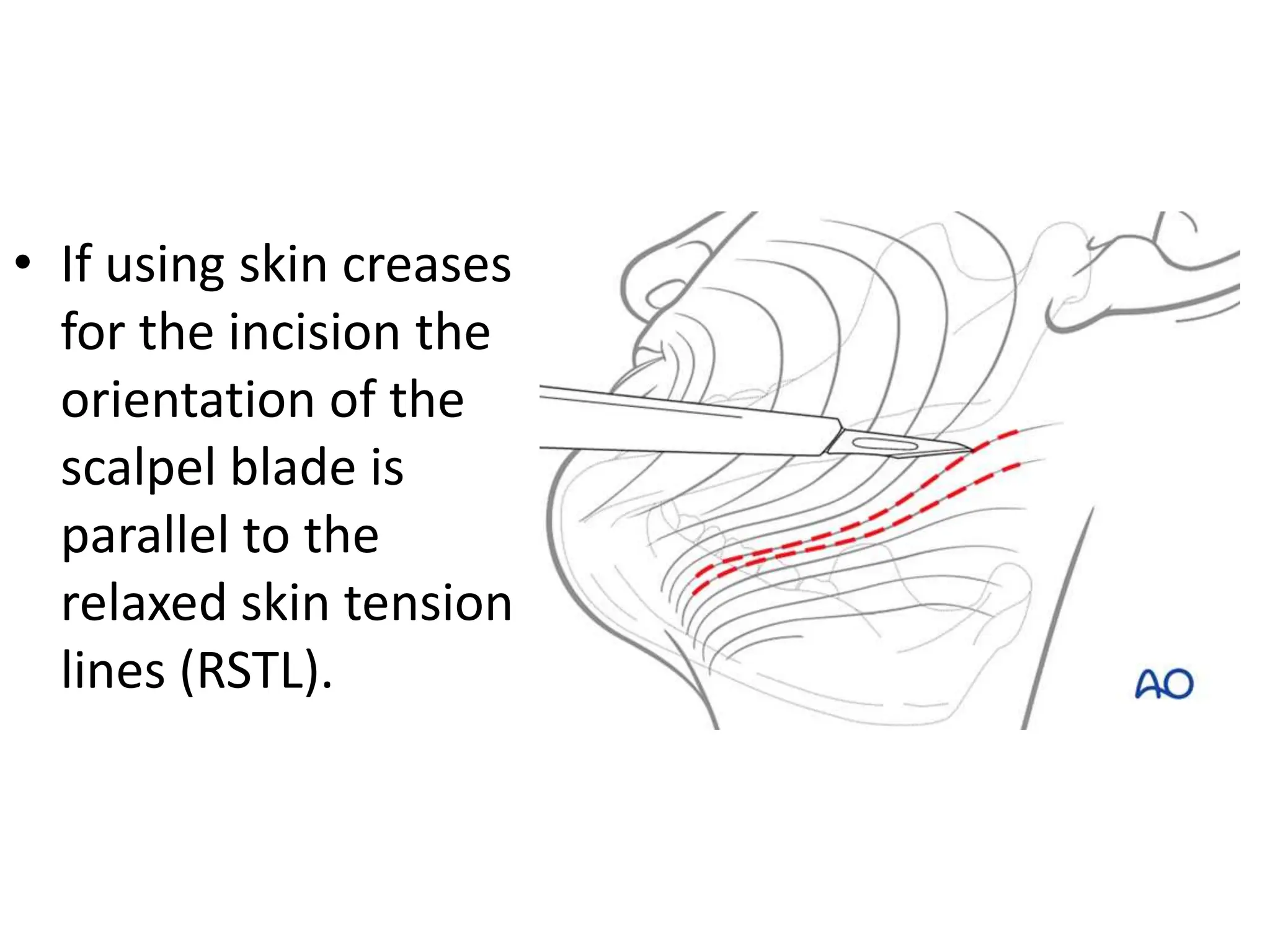
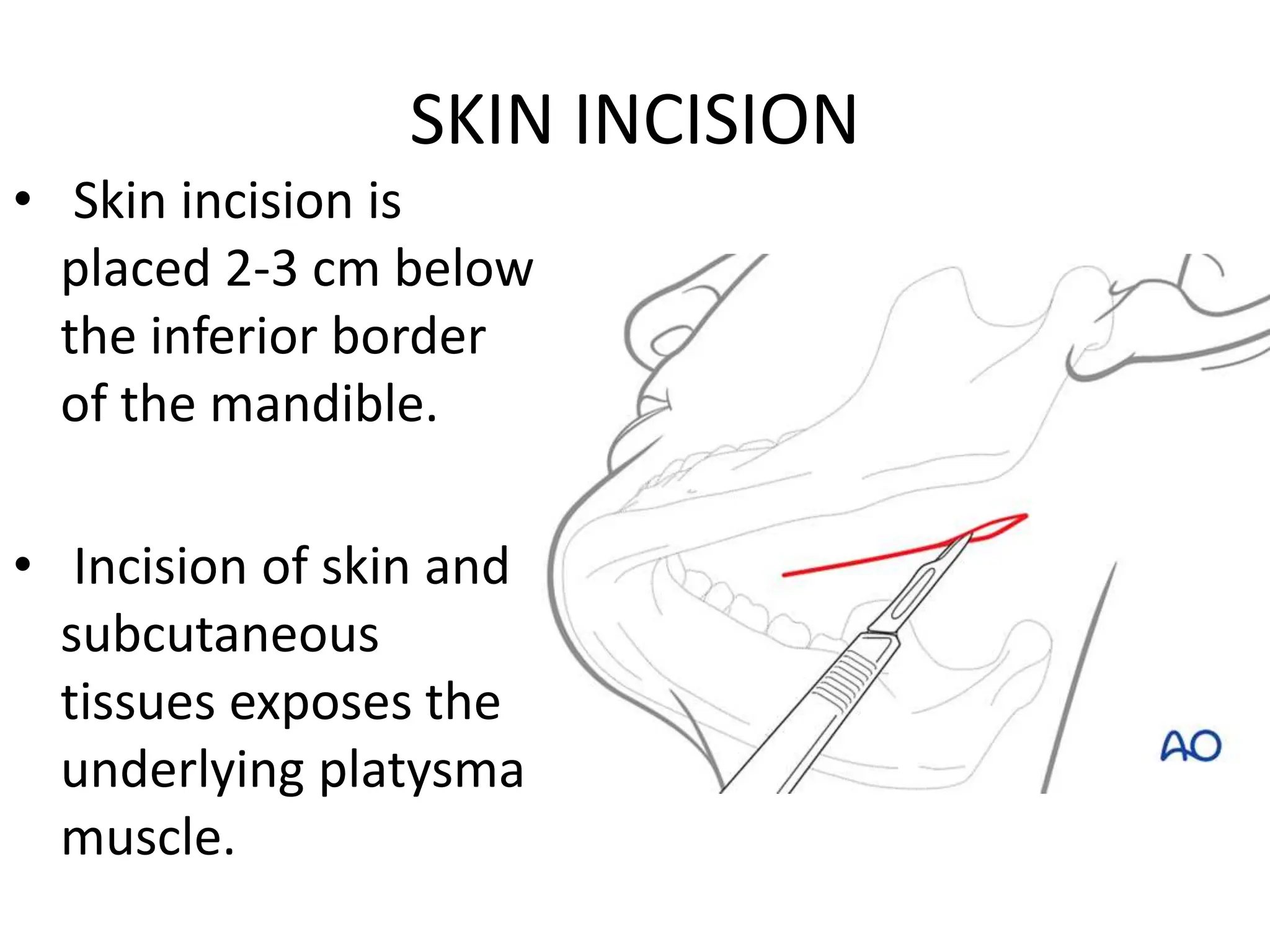
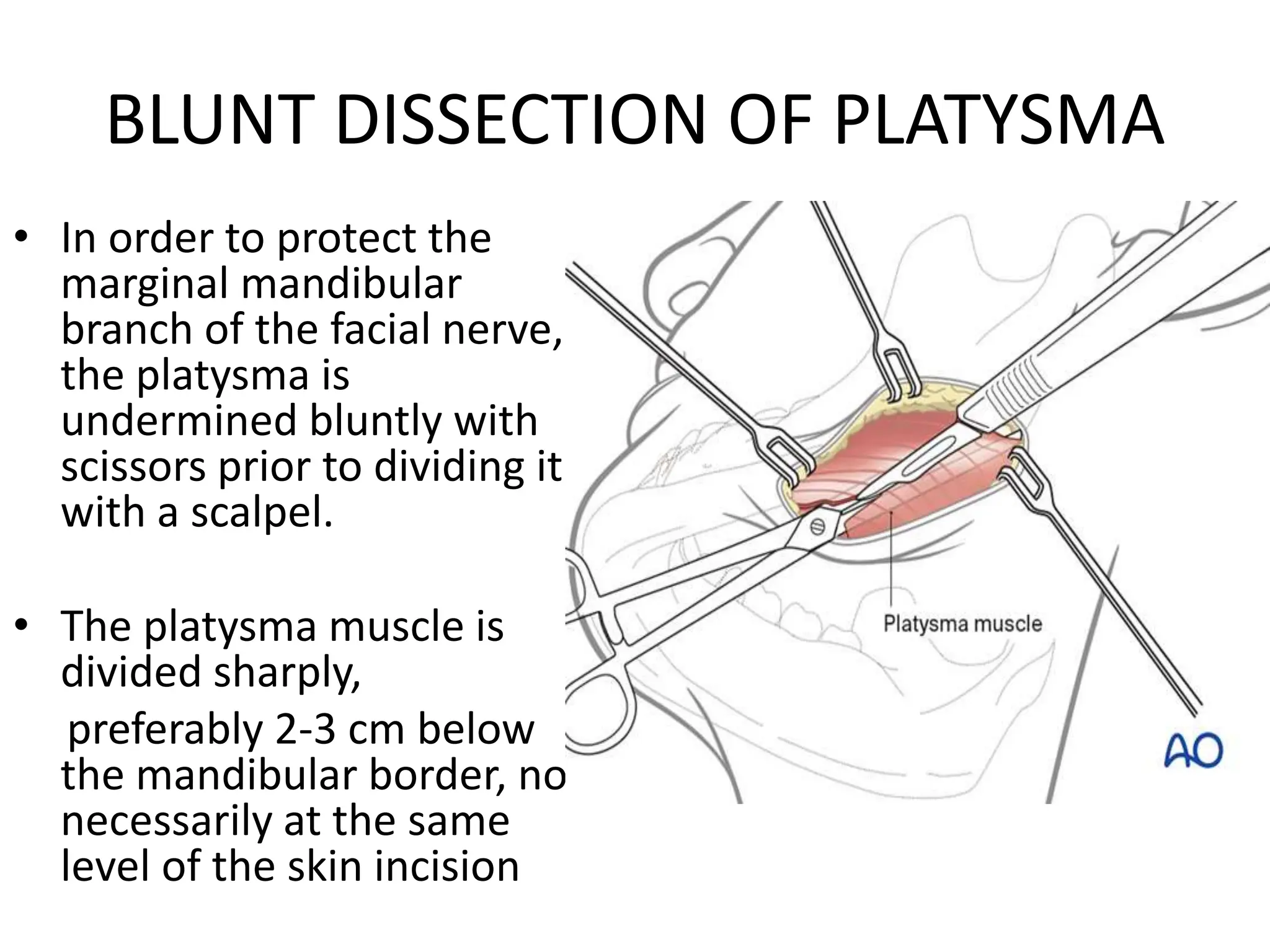
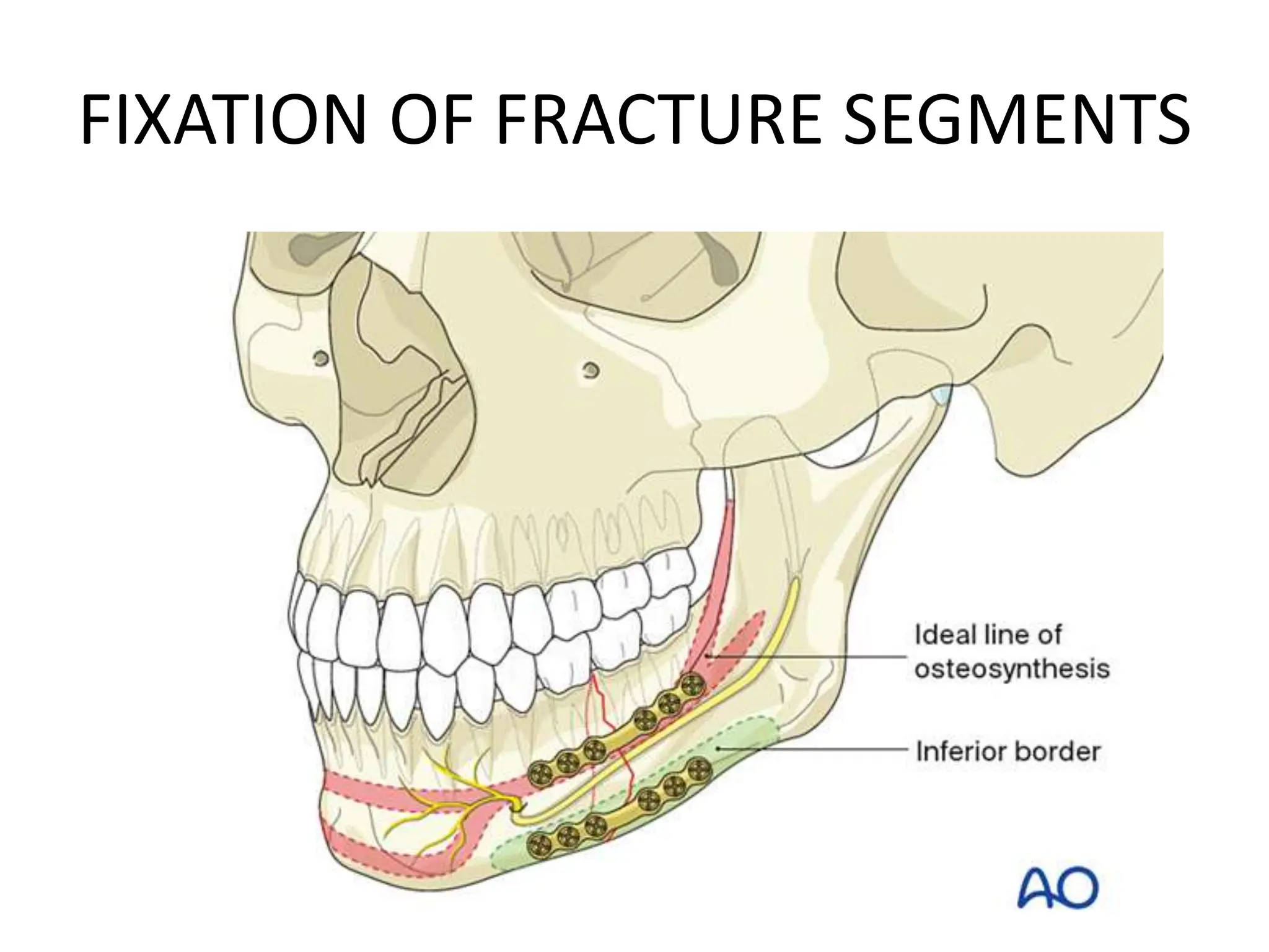
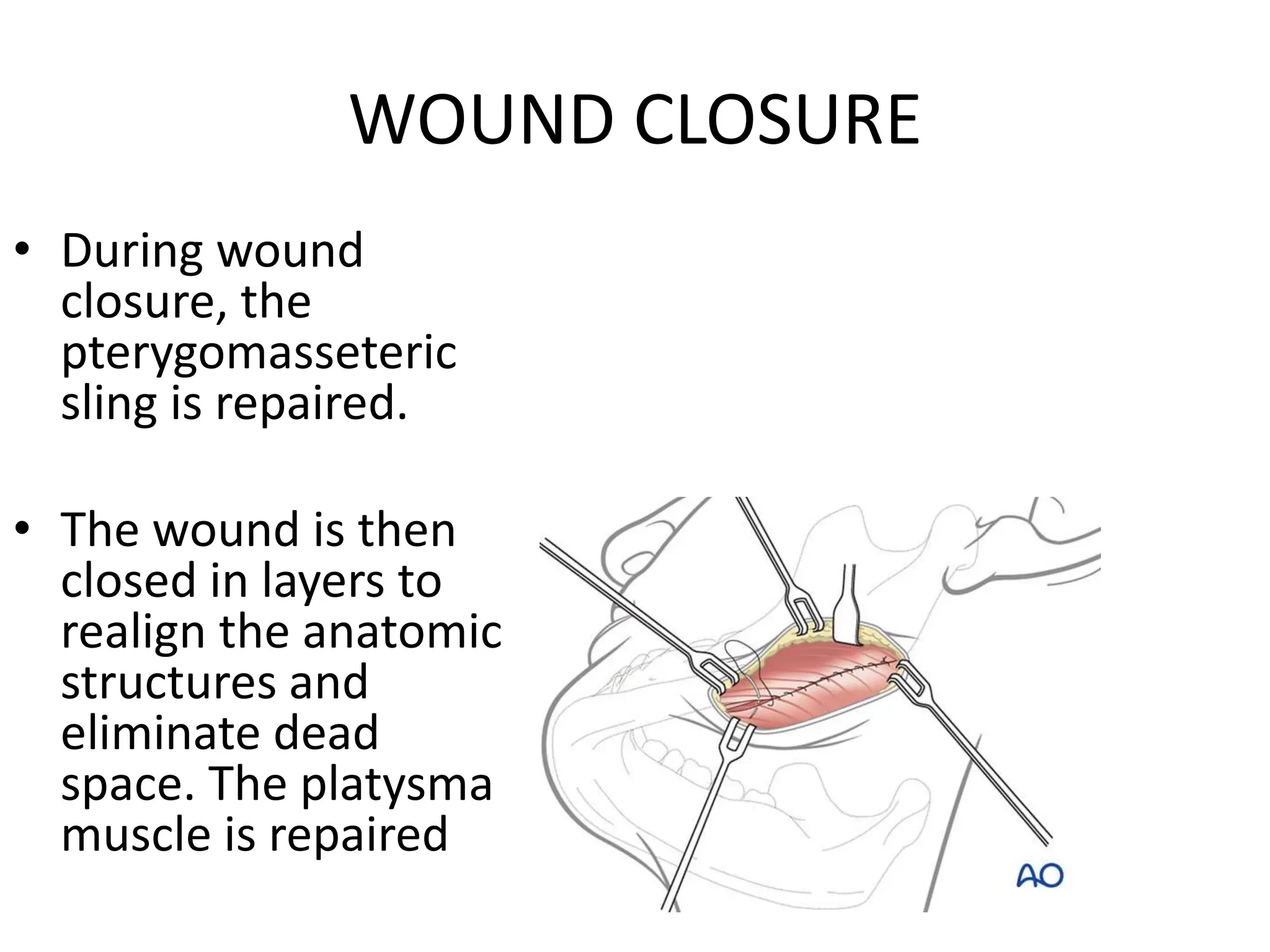
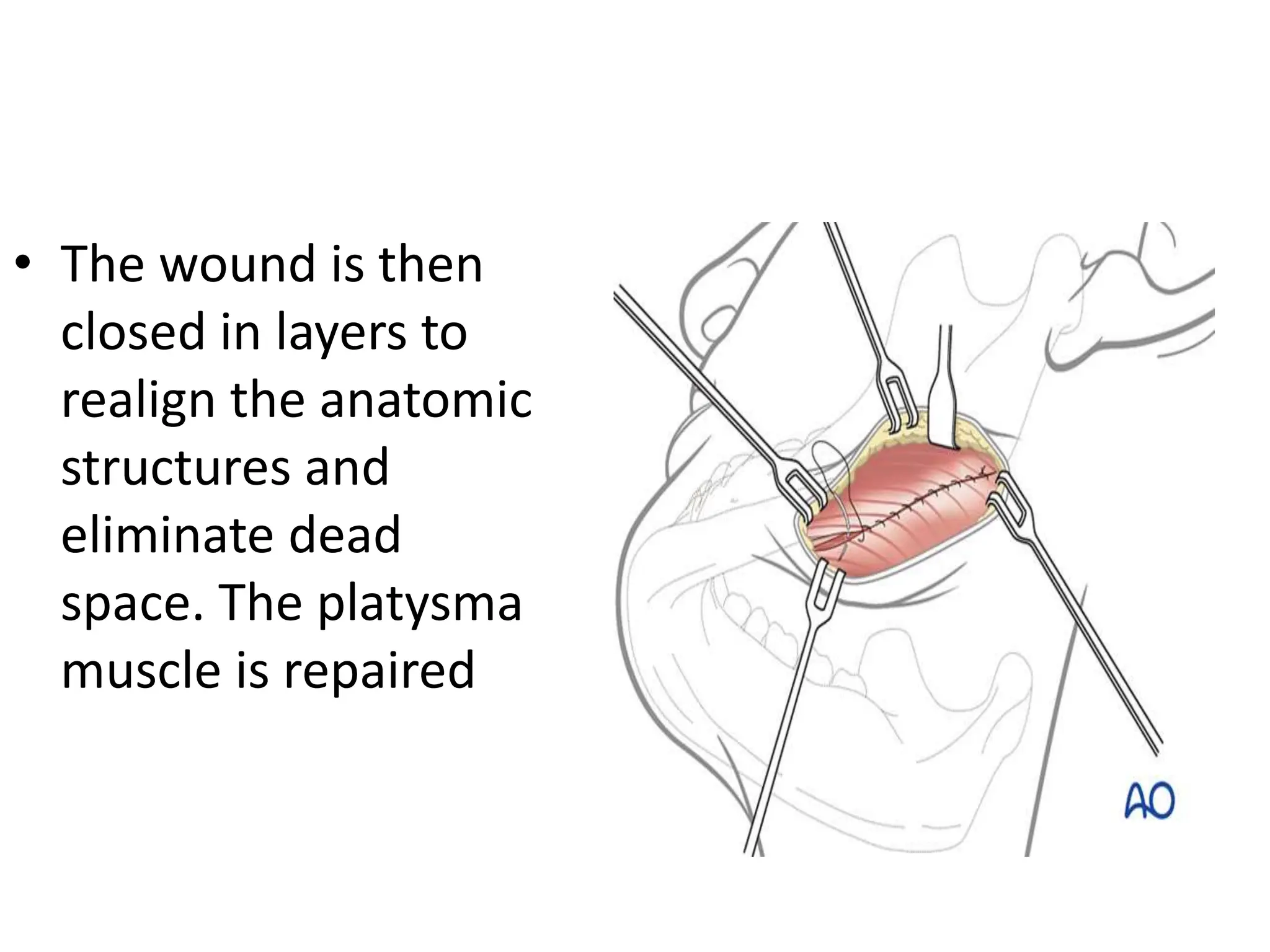
SUBMANDIBULAR
APPROACH
Presented by – Dr. Ashish ( R1 )
INDICATIONS
• This approach is selected for fractures of the
mandibular body and angle regions unsuitable
for intraoral treatment.
• This applies to more difficult fracture patterns
such as comminuted, atrophic, and defect
fractures to allow optimal manipulation of the
fragments, good control of the lingual cortex
and inferior border
Exposure offered by the
submandibular approach.
LAYERS IN SUBMANDIBULAR REGION
SKIN
SUCUTANEOUS TISSUE
PLATYSMA
SUPERFICIAL LAYER OF
DEEP CERVICAL FASCIA
PTERYGOMASSETRIC
SLING
PERIOSTEUM
VITAL STRUCTURES
SKIN INCISION
• The incision can either
be parallel to the
inferior border of the
mandible (A) or
• be placed in an
existing skin crease (B)
for maximum cosmetic
benefit (RSTL)
• If using skin creases
for the incision the
orientation of the
scalpel blade is
parallel to the
relaxed skin tension
lines (RSTL).
SKIN INCISION
• Skin incision is
placed 2-3 cm below
the inferior border
of the mandible.
• Incision of skin and
subcutaneous
tissues exposes the
underlying platysma
muscle.
BLUNT DISSECTION OF PLATYSMA
• In order to protect the
marginal mandibular
branch of the facial nerve,
the platysma is
undermined bluntly with
scissors prior to dividing it
with a scalpel.
• The platysma muscle is
divided sharply,
preferably 2-3 cm below
the mandibular border, not
necessarily at the same
level of the skin incision
EXPOSURE AND SPLITTING OF
PTERYGOMASSETRIC SLING
• Superior subplatysmal
dissection would
expose the underlying
marginal mandibular
branch of the facial
nerve (CN VII)
• Divide the
pterygomasseteric sling
and incise the
periosteum at the
inferior border to
expose the ramus.
FIXATION OF FRACTURE SEGMENTS
WOUND CLOSURE
• During wound
closure, the
pterygomasseteric
sling is repaired.
• The wound is then
closed in layers to
realign the anatomic
structures and
eliminate dead
space. The platysma
muscle is repaired
• The wound is then
closed in layers to
realign the anatomic
structures and
eliminate dead
space. The platysma
muscle is repaired
THANK YOU

SUBMANDIBULAR APPROACH PPT for mandibular fractures.pptx

  • 1.
  • 2.
    INDICATIONS • This approachis selected for fractures of the mandibular body and angle regions unsuitable for intraoral treatment. • This applies to more difficult fracture patterns such as comminuted, atrophic, and defect fractures to allow optimal manipulation of the fragments, good control of the lingual cortex and inferior border
  • 3.
    Exposure offered bythe submandibular approach.
  • 4.
    LAYERS IN SUBMANDIBULARREGION SKIN SUCUTANEOUS TISSUE PLATYSMA SUPERFICIAL LAYER OF DEEP CERVICAL FASCIA PTERYGOMASSETRIC SLING PERIOSTEUM
  • 5.
  • 6.
    SKIN INCISION • Theincision can either be parallel to the inferior border of the mandible (A) or • be placed in an existing skin crease (B) for maximum cosmetic benefit (RSTL)
  • 7.
    • If usingskin creases for the incision the orientation of the scalpel blade is parallel to the relaxed skin tension lines (RSTL).
  • 8.
    SKIN INCISION • Skinincision is placed 2-3 cm below the inferior border of the mandible. • Incision of skin and subcutaneous tissues exposes the underlying platysma muscle.
  • 12.
    BLUNT DISSECTION OFPLATYSMA • In order to protect the marginal mandibular branch of the facial nerve, the platysma is undermined bluntly with scissors prior to dividing it with a scalpel. • The platysma muscle is divided sharply, preferably 2-3 cm below the mandibular border, not necessarily at the same level of the skin incision
  • 13.
    EXPOSURE AND SPLITTINGOF PTERYGOMASSETRIC SLING • Superior subplatysmal dissection would expose the underlying marginal mandibular branch of the facial nerve (CN VII) • Divide the pterygomasseteric sling and incise the periosteum at the inferior border to expose the ramus.
  • 14.
  • 15.
    WOUND CLOSURE • Duringwound closure, the pterygomasseteric sling is repaired. • The wound is then closed in layers to realign the anatomic structures and eliminate dead space. The platysma muscle is repaired
  • 16.
    • The woundis then closed in layers to realign the anatomic structures and eliminate dead space. The platysma muscle is repaired
  • 17.