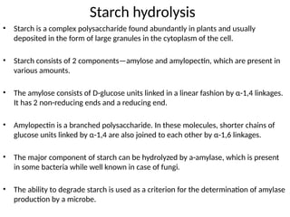
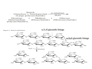
Starch hydrolysis
• Starchis a complex polysaccharide found abundantly in plants and usually
deposited in the form of large granules in the cytoplasm of the cell.
• Starch consists of 2 components—amylose and amylopectin, which are present in
various amounts.
• The amylose consists of D-glucose units linked in a linear fashion by α-1,4 linkages.
It has 2 non-reducing ends and a reducing end.
• Amylopectin is a branched polysaccharide. In these molecules, shorter chains of
glucose units linked by α-1,4 are also joined to each other by α-1,6 linkages.
• The major component of starch can be hydrolyzed by a-amylase, which is present
in some bacteria while well known in case of fungi.
• The ability to degrade starch is used as a criterion for the determination of amylase
production by a microbe.
• Objective:
• Todetermine the ability of an organism to hydrolyze starch
• To differentiate organism based on their α- amylase enzyme activity
• Principle:
• Many bacteria produce extracellular enzymes used to catalyze chemical reactions outside of the
cell.
• In this manner, nutrient sources, such as starch, that are too large to be absorbed through the cell
membrane can be broken down into smaller molecules and transported into the cell via diffusion.
• In the starch hydrolysis test, the test bacteria are grown on agar plates containing starch.
• If the bacteria have the ability to hydrolyze starch, it does so in the medium, particularly in the
areas surrounding their growth while the rest of the area of the plate still contain non-hydrolysed
starch.
• Since no color change occurs in the medium when organisms hydrolyze starch, iodine solution is
added as an indicator to the plate after incubation.
• While the non-hydrolysed starch forms dark blue color with iodine, its hydrolyzed end products do
not acquire such dark blue color with iodine.
• Consequently, transparent clear zones are formed around the colonies that hydrolyze starch while
the rest of the plate show a dark blue coloration as iodine forms the colored complex with starch.
5.
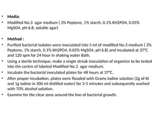
• Media:
• ModifiedNo.3 agar medium ( 3% Peptone, 1% starch, 0.1% KH2PO4, 0.05%
MgSO4, pH 6.8, soluble agar)
• Method :
• Purified bacterial isolates were inoculated into 5 ml of modified No.3 medium ( 3%
Peptone, 1% starch, 0.1% KH2PO4, 0.05% MgSO4, pH 6.8) and incubated at 37°C
and 120 spm for 24 hour in shaking water Bath.
• Using a sterile technique, make a single streak inoculation of organism to be tested
into the centre of labeled Modified No.3 agar medium.
• Incubate the bacterial inoculated plates for 48 hours at 37°C.
• After proper incubation, plates were flooded with Grams iodine solution (2g of KI
and 1g iodine in 300 ml distilled water) for 3-5 minutes and subsequently washed
with 70% alcohol solution.
• Examine for the clear zone around the line of bacterial growth.
6.
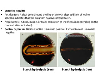
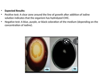
• Expected Results:
•Positive test: A clear zone around the line of growth after addition of iodine
solution indicates that the organism has hydrolyzed starch.
• Negative test: A blue, purple, or black coloration of the medium (depending on the
concentration of iodine).
• Control organism: Bacillus subtilis is amylase positive; Escherichia coli is amylase
negative
7.
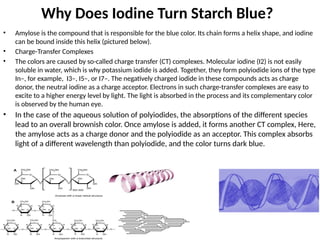
Why Does IodineTurn Starch Blue?
• Amylose is the compound that is responsible for the blue color. Its chain forms a helix shape, and iodine
can be bound inside this helix (pictured below).
• Charge-Transfer Complexes
• The colors are caused by so-called charge transfer (CT) complexes. Molecular iodine (I2) is not easily
soluble in water, which is why potassium iodide is added. Together, they form polyiodide ions of the type
In–, for example, I3–, I5–, or I7–. The negatively charged iodide in these compounds acts as charge
donor, the neutral iodine as a charge acceptor. Electrons in such charge-transfer complexes are easy to
excite to a higher energy level by light. The light is absorbed in the process and its complementary color
is observed by the human eye.
• In the case of the aqueous solution of polyiodides, the absorptions of the different species
lead to an overall brownish color. Once amylose is added, it forms another CT complex, Here,
the amylose acts as a charge donor and the polyiodide as an acceptor. This complex absorbs
light of a different wavelength than polyiodide, and the color turns dark blue.
8.
Casein Hydrolysis Test
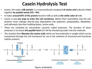
•Casein, the major milk protein, is a macromolecule composed of amino acid subunits linked
together by peptide bonds (CO—NH).
• It makes around 85% of the protein found in milk as well as the white color of milk.
• Casein is way too large to enter the cell membrane. Before their assimilation into the cell,
proteins must undergo step-by-step degradation into peptones, polypeptides, dipeptides,
and ultimately into their building blocks, amino acids.
• These are mediated by extracellular enzymes called proteases. The function of these
proteases is to cleave the peptide bond CO–NH by introducing water into the molecule.
• The reaction then liberates the amino acids which are low-molecular in weight which can be
transported through the cell membrane for use in the synthesis of structural and functional
cellular proteins.
Figure: proteolysis
9.
• Objectives:
• Todetermine the ability of the organism to degrade the casein protein.
• To differentiate the organism on the basis of production of exoenzyme proteinase
(caseinase)
10.
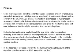
• Principle:
• Somemicroorganisms have the ability to degrade the casein protein by producing
proteolytic exoenzyme, called proteinase (caseinase). For demonstration of such an
activity, in the lab, milk agar is used. The medium is composed of nutrient agar
supplemented with milk that contains the protein substrate casein. Similar to other
proteins, milk protein is a colloidal suspension that gives the medium its color and
opacity because it deflects light rays rather than transmitting them.
• Following inoculation and incubation of the agar plate cultures, organisms
secreting proteases will exhibit a zone of proteolysis, which is demonstrated by a
clear area surrounding the bacterial growth. This loss of opacity is the result of a
hydrolytic reaction yielding soluble, non-colloidal amino acids, and it represents a
positive reaction.
• In the absence of protease activity, the medium surrounding the growth of the
organism remains opaque, which is a negative reaction.)
11.
• Media:
• Skimmilk agar
• SM powder 28.0gm/L, Tryptone 5.0gm/L, Yeast extract 2.50gm/L, Dextrose
(Glucose) 1.0gm/L, Agar 15.0gm/L.
• Method:
• Purified bacterial isolates were inoculated into 5 ml of Skim milk media (SM
powder 28.0gm/L, Tryptone 5.0gm/L, Yeast extract 2.50gm/L, Dextrose (Glucose)
1.0gm/L) and incubated at 37°C and 120 spm for 24 hour in shaking water Bath.
• Using a sterile technique, make a single streak inoculation of organism to be tested
into the centre of labeled Skim milk agar plate
• Incubate the bacterial inoculated plates for 48 hours at 37°C.
• Examine the milk agar plate cultures for the presence or absence of a clear area, or
zone of proteolysis, surrounding the growth of each of the bacterial test organisms.
12.
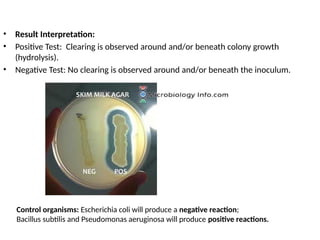
• Result Interpretation:
•Positive Test: Clearing is observed around and/or beneath colony growth
(hydrolysis).
• Negative Test: No clearing is observed around and/or beneath the inoculum.
Control organisms: Escherichia coli will produce a negative reaction;
Bacillus subtilis and Pseudomonas aeruginosa will produce positive reactions.
13.
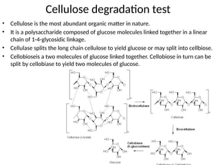
Cellulose degradation test
•Cellulose is the most abundant organic matter in nature.
• It is a polysaccharide composed of glucose molecules linked together in a linear
chain of 1-4-glycosidic linkage.
• Cellulase splits the long chain cellulose to yield glucose or may split into cellbiose.
• Cellobioseis a two molecules of glucose linked together. Cellobiose in turn can be
split by cellobiase to yield two molecules of glucose.
14.
• Media:
• ModifiedNo.3 agar medium ( 3% Peptone, 1% CMC (carboxy methyl cellulose),
0.1% KH2PO4, 0.05% MgSO4, pH 6.8, soluble agar)
• Carboxymethyl cellulose (CMC) is a sodium salt derivative of cellulose. Unlike
cellulose, it is water soluble.
• Method :
• Purified bacterial isolates were inoculated into 5 ml of modified No.3 medium ( 3%
Peptone, 1% CMC, 0.1% KH2PO4, 0.05% MgSO4, pH 6.8) and incubated at 37°C and
120 spm for 24 hour in shaking water Bath.
• Using a sterile technique, make a single streak inoculation of organism to be tested
into the centre of labeled Modified No.3 agar medium.
• Incubate the bacterial inoculated plates for 48 hours at 37°C.
• After proper incubation, plates were flooded with Grams iodine solution (2g of KI
and 1g iodine in 300 ml distilled water) for 3-5 minutes and subsequently washed
with 70% alcohol solution.
• Examine for the clear zone around the line of bacterial growth.
15.
• Expected Results:
•Positive test: A clear zone around the line of growth after addition of iodine
solution indicates that the organism has hydrolyzed CMC.
• Negative test: A blue, purple, or black coloration of the medium (depending on the
concentration of iodine).