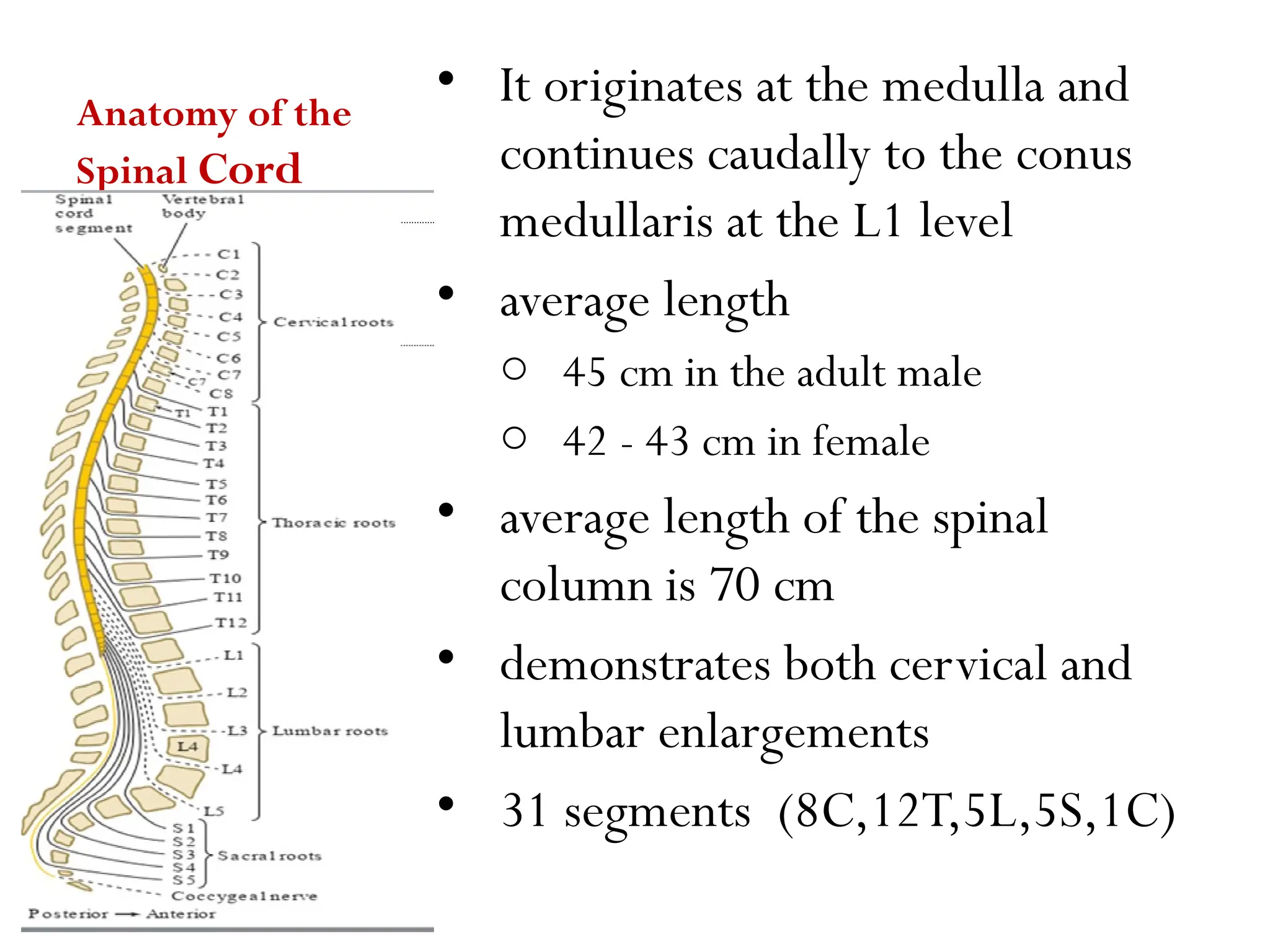
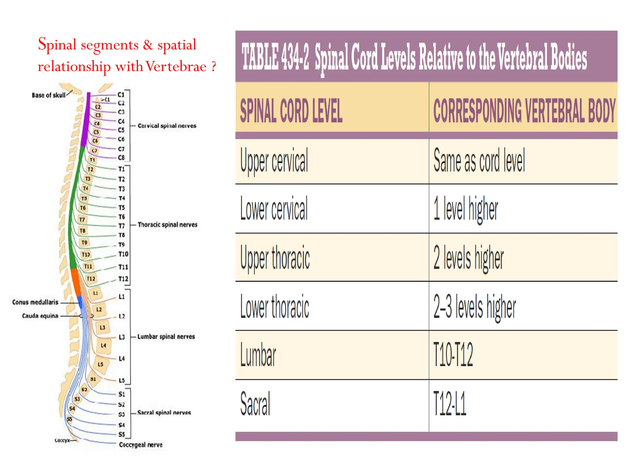
Spinal Cord Disorders
( neurolocalization)
Anatomy of the
Spinal Cord
• It originates at the medulla and
continues caudally to the conus
medullaris at the L1 level
• average length
o 45 cm in the adult male
o 42 - 43 cm in female
• average length of the spinal
column is 70 cm
• demonstrates both cervical and
lumbar enlargements
• 31 segments (8C,12T,5L,5S,1C)
Spinal segments & spatial
relationship withVertebrae ?
Cross-Sectional Anatomy of the Spinal Cord
• centrally placed gray
matter surrounded by
white matter.
• two lateral columns
joined by a transverse
commissure
• The central canal of the
spinal cord is located in
the center of the gray
commissure
Major Ascending and DescendingTracts of
the Spinal Cord
AscendingTracts
• Sensory afferent input to the SC enters by
way of the dorsal roots
• The central end of a dorsal root splits into
lateral and medial bundles
• The axons of second-order neurons,
decussate over several segments by way of
the ventral white commissure;
• They proceed to the ventrolateral quadrant
of the spinal cord and ascend as the lateral
spinothalamic tract to reach the thalamus
• These fibers convey pain and temperature
sensation
o Fibers carrying information from cervical
regions lie dorsomedially
o and those from sacral regions lie
ventrolaterally.
• The medial bundle of the dorsal root pass over
the dorsal horn into the dorsal funiculus
• Fibers from the lowermost part of the body are
located more medially and constitute the
fasciculus gracilis
• whereas those coming from the upper part of
the body occupy a more lateral position and
constitute the fasciculus cuneatus
• Axons of these two nuclei decussate in the
lower medulla and ascend to the thalamus.
• These fibers carry information concerning
discriminative senses (position sense, vibration
sense, weight perception, discriminative touch,
pressure touch, two-point discrimination,
stereognosis, and shape and movement
awareness).
DescendingTracts
CorticospinalTract
• Fibers arise mainly from
somatotopically organized areas from
the primary motor cortex, lateral
premotor cortex, and supplementary
motor cortex of the contralateral
hemisphere.
• These fibers descend through the corona
radiata posterior limb of the internal
capsule, and ventral portion of the
mesencephalon and pons down to the
ventral portion of the medulla, where
they form two large pyramids
• approximately 90% of fibers of each
pyramid cross over to descend as the
lateral corticospinal tract
• The remainder 10% of fibers that do not
decussate, desend in the ipsilateral
ventral funiculus as CST
Arterial Supply to
the Spinal Cord
• The main arterial supply to
the spinal cord arises from the
o anterior spinal artery
o the paired posterior
(posterolateral) spinal arteries
Venous Drainage
of the Spinal Cord
• Veins draining the spinal cord
have a distribution similar to
arteries.
• There is longitudinally and
circumferentially arranged
network of valveless veins,
known as the internal venous
plexus
• The vertebral venous plexus is
often referred to as Batson
plexus
Lesions of the Spinal Cord
• Spinal cord syndromes may develop
o acutely (minutes or hours)
o subacutely (days or weeks),
o or chronically (months or even years).
• Spinal cord syndromes may be further classified as
o complete or
o incomplete
Signs of spinal cord involvement
• Sensory level
• Autonomic dysfunction, sweat level
• Paraspinal muscle weakness
• Segmental signs at the lesion LMN and UMN signs
below the lesion
• a 60 - year - old male who presented with a 2 - month
history of new onset back pain. which did not improve
despite regular analgesics.
• He reports that the pain is in the middle of his back, just
below the bottom of his rib cage, and that it does not radiate
anywhere else but has been becoming progressively more
severe over the last month.
• He used to smoke 20 cigarettes per day for the past 20 yrs
• He has been constipated and he has difficulty to empty his
bladder, he has not been able to pass more than a few drops
of urine and still feels that his bladder is not completely
empty
• over the past 10 days he has noticed some tingling in his
feet on both sides and he is now unable to walk
PE
• There is a palpable bladder
• His cranial nerves and upper limbs are entirely normal.
• In the legs, there is no wasting or fasciculations, and tone is increased
bilaterally.
• power 1/5 bilaterally lower extremities
• The reflexes were brisk and symmetrical and there was clonus at the ankle
bilaterally.
• The plantars were bilaterally and symmetrically up going,
• and there was sensory loss to all modalities up to the level of the umbilicus.
• He had pain on percussion of his back in the mid thoracic region.
a. Based on the symptoms and signs where is the lesion?
b. What is the most likely diagnosis? DDX?
Complete Spinal
CordTransection
(Transverse
Myelopathy)
• With complete cord
transection (, all ascending
tracts from below the level of
the lesion, and all descending
tracts from above the level of
the lesion are interrupted
• Therefore, there is
impairment of all motor and
sensory functions below the
level of spinal cord damage.
• Bilateral sensory loss below the
lesion
• Bilateral spastic paralysis below
the lesion
• Bilateral LMN signs at the
segment of the lesion.
• Autonomic disturbances ( rectal
& bladder sphincter dysfunction,
anhidrosis, tropic skin changes,
vasomotor instability with
orthostasis, sexual dysfunction)
• Acute onset of transverse
myelopathy is often the result of
o traumatic spine injuries
o tumors
o multiple sclerosis,
o or vascular disorders
• Less commonly, it may result from
o spinal epidural hematomas or
o abscesses
o paraneoplastic myelopathy,
o autoimmune disorders,
o herniated intervertebral disc
Case 2
• A 54 year old male patient has been brought in by
ambulance after he sustained stab injury to his
back.
• He complains of immediate onset of back pain and
right leg weakness following the injury.
• On examination there is weakness of all muscle
groups of right hip and leg. There is loss of
proprioception and vibration sense in the
distribution of L1-S5 of the right hip and lower
limb.
• There is loss of pain and temperature sensation in
the distribution of L1 –S5 of the left hip and lower
limb What is the most likely diagnosis? DDX?
Hemisection of the Spinal Cord(Brown-
Sequard Syndrome)
• This consists of ipsilateral
• weakness and loss of joint position
and vibratory sense
• contralateral loss of pain and
temperature sense one or two levels
below
• Segmental LMN signs & anesthesia at
the level of the lesion.
• Ipsilateral loss of sweating caudal to
the lesion
• ipsilateral hemidiaphragmatic paralysis if
the lesion is high cervical.
Case 3
• A 46-year-old man sustained a motor
vehicle accident at age 18 years,
sustaining C2 and C3 fractures. These
fractures caused quadriparesis that
gradually improved. In recent years,
however, he has had increasing difficulty
walking. He has also developed pain and
numbness in the shoulders and arms, more
severe on the left side. Exam was notable for
increased tone in all extremities and 3/5 to
4/5 power throughout. These exam findings
were slightly worse than in previous years.
He had brisk reflexes and bilateral upgoing
toes. Gait was shuffling and slow. He had
decreased pinprick sensation in the left arm
from the shoulder down, and over the right
shoulder. Vibration sense was intact.
What is the most likely diagnosis? DDX?
Lesions Affecting the
Spinal Cord Centrally
• This syndrome results from selective
damage to the gray matter nerve cells
and crossing spinothalamic tracts
surrounding the central canal.
• In the cervical cord, the central cord
syndrome produces arm weakness out of
proportion to leg weakness
• and a “dissociated” sensory loss, meaning
loss of pain and temperature sensations
over the shoulders, lower neck, and
upper trunk (cape distribution),
• in contrast to preservation of light touch,
joint position, and vibration sense in
these regions
causes
• Syringomyelia
• Hydromyelia
• hematomyelia,
• and intramedullary spinal
cord tumor
• Spinal trauma,
• An acute cervical central spinal cord
syndrome may follow severe
hyperextension neck injuries
• Patients may become quadriplegic, but
often regain strength in the legs within
hours or even minutes.
• There is urinary retention, patchy and
variable sensory loss below the lesion, and
UMN weakness more pronounced in the
arms (“man-in-a-barrel syndrome”); the
muscle stretch reflexes are initially absent.
• This syndrome results from damage to the
central gray matter and lateral
corticospinal tracts at the cervical spinal
cord enlargement.
Posterolateral
Column Disease
• The posterior and lateral
columns in the upper spinal
cord may be selectively
damaged in cases of subacute
combined degeneration due to
vitamin B12 deficiency
• Neurologic manifestations
reflect myelin degeneration of
the dorsal and lateral columns
of the spinal cord, peripheral
nerve dysfunction, and
cerebral dysfunction
…
oLoss ofVibration & Position senses,
oSpastic paralysis
oReflexes may be decreased/ absent
oSensory ataxia
• Posterior and lateral spinal cord involvement is also seen in
cases of
 vacuolar myelopathy associated with the late stages ofAIDS
 humanTlymphotropic virus type 1 (HTLV-1) associated
myelopathy (HAM)
 extrinsic spinal cord compression (e.g., cervical
spondylosis),
 copper deficiency myelopathy,
 and a variety of spinocerebellar ataxias.
Case 4
• A 37-year-old woman came to the emergency room
complaining of tingling and numbness of her arms,
legs, and trunk of 1 week’s duration.
• She also complained occasional, brief, electricity-
like sensation that she felt beginning in her spine
and running down her arms and legs.
• This unusual sensation was brought on by neck
movement. Exam was notable for dysmetria on
finger-to-nose testing, which was markedly worse
with the eyes closed
• . Tandem gait was somewhat unsteady. Romberg
sign was present.
• There was profound loss of vibration sense, absent in
the toes, ankles, and knee Joint position sense was
moderately decreased in the toes and slightly
decreased in the fingers.
• Pinprick and temperature sensation were intact
 where is the lesion?
 What is the most likely diagnosis?
 What are some other possibilities?
Posterior
Column Disease
Tabes Dorsalis
• The posterior columns are selectively damaged by tabes
dorsalis, a form of tertiary neurosyphilis
• Classic signs (triad of 3As) seen in tabes dorsalis are
o Argyll Robertson pupils
 (small, miotic, and irregular pupils, unreactive to
light, but normally reactive to accommodation)
o areflexia,
o and ataxia
• Abadie’s sign: diminished pain sensation while
applying pressure over achilles tendon.
• Lhermitte’s sign or barber’s chain syndrom
Anterior Horn
Cell Syndromes
• A number of clinically and
pathologically heterogeneous
disease processes may selectively
damage the anterior horn cells of
the spinal cord
With extensive anterior horn
cell damage, there
is diffuse weakness, atrophy, and
fasciculations in muscles of the
trunk and
extremities.
Muscle tone is usually
decreased, and muscle stretch
reflexes are depressed or absent
Case 5
• A 64 years old male presented with 8 month
history of generalized body weakness,
• on examination he has
 fasciculation and weakness in both arms
with absent reflex.
 Lower limb increased tone with brisk reflex
 Sensation was normal
• What is the most likely Dx
Combined Anterior
Horn Cell and
PyramidalTract
Disease
• This syndrome characterizes ALS
characterized by degenerative changes of
the UMNs and LMNs
• Progressive diffuse LMN signs are
superimposed to signs and symptoms of
Syndromes UMN dysfunction.
• Sensory abnormalities are usually absent
Vascular Disorders of the Spinal Cord and
Spinal Canal
• Arterial Spinal Cord Infarction
o Spinal cord infarction accounts for only 1% to
2% of all strokes; most result from involvement
of the anterior spinal artery territory.
o Any spinal cord segment can be affected; the
lower thoracic segment and conus medullaris
are most frequently involved
Case 6
• A 26-year-old woman was pushing a shopping
cart and suddenly developed neck pain, arm
pain, and bilateral hand weakness.
• Shortly afterward she found that she had
urinary retention, being unable to void
voluntarily, as well as fecal incontinence.
• PE
 upper extremities with bilateral decreased tone,
 3/5 triceps strength (C7),
 and 0/5 grip and finger extensors bilaterally
(C8–T1), with absent triceps reflexes.
 she had a sensory level to pinprick and
temperature sensation as shown in the Figure
with preserved vibration and joint position
sense.
 Rectal tone was absent, and she required
urinary catheterization to void.
 where is the patient’s lesion?
 What are some possible causes?
Anterior SpinalArtery Syndrome
• Infarction of the cord is generally the result of
occlusion or diminished flow in this artery.
• The result is bilateral tissue destruction at several
contiguous levels that spares the posterior columns.
• All spinal cord functions—motor, sensory, and
autonomic—are lost below the level of the lesion,
with the striking exception of retained vibration
and position sensation.
Extramedullary Cord Lesions andTheir
Differentiation from Intramedullary Cord Lesions
• It is useful to differentiate
• intramedullary processes,
arising within the
substance of the cord,
• from extramedullary ones
that lie outside the cord
andcompress the spinal
cord or its vascular
supply.
Pain
• Pain is an important early manifestation of cord compression and
may be categorized as:
o Radicular pain characterized by unilateral, lancinating,
dermatomal pain often exacerbated by cough, sneeze, or
Valsalva maneuver.
 Common with extradural lesions and rare with
intramedullary lesions,
 root pain may occur in isolation before development
of dermatomal hypesthesia, segmental paresis,
amyotrophy, or fasciculations
• Vertebral pain characterized by aching localized pain, is often
accompanied by point tenderness.
o Spinal pain is more commonly observed with neoplastic
or inflammatory extradural lesions
• Funicular (central) pain, common with intramedullary lesions,
is characterized by deep and ill-defined painful dysesthesias,
o usually remote from the affected spinal cord level, and
probably related to spinothalamic tract or posterior
column dysfunction.
Disturbances of Motor Function
• Motor dysfunction may result from involvement of the LMN,
UMN, or both.
 Gradually compressive corticospinal tract lesions often
result in spasticity;
 whereas acute lesions result in flaccid paresis.
• UMN signs may present late with intramedullary lesions, and
early with extramedullary lesions.
• Coexistence of UMN and LMN signs suggests an
intramedullary lesion, but do not exclude intradural-
extramedullary pathology
Sensory Disturbances
• Paresthesias may follow a radicular or funicular
distribution. Patients may experience distal “pins-
and- needles” sensation, resembling a
polyneuropathy.
o Descending progression of paresthesias suggests
intramedullary lesions;
o conversely, ascending progression of paresthesias
suggests extramedullary lesions.
• A Brown-Séquard syndrome while more commonly observed with
extramedullary lesions, may also occur with intramedullary pathology
• Dissociated sensory loss and sacral sparing is characteristic of
intramedullary cord involvement.
• greater impairment of vibratory than position sense characterizes
intramedullary lesions
• Sphincter Function
o Early- intramedullary, & late- extramedullary
• Autonomic Function
o Are no of clinical significance in distinguishing intra-medullary & extra-
medullary lesions.
Foramen Magnum Syndrome and Lesions of the Upper
Cervical Cord
• Compressive lesions near the foramen magnum may
produce weakness of the ipsilateral shoulder and arm
followed by weakness of the ipsilateral leg,
• then the contralateral leg, and finally the contralateral arm,
an “around the clock” pattern that may begin in any of the
four limbs
o Causes: tumors, Cervical Spondylosis, syrinx, atlanto-
axial subluxation, MS, Chaire I malformation, basilar
invagination in Paget’s disease
• A lesion at the cervicomedullary junction may result in the
unusual combination of contralateral upper extremity
paresis and ipsilateral lower extremity paresis (hemiplegia
cruciata).
• Compressive lesions of the upper cervical cord (C1–C4)
segments may compromise cranial nerve XI function, resulting in
o anomalous head position
o inadequate contraction and atrophy of the
sternocleidomastoid muscle,
o and impaired elevation of the shoulder toward the ipsilateral
ear
• Diaphragmatic paralysis may result from lesions involving the
C3–C5 cord segments, leading to limited lateral expansion of the
lower rib cage during inspiration.
Lesions of the Fifth and Sixth
Cervical Segments
• Compression of the lower cervical spinal cord results in
o LMN signs at the corresponding segmental levels,
o and UMN signs below the lesion (e.g., spastic paraplegia).
• Lesions affecting the C5 and C6 segments cause LMN signs
especially involving the deltoid, biceps, brachioradialis,
brachialis, pectorals, latissimus dorsi, triceps, and extensor
carpi radialis muscles, among others.
• Spastic paraparesis of the lower extremities is often present.
• With C5 segment lesions,
o the biceps (C5–C6) and the brachioradialis (C5–C6)
reflexes are absent or diminished,
o the triceps reflex (C7–C8) and the finger flexor reflex
(C8–T1) are exaggerated
• With C6 segmental lesions,
o the biceps, brachioradialis, and triceps reflexes are
hypoactive or absent,
o the finger flexor reflex is exaggerated.
• With complete C5 segment lesions
o sensory loss occurs over the entire body below the
neck and anterior shoulder;
Lesions of the Seventh Cervical Segment
 There is paresis of the flexors and extensors of
the wrists and fingers.
 The biceps and brachioradialis reflexes (C5–C6)
are preserved,
 whereas the finger flexor reflex is exaggerated.
Lesions of the Eighth Cervical and
FirstThoracic Segments
• Lesions at the C8 andT1 segments level result in weakness
predominantly involving small hand muscles.There is
associated spastic paraparesis.
• With C8 lesions, the triceps reflex and the finger flexor
reflex are hypoactive or absent
• WithT1 lesions, the triceps reflex is preserved, but the
finger flexor reflex is hypoactive.
• Sensory loss involves the fifth digit and the medial aspect of
the forearm and arm, as well as the rest of the body below
the lesion.
Lesions of theThoracic Segments
• These are characterized by root pain or paresthesias
mimicking intercostal neuralgia.
• Establishing the presence of segmental LMN
involvement is challenging.
• Paraplegia, sensory loss below the involved level, and
bladder, bowel, or sexual dysfunction may occur.
• Lesions aboveT5 may impair vasomotor control,
resulting in syncope upon arising.
• Autonomic dysreflexia may occur with lesions rostral to
the splanchnic sympathetic outflow (above theT6 level) in
which a stimulus (usually bladder or rectal distention)
results in a sympathetic over-response characterized by
o excessive sweating below level of injury,
o severe hypertension,
o reflex bradycardia
o pounding headaches, blurred vision, nausea, blockage
of nasal passages, and cutaneous flushing.
• Untreated, autonomic dysreflexia may result in intracranial
hemorrhages, seizures, retinal hemorrhages, cardiac
arrhythmias, myocardial infarction, and death.
• With cord lesions at theT10 level
o the upper abdominal muscular function is preserved,
o the lower abdominal muscles are weak.
• Lesions atT9-T10 paralyze the lower—but not the upper—abdominal
muscles, resulting in upward movement of the umbilicus when the
abdominal wall contracts (Beevor’s sign).
 With lesions aboveT6, the superficial abdominal reflexes
cannot be elicited.
 With lesions at or belowT10, the upper and middle superficial
abdominal reflexes are present;
 if the lesion is belowT12, all abdominal reflexes are present.
Lesions of the First Lumbar Segment
 With L1 segmental cord lesions, all muscles of the
lower extremities are weak.
 Lower abdominal paresis (internal oblique, transversus,
and abdominis muscles) may be difficult to demonstrate.
 Sensory loss involves both the lower extremities up to
the groins and the back up to a level above the buttocks.
 With longstanding lesions there is patellar and ankle
hyperreflexia.
Lesions of the Second Lumbar Segment
 There is spastic paraparesis, but no weakness of
the abdominal muscles.
 The cremasteric reflex (L2) is not elicitable,
 the patellar reflexes may be hypoactive,
 and the ankle reflexes are hyperactive.
 There is normal sensation on the upper anterior
aspect of the thighs
Lesions of theThird Lumbar Segment
 The patellar reflexes are hypoactive or absent.
 The ankle reflexes are hyperactive.
 Sensation is normal on the upper anterior aspect
of the thighs.
Lesions of the Fourth Lumbar Segment
 There is patellar areflexia.
 The ankle reflexes are hyperactive.
 Sensation is normal on the anterior aspect of the
thighs and superomedial aspects of the knees.
Lesions of the Fifth Lumbar Segment
 The patellar reflexes are present.
 The ankle reflexes are hyperactive.
 Sensory function is preserved on the anterior
aspect of the thighs and the medial aspects of the
legs, ankles, and soles.
Lesions of the First and Second Sacral
Segments
 The ankle reflexes are absent,
 patellar reflexes are preserved.
 There is complete sensory loss over the sole, heel, and
outer aspect of the foot and ankle.
 The medial aspects of the calf and posterior thigh and
the outer aspect of the “saddle” area are also anesthetic.
Case 7
• A 39-year-old man came to the emergency room with 10
days of bilateral gluteal pain, numbness, and sphincteric
dysfuncion Ten days prior to admission the patient was
doing heavy labor with concrete when he coughed and felt a
sudden “pop” followed by sharp pain in the gluteal region
bilaterally.
• The pain was only partly relieved by over-the-counter pain
medications. During the following days he noticed that he
had no erections, even upon awakening. In addition, he
noticed a loss of sensation over his genitals and buttocks.
• When he sat down it felt as though he was “on air” because
he could not feel the seat. He also became constipated and
did not have any bowel movements for 10 days, despite
frequent attempts. Urination was also difficult, and when
he felt discomfort from bladder distention, he applied
pressure over his lower abdomen to initiate flow.
• PHYSICAL EXAMINATION
• Firm, distended bladder
• Rectal tone flaccid
• No anal wink. Only trace
bulbocavernosus reflex
• Decreased pinprick and light
touch sensation in a saddle
distribution, including the
genitals, perianal area,
buttocks, and upper posterior
thighs
• What is the most likely
diagnosis and DdX???
Conus Medullaris Lesions
• The distinctive conus syndrome consists of
o bilateral saddle anesthesia (S3-S5),
o prominent bladder and bowel dysfunction
(urinary retention and incontinence with lax
anal tone), and impotence.
o The bulbocavernosus (S2-S4) and anal (S4-S5)
reflexes are absent
Cauda Equina
Lesions
• characterized by
o low back and radicular pain,
o asymmetric leg weakness and
sensory loss,
o Variable areflexia in the lower
extremities,
o and relative sparing of bowel
and bladder function
Conus Medullaris vs. Cauda Equina Lesion
Finding Conus Cauda Equina
Motor Symmetric Asymmetric
Sensory loss Saddle Saddle
Asymmetric
Pain late early
Reflexes Increased Decreased
Bowel/bladder Common Uncommon(late)

Spinal cord injury2 ,disease and other chronic lower extremity deformity of bones and spinal vertebrae management and diagnosis.pptx

  • 1.
    Spinal Cord Disorders (neurolocalization)
  • 2.
    Anatomy of the SpinalCord • It originates at the medulla and continues caudally to the conus medullaris at the L1 level • average length o 45 cm in the adult male o 42 - 43 cm in female • average length of the spinal column is 70 cm • demonstrates both cervical and lumbar enlargements • 31 segments (8C,12T,5L,5S,1C)
  • 3.
    Spinal segments &spatial relationship withVertebrae ?
  • 4.
    Cross-Sectional Anatomy ofthe Spinal Cord • centrally placed gray matter surrounded by white matter. • two lateral columns joined by a transverse commissure • The central canal of the spinal cord is located in the center of the gray commissure
  • 5.
    Major Ascending andDescendingTracts of the Spinal Cord
  • 6.
    AscendingTracts • Sensory afferentinput to the SC enters by way of the dorsal roots • The central end of a dorsal root splits into lateral and medial bundles • The axons of second-order neurons, decussate over several segments by way of the ventral white commissure; • They proceed to the ventrolateral quadrant of the spinal cord and ascend as the lateral spinothalamic tract to reach the thalamus • These fibers convey pain and temperature sensation o Fibers carrying information from cervical regions lie dorsomedially o and those from sacral regions lie ventrolaterally.
  • 7.
    • The medialbundle of the dorsal root pass over the dorsal horn into the dorsal funiculus • Fibers from the lowermost part of the body are located more medially and constitute the fasciculus gracilis • whereas those coming from the upper part of the body occupy a more lateral position and constitute the fasciculus cuneatus • Axons of these two nuclei decussate in the lower medulla and ascend to the thalamus. • These fibers carry information concerning discriminative senses (position sense, vibration sense, weight perception, discriminative touch, pressure touch, two-point discrimination, stereognosis, and shape and movement awareness).
  • 8.
  • 9.
    CorticospinalTract • Fibers arisemainly from somatotopically organized areas from the primary motor cortex, lateral premotor cortex, and supplementary motor cortex of the contralateral hemisphere. • These fibers descend through the corona radiata posterior limb of the internal capsule, and ventral portion of the mesencephalon and pons down to the ventral portion of the medulla, where they form two large pyramids • approximately 90% of fibers of each pyramid cross over to descend as the lateral corticospinal tract • The remainder 10% of fibers that do not decussate, desend in the ipsilateral ventral funiculus as CST
  • 10.
    Arterial Supply to theSpinal Cord • The main arterial supply to the spinal cord arises from the o anterior spinal artery o the paired posterior (posterolateral) spinal arteries
  • 11.
    Venous Drainage of theSpinal Cord • Veins draining the spinal cord have a distribution similar to arteries. • There is longitudinally and circumferentially arranged network of valveless veins, known as the internal venous plexus • The vertebral venous plexus is often referred to as Batson plexus
  • 13.
    Lesions of theSpinal Cord • Spinal cord syndromes may develop o acutely (minutes or hours) o subacutely (days or weeks), o or chronically (months or even years). • Spinal cord syndromes may be further classified as o complete or o incomplete
  • 14.
    Signs of spinalcord involvement • Sensory level • Autonomic dysfunction, sweat level • Paraspinal muscle weakness • Segmental signs at the lesion LMN and UMN signs below the lesion
  • 15.
    • a 60- year - old male who presented with a 2 - month history of new onset back pain. which did not improve despite regular analgesics. • He reports that the pain is in the middle of his back, just below the bottom of his rib cage, and that it does not radiate anywhere else but has been becoming progressively more severe over the last month. • He used to smoke 20 cigarettes per day for the past 20 yrs • He has been constipated and he has difficulty to empty his bladder, he has not been able to pass more than a few drops of urine and still feels that his bladder is not completely empty • over the past 10 days he has noticed some tingling in his feet on both sides and he is now unable to walk
  • 16.
    PE • There isa palpable bladder • His cranial nerves and upper limbs are entirely normal. • In the legs, there is no wasting or fasciculations, and tone is increased bilaterally. • power 1/5 bilaterally lower extremities • The reflexes were brisk and symmetrical and there was clonus at the ankle bilaterally. • The plantars were bilaterally and symmetrically up going, • and there was sensory loss to all modalities up to the level of the umbilicus. • He had pain on percussion of his back in the mid thoracic region. a. Based on the symptoms and signs where is the lesion? b. What is the most likely diagnosis? DDX?
  • 17.
    Complete Spinal CordTransection (Transverse Myelopathy) • Withcomplete cord transection (, all ascending tracts from below the level of the lesion, and all descending tracts from above the level of the lesion are interrupted • Therefore, there is impairment of all motor and sensory functions below the level of spinal cord damage.
  • 18.
    • Bilateral sensoryloss below the lesion • Bilateral spastic paralysis below the lesion • Bilateral LMN signs at the segment of the lesion. • Autonomic disturbances ( rectal & bladder sphincter dysfunction, anhidrosis, tropic skin changes, vasomotor instability with orthostasis, sexual dysfunction)
  • 19.
    • Acute onsetof transverse myelopathy is often the result of o traumatic spine injuries o tumors o multiple sclerosis, o or vascular disorders • Less commonly, it may result from o spinal epidural hematomas or o abscesses o paraneoplastic myelopathy, o autoimmune disorders, o herniated intervertebral disc
  • 20.
    Case 2 • A54 year old male patient has been brought in by ambulance after he sustained stab injury to his back. • He complains of immediate onset of back pain and right leg weakness following the injury. • On examination there is weakness of all muscle groups of right hip and leg. There is loss of proprioception and vibration sense in the distribution of L1-S5 of the right hip and lower limb. • There is loss of pain and temperature sensation in the distribution of L1 –S5 of the left hip and lower limb What is the most likely diagnosis? DDX?
  • 21.
    Hemisection of theSpinal Cord(Brown- Sequard Syndrome) • This consists of ipsilateral • weakness and loss of joint position and vibratory sense • contralateral loss of pain and temperature sense one or two levels below • Segmental LMN signs & anesthesia at the level of the lesion. • Ipsilateral loss of sweating caudal to the lesion • ipsilateral hemidiaphragmatic paralysis if the lesion is high cervical.
  • 23.
    Case 3 • A46-year-old man sustained a motor vehicle accident at age 18 years, sustaining C2 and C3 fractures. These fractures caused quadriparesis that gradually improved. In recent years, however, he has had increasing difficulty walking. He has also developed pain and numbness in the shoulders and arms, more severe on the left side. Exam was notable for increased tone in all extremities and 3/5 to 4/5 power throughout. These exam findings were slightly worse than in previous years. He had brisk reflexes and bilateral upgoing toes. Gait was shuffling and slow. He had decreased pinprick sensation in the left arm from the shoulder down, and over the right shoulder. Vibration sense was intact. What is the most likely diagnosis? DDX?
  • 24.
    Lesions Affecting the SpinalCord Centrally • This syndrome results from selective damage to the gray matter nerve cells and crossing spinothalamic tracts surrounding the central canal. • In the cervical cord, the central cord syndrome produces arm weakness out of proportion to leg weakness • and a “dissociated” sensory loss, meaning loss of pain and temperature sensations over the shoulders, lower neck, and upper trunk (cape distribution), • in contrast to preservation of light touch, joint position, and vibration sense in these regions
  • 25.
    causes • Syringomyelia • Hydromyelia •hematomyelia, • and intramedullary spinal cord tumor • Spinal trauma,
  • 26.
    • An acutecervical central spinal cord syndrome may follow severe hyperextension neck injuries • Patients may become quadriplegic, but often regain strength in the legs within hours or even minutes. • There is urinary retention, patchy and variable sensory loss below the lesion, and UMN weakness more pronounced in the arms (“man-in-a-barrel syndrome”); the muscle stretch reflexes are initially absent. • This syndrome results from damage to the central gray matter and lateral corticospinal tracts at the cervical spinal cord enlargement.
  • 27.
    Posterolateral Column Disease • Theposterior and lateral columns in the upper spinal cord may be selectively damaged in cases of subacute combined degeneration due to vitamin B12 deficiency • Neurologic manifestations reflect myelin degeneration of the dorsal and lateral columns of the spinal cord, peripheral nerve dysfunction, and cerebral dysfunction
  • 28.
    … oLoss ofVibration &Position senses, oSpastic paralysis oReflexes may be decreased/ absent oSensory ataxia
  • 29.
    • Posterior andlateral spinal cord involvement is also seen in cases of  vacuolar myelopathy associated with the late stages ofAIDS  humanTlymphotropic virus type 1 (HTLV-1) associated myelopathy (HAM)  extrinsic spinal cord compression (e.g., cervical spondylosis),  copper deficiency myelopathy,  and a variety of spinocerebellar ataxias.
  • 30.
    Case 4 • A37-year-old woman came to the emergency room complaining of tingling and numbness of her arms, legs, and trunk of 1 week’s duration. • She also complained occasional, brief, electricity- like sensation that she felt beginning in her spine and running down her arms and legs. • This unusual sensation was brought on by neck movement. Exam was notable for dysmetria on finger-to-nose testing, which was markedly worse with the eyes closed
  • 31.
    • . Tandemgait was somewhat unsteady. Romberg sign was present. • There was profound loss of vibration sense, absent in the toes, ankles, and knee Joint position sense was moderately decreased in the toes and slightly decreased in the fingers. • Pinprick and temperature sensation were intact  where is the lesion?  What is the most likely diagnosis?  What are some other possibilities?
  • 32.
  • 33.
    Tabes Dorsalis • Theposterior columns are selectively damaged by tabes dorsalis, a form of tertiary neurosyphilis • Classic signs (triad of 3As) seen in tabes dorsalis are o Argyll Robertson pupils  (small, miotic, and irregular pupils, unreactive to light, but normally reactive to accommodation) o areflexia, o and ataxia • Abadie’s sign: diminished pain sensation while applying pressure over achilles tendon. • Lhermitte’s sign or barber’s chain syndrom
  • 34.
    Anterior Horn Cell Syndromes •A number of clinically and pathologically heterogeneous disease processes may selectively damage the anterior horn cells of the spinal cord With extensive anterior horn cell damage, there is diffuse weakness, atrophy, and fasciculations in muscles of the trunk and extremities. Muscle tone is usually decreased, and muscle stretch reflexes are depressed or absent
  • 35.
    Case 5 • A64 years old male presented with 8 month history of generalized body weakness, • on examination he has  fasciculation and weakness in both arms with absent reflex.  Lower limb increased tone with brisk reflex  Sensation was normal • What is the most likely Dx
  • 36.
    Combined Anterior Horn Celland PyramidalTract Disease • This syndrome characterizes ALS characterized by degenerative changes of the UMNs and LMNs • Progressive diffuse LMN signs are superimposed to signs and symptoms of Syndromes UMN dysfunction. • Sensory abnormalities are usually absent
  • 37.
    Vascular Disorders ofthe Spinal Cord and Spinal Canal • Arterial Spinal Cord Infarction o Spinal cord infarction accounts for only 1% to 2% of all strokes; most result from involvement of the anterior spinal artery territory. o Any spinal cord segment can be affected; the lower thoracic segment and conus medullaris are most frequently involved
  • 38.
    Case 6 • A26-year-old woman was pushing a shopping cart and suddenly developed neck pain, arm pain, and bilateral hand weakness. • Shortly afterward she found that she had urinary retention, being unable to void voluntarily, as well as fecal incontinence. • PE  upper extremities with bilateral decreased tone,  3/5 triceps strength (C7),  and 0/5 grip and finger extensors bilaterally (C8–T1), with absent triceps reflexes.  she had a sensory level to pinprick and temperature sensation as shown in the Figure with preserved vibration and joint position sense.  Rectal tone was absent, and she required urinary catheterization to void.  where is the patient’s lesion?  What are some possible causes?
  • 39.
    Anterior SpinalArtery Syndrome •Infarction of the cord is generally the result of occlusion or diminished flow in this artery. • The result is bilateral tissue destruction at several contiguous levels that spares the posterior columns. • All spinal cord functions—motor, sensory, and autonomic—are lost below the level of the lesion, with the striking exception of retained vibration and position sensation.
  • 40.
    Extramedullary Cord LesionsandTheir Differentiation from Intramedullary Cord Lesions • It is useful to differentiate • intramedullary processes, arising within the substance of the cord, • from extramedullary ones that lie outside the cord andcompress the spinal cord or its vascular supply.
  • 41.
    Pain • Pain isan important early manifestation of cord compression and may be categorized as: o Radicular pain characterized by unilateral, lancinating, dermatomal pain often exacerbated by cough, sneeze, or Valsalva maneuver.  Common with extradural lesions and rare with intramedullary lesions,  root pain may occur in isolation before development of dermatomal hypesthesia, segmental paresis, amyotrophy, or fasciculations
  • 42.
    • Vertebral paincharacterized by aching localized pain, is often accompanied by point tenderness. o Spinal pain is more commonly observed with neoplastic or inflammatory extradural lesions • Funicular (central) pain, common with intramedullary lesions, is characterized by deep and ill-defined painful dysesthesias, o usually remote from the affected spinal cord level, and probably related to spinothalamic tract or posterior column dysfunction.
  • 43.
    Disturbances of MotorFunction • Motor dysfunction may result from involvement of the LMN, UMN, or both.  Gradually compressive corticospinal tract lesions often result in spasticity;  whereas acute lesions result in flaccid paresis. • UMN signs may present late with intramedullary lesions, and early with extramedullary lesions. • Coexistence of UMN and LMN signs suggests an intramedullary lesion, but do not exclude intradural- extramedullary pathology
  • 44.
    Sensory Disturbances • Paresthesiasmay follow a radicular or funicular distribution. Patients may experience distal “pins- and- needles” sensation, resembling a polyneuropathy. o Descending progression of paresthesias suggests intramedullary lesions; o conversely, ascending progression of paresthesias suggests extramedullary lesions.
  • 45.
    • A Brown-Séquardsyndrome while more commonly observed with extramedullary lesions, may also occur with intramedullary pathology • Dissociated sensory loss and sacral sparing is characteristic of intramedullary cord involvement. • greater impairment of vibratory than position sense characterizes intramedullary lesions • Sphincter Function o Early- intramedullary, & late- extramedullary • Autonomic Function o Are no of clinical significance in distinguishing intra-medullary & extra- medullary lesions.
  • 47.
    Foramen Magnum Syndromeand Lesions of the Upper Cervical Cord • Compressive lesions near the foramen magnum may produce weakness of the ipsilateral shoulder and arm followed by weakness of the ipsilateral leg, • then the contralateral leg, and finally the contralateral arm, an “around the clock” pattern that may begin in any of the four limbs o Causes: tumors, Cervical Spondylosis, syrinx, atlanto- axial subluxation, MS, Chaire I malformation, basilar invagination in Paget’s disease • A lesion at the cervicomedullary junction may result in the unusual combination of contralateral upper extremity paresis and ipsilateral lower extremity paresis (hemiplegia cruciata).
  • 48.
    • Compressive lesionsof the upper cervical cord (C1–C4) segments may compromise cranial nerve XI function, resulting in o anomalous head position o inadequate contraction and atrophy of the sternocleidomastoid muscle, o and impaired elevation of the shoulder toward the ipsilateral ear • Diaphragmatic paralysis may result from lesions involving the C3–C5 cord segments, leading to limited lateral expansion of the lower rib cage during inspiration.
  • 49.
    Lesions of theFifth and Sixth Cervical Segments • Compression of the lower cervical spinal cord results in o LMN signs at the corresponding segmental levels, o and UMN signs below the lesion (e.g., spastic paraplegia). • Lesions affecting the C5 and C6 segments cause LMN signs especially involving the deltoid, biceps, brachioradialis, brachialis, pectorals, latissimus dorsi, triceps, and extensor carpi radialis muscles, among others. • Spastic paraparesis of the lower extremities is often present.
  • 50.
    • With C5segment lesions, o the biceps (C5–C6) and the brachioradialis (C5–C6) reflexes are absent or diminished, o the triceps reflex (C7–C8) and the finger flexor reflex (C8–T1) are exaggerated • With C6 segmental lesions, o the biceps, brachioradialis, and triceps reflexes are hypoactive or absent, o the finger flexor reflex is exaggerated. • With complete C5 segment lesions o sensory loss occurs over the entire body below the neck and anterior shoulder;
  • 51.
    Lesions of theSeventh Cervical Segment  There is paresis of the flexors and extensors of the wrists and fingers.  The biceps and brachioradialis reflexes (C5–C6) are preserved,  whereas the finger flexor reflex is exaggerated.
  • 52.
    Lesions of theEighth Cervical and FirstThoracic Segments • Lesions at the C8 andT1 segments level result in weakness predominantly involving small hand muscles.There is associated spastic paraparesis. • With C8 lesions, the triceps reflex and the finger flexor reflex are hypoactive or absent • WithT1 lesions, the triceps reflex is preserved, but the finger flexor reflex is hypoactive. • Sensory loss involves the fifth digit and the medial aspect of the forearm and arm, as well as the rest of the body below the lesion.
  • 53.
    Lesions of theThoracicSegments • These are characterized by root pain or paresthesias mimicking intercostal neuralgia. • Establishing the presence of segmental LMN involvement is challenging. • Paraplegia, sensory loss below the involved level, and bladder, bowel, or sexual dysfunction may occur. • Lesions aboveT5 may impair vasomotor control, resulting in syncope upon arising.
  • 54.
    • Autonomic dysreflexiamay occur with lesions rostral to the splanchnic sympathetic outflow (above theT6 level) in which a stimulus (usually bladder or rectal distention) results in a sympathetic over-response characterized by o excessive sweating below level of injury, o severe hypertension, o reflex bradycardia o pounding headaches, blurred vision, nausea, blockage of nasal passages, and cutaneous flushing. • Untreated, autonomic dysreflexia may result in intracranial hemorrhages, seizures, retinal hemorrhages, cardiac arrhythmias, myocardial infarction, and death.
  • 55.
    • With cordlesions at theT10 level o the upper abdominal muscular function is preserved, o the lower abdominal muscles are weak. • Lesions atT9-T10 paralyze the lower—but not the upper—abdominal muscles, resulting in upward movement of the umbilicus when the abdominal wall contracts (Beevor’s sign).  With lesions aboveT6, the superficial abdominal reflexes cannot be elicited.  With lesions at or belowT10, the upper and middle superficial abdominal reflexes are present;  if the lesion is belowT12, all abdominal reflexes are present.
  • 56.
    Lesions of theFirst Lumbar Segment  With L1 segmental cord lesions, all muscles of the lower extremities are weak.  Lower abdominal paresis (internal oblique, transversus, and abdominis muscles) may be difficult to demonstrate.  Sensory loss involves both the lower extremities up to the groins and the back up to a level above the buttocks.  With longstanding lesions there is patellar and ankle hyperreflexia.
  • 57.
    Lesions of theSecond Lumbar Segment  There is spastic paraparesis, but no weakness of the abdominal muscles.  The cremasteric reflex (L2) is not elicitable,  the patellar reflexes may be hypoactive,  and the ankle reflexes are hyperactive.  There is normal sensation on the upper anterior aspect of the thighs
  • 58.
    Lesions of theThirdLumbar Segment  The patellar reflexes are hypoactive or absent.  The ankle reflexes are hyperactive.  Sensation is normal on the upper anterior aspect of the thighs.
  • 59.
    Lesions of theFourth Lumbar Segment  There is patellar areflexia.  The ankle reflexes are hyperactive.  Sensation is normal on the anterior aspect of the thighs and superomedial aspects of the knees.
  • 60.
    Lesions of theFifth Lumbar Segment  The patellar reflexes are present.  The ankle reflexes are hyperactive.  Sensory function is preserved on the anterior aspect of the thighs and the medial aspects of the legs, ankles, and soles.
  • 61.
    Lesions of theFirst and Second Sacral Segments  The ankle reflexes are absent,  patellar reflexes are preserved.  There is complete sensory loss over the sole, heel, and outer aspect of the foot and ankle.  The medial aspects of the calf and posterior thigh and the outer aspect of the “saddle” area are also anesthetic.
  • 62.
    Case 7 • A39-year-old man came to the emergency room with 10 days of bilateral gluteal pain, numbness, and sphincteric dysfuncion Ten days prior to admission the patient was doing heavy labor with concrete when he coughed and felt a sudden “pop” followed by sharp pain in the gluteal region bilaterally. • The pain was only partly relieved by over-the-counter pain medications. During the following days he noticed that he had no erections, even upon awakening. In addition, he noticed a loss of sensation over his genitals and buttocks. • When he sat down it felt as though he was “on air” because he could not feel the seat. He also became constipated and did not have any bowel movements for 10 days, despite frequent attempts. Urination was also difficult, and when he felt discomfort from bladder distention, he applied pressure over his lower abdomen to initiate flow.
  • 63.
    • PHYSICAL EXAMINATION •Firm, distended bladder • Rectal tone flaccid • No anal wink. Only trace bulbocavernosus reflex • Decreased pinprick and light touch sensation in a saddle distribution, including the genitals, perianal area, buttocks, and upper posterior thighs • What is the most likely diagnosis and DdX???
  • 64.
    Conus Medullaris Lesions •The distinctive conus syndrome consists of o bilateral saddle anesthesia (S3-S5), o prominent bladder and bowel dysfunction (urinary retention and incontinence with lax anal tone), and impotence. o The bulbocavernosus (S2-S4) and anal (S4-S5) reflexes are absent
  • 65.
    Cauda Equina Lesions • characterizedby o low back and radicular pain, o asymmetric leg weakness and sensory loss, o Variable areflexia in the lower extremities, o and relative sparing of bowel and bladder function
  • 66.
    Conus Medullaris vs.Cauda Equina Lesion Finding Conus Cauda Equina Motor Symmetric Asymmetric Sensory loss Saddle Saddle Asymmetric Pain late early Reflexes Increased Decreased Bowel/bladder Common Uncommon(late)

Editor's Notes

  • #2  During embryologic development, growth of the cord lags behind that of the vertebral column, and the mature spinal cord ends at approximately the first lumbar vertebral body. The lower spinal nerves take an increasingly downward course to exit via intervertebral foramina
  • #3 The first seven pairs of cervical spinal nerves exit above the same-numbered vertebral bodies, whereas all the subsequent nerves exit below the same-numbered vertebral bodies because of the presence of eight cervical spinal cord segments but only seven cervical vertebrae
  • #5  Transverse section through the spinal cord, composite representation, illustrating the principal ascending (left) and descending (right) pathways. The lateral and ventral spinothalamic tracts ascend contralateral to the side of the body that is innervated. C, cervical; D, distal; E, extensors; F, flexors; L, lumbar; P, proximal; S, sacral; T, thoracic
  • #14 @ The uppermost level of a spinal cord lesion can also be localized by attention to the segmental signs corresponding to disturbed motor or sensory innervation by an individual cord segment. A band of altered sensation (hyperalgesia or hyperpathia) at the upper end of the sensory disturbance, fasciculations or atrophy in muscles innervated by one or several segments, or a muted or absent deep tendon reflex may be noted at this level
  • #21  Segmental signs, such as radicular pain, muscle atrophy, or loss of a deep tendon reflex, are unilateral @ light touch –crossover to the opposite side through the ventral white commissure & ascend as ventral spinothalamic tract
  • #28 @ Decreased reflexes may be due to superimposed peripheral Neuropathy.
  • #33 @ dorsal column & roots @ the longest latent period b/n primary infection and onset of symptoms of all forms of neurosyphilis. @ Characterized by sensory ataxia, lancinating pain, less common symptoms are paresthesias and gastric crises characterized by recurrent epigastric pain, nausea & vomiting. @ pupillary irregularities seen in 50%. @ Other findings seen with tabes dorsalis include absent lower extremity reflexes, impaired vibratory and position sensation, and, less commonly, impaired touch and pain, sensory ataxia, and optic atrophy. The CSF may be completely normal in tabes dorsalis, or may show a mild lymphocytic pleocytosis with 10 to 50 cells/microL and protein concentrations of 45 to 75 mg/dL. In this form of neurosyphilis, as many as one-quarter of the CSF-VDRL tests are nonreactive.
  • #48  Horner’s syndrome (miosis, ptosis, and facial hypohidrosis) may accompany a cervical cord lesion at any level