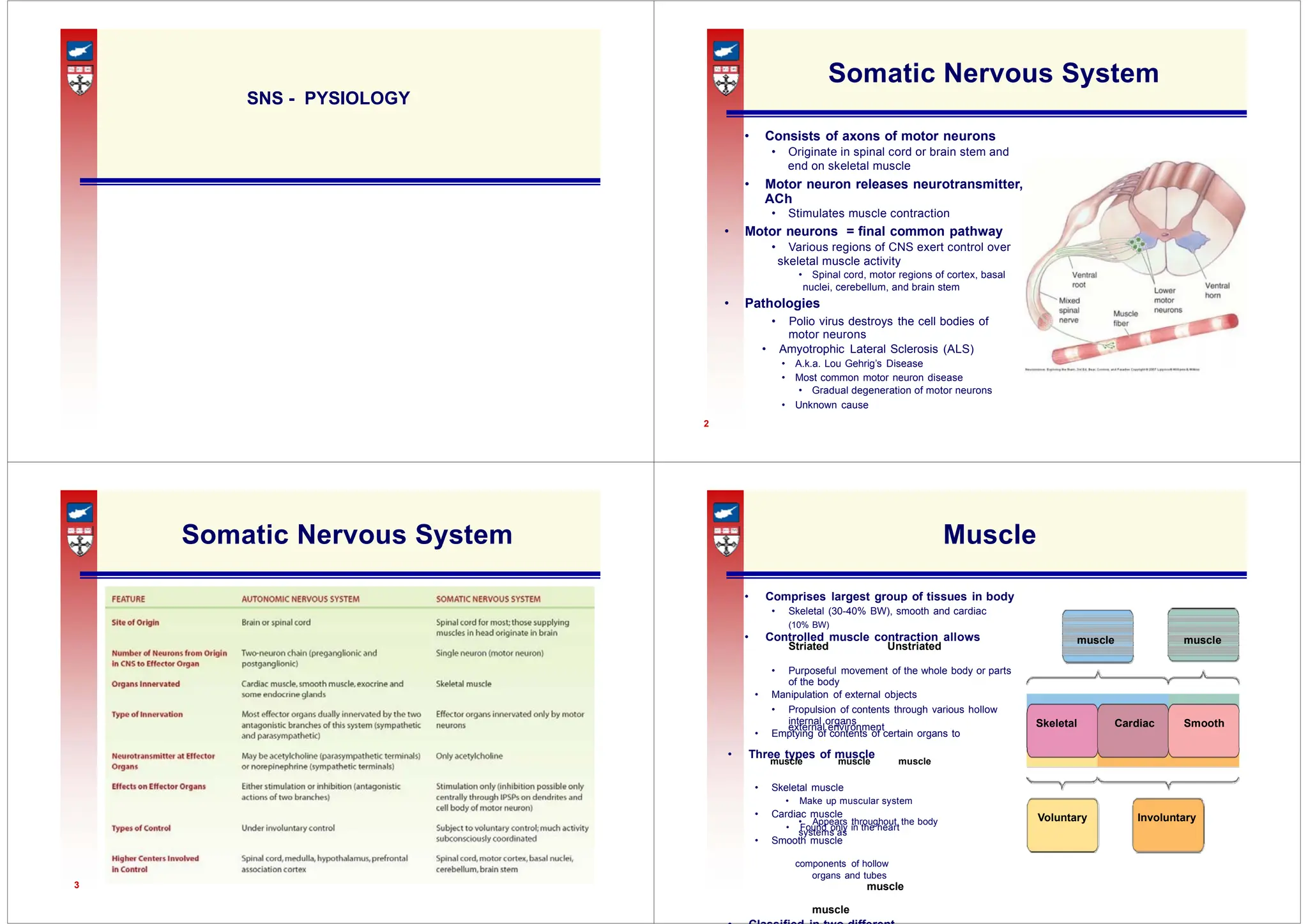
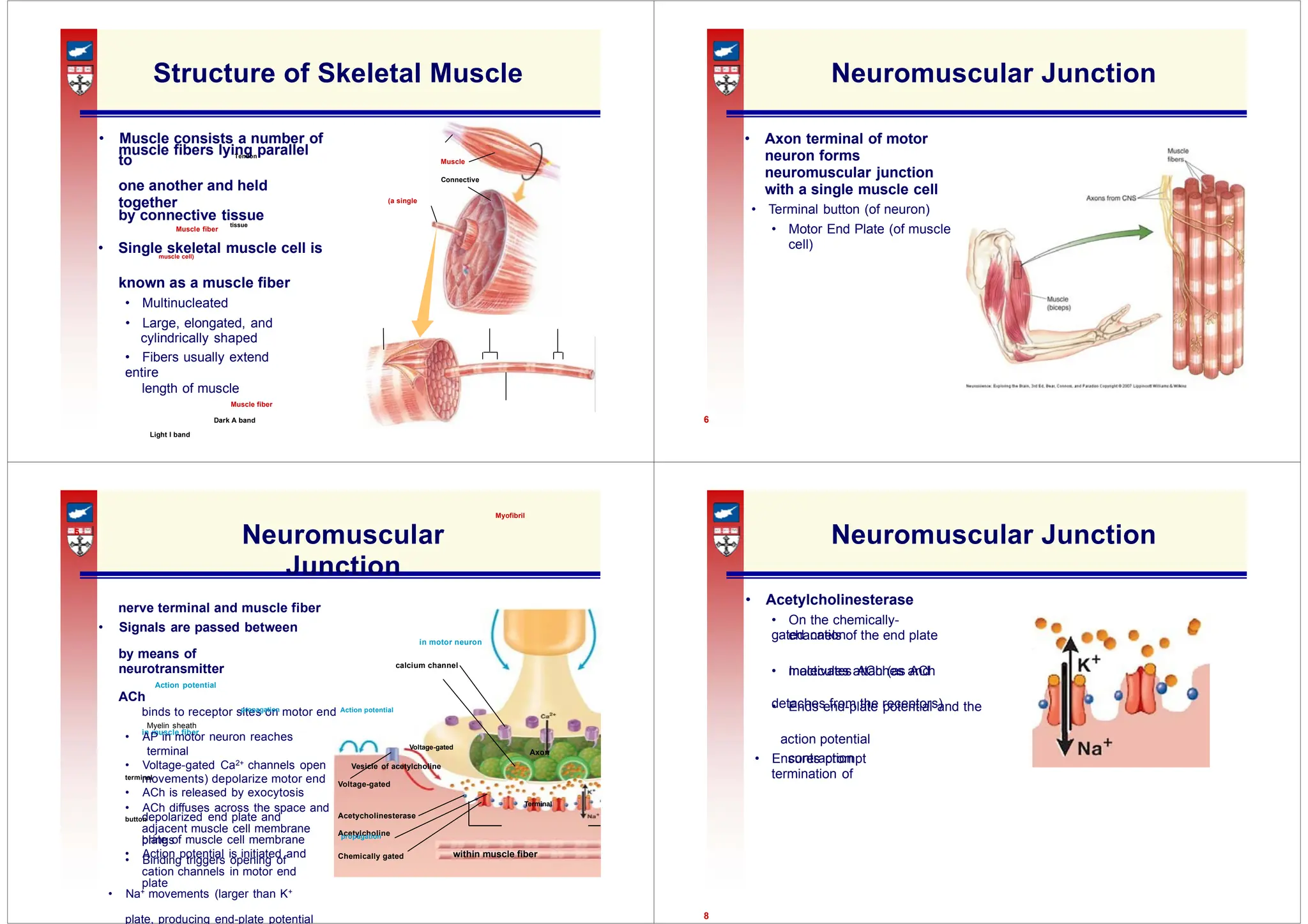
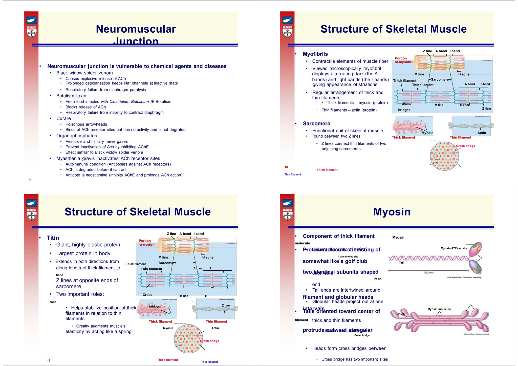
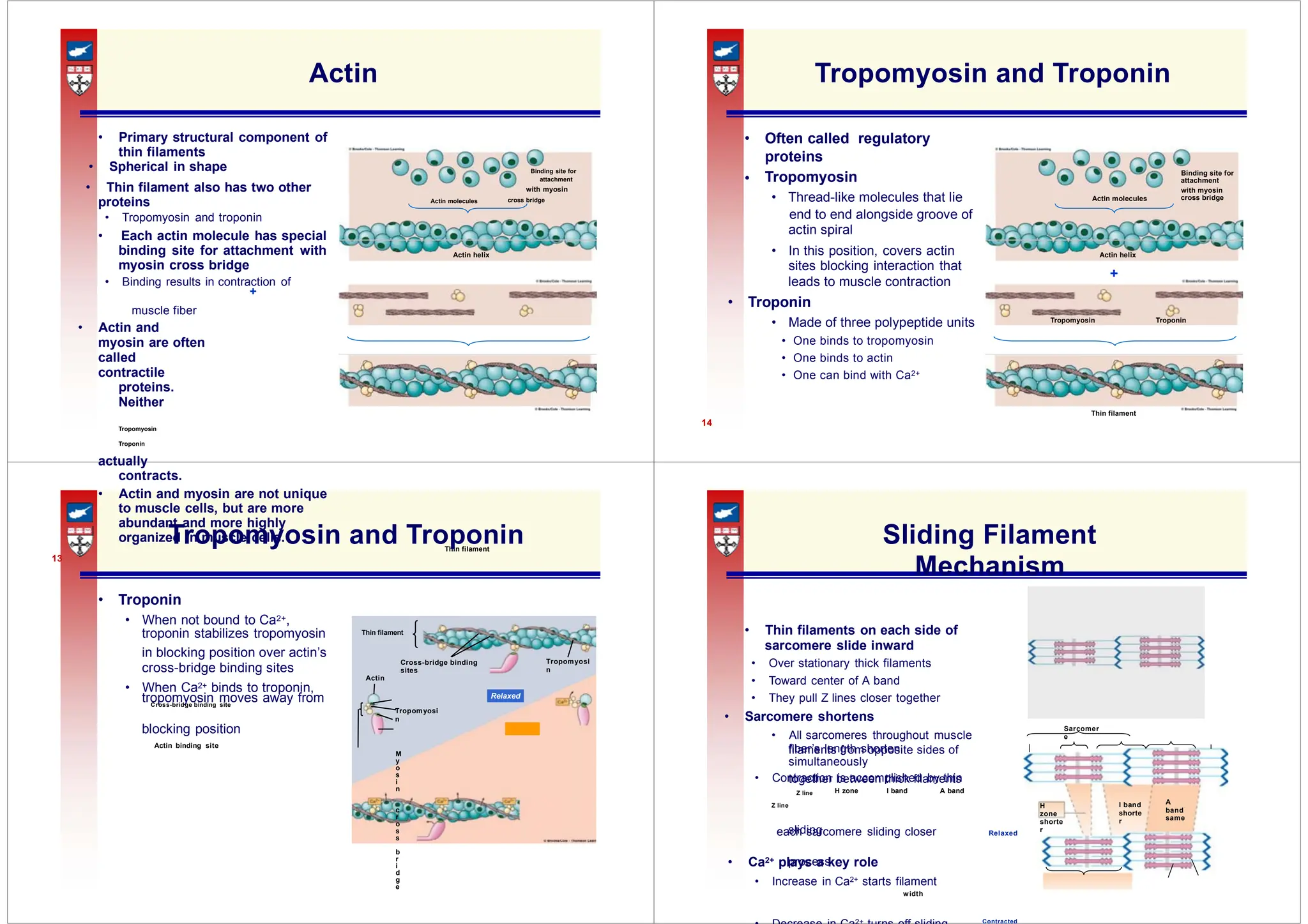
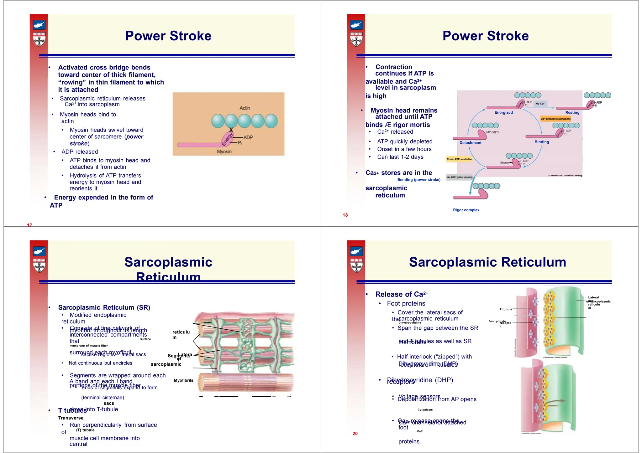
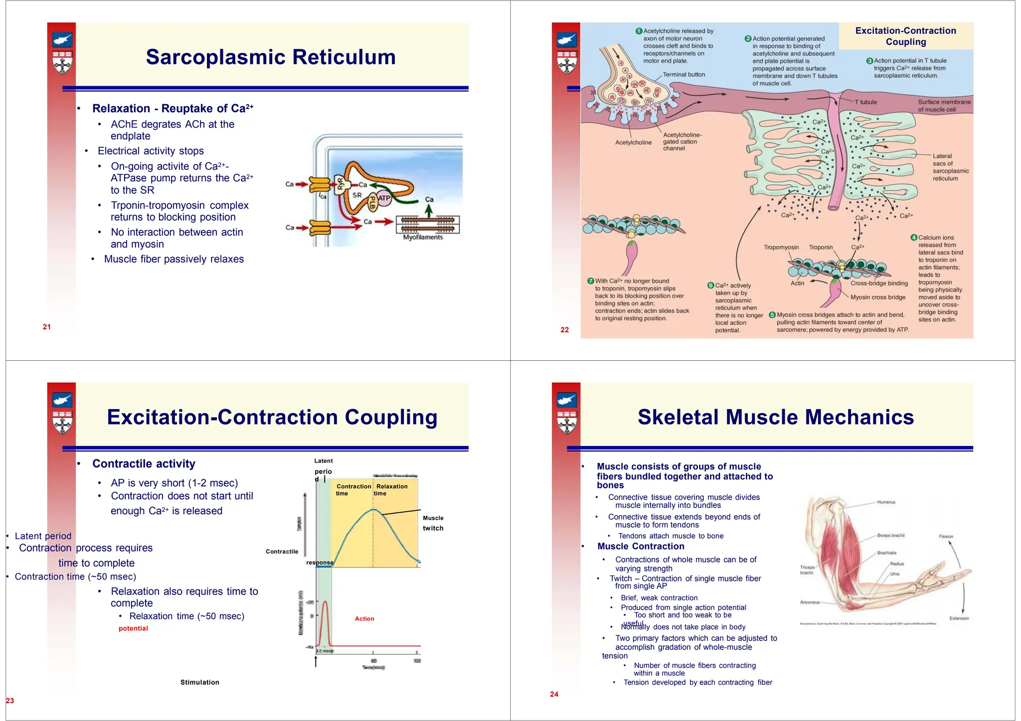
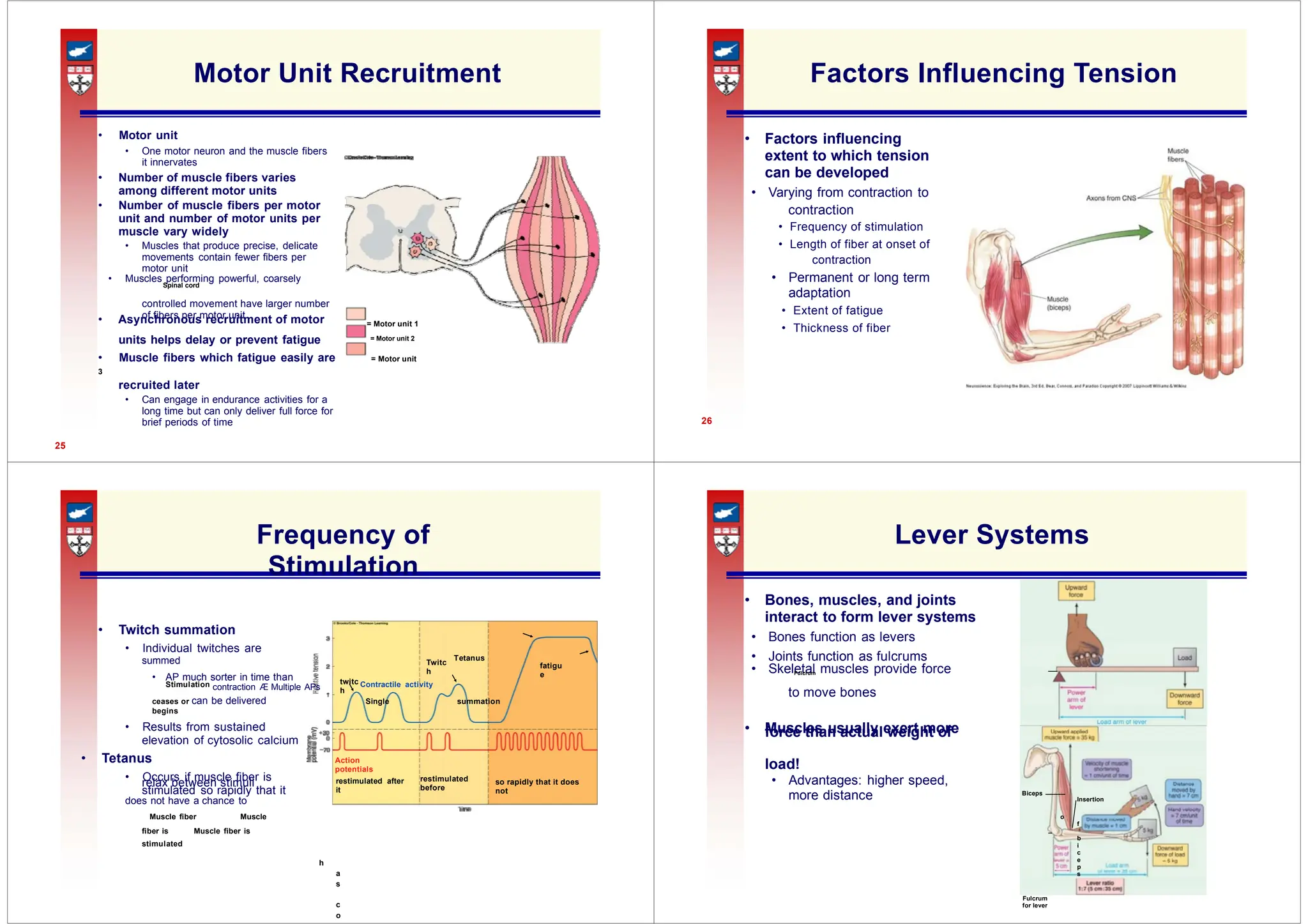
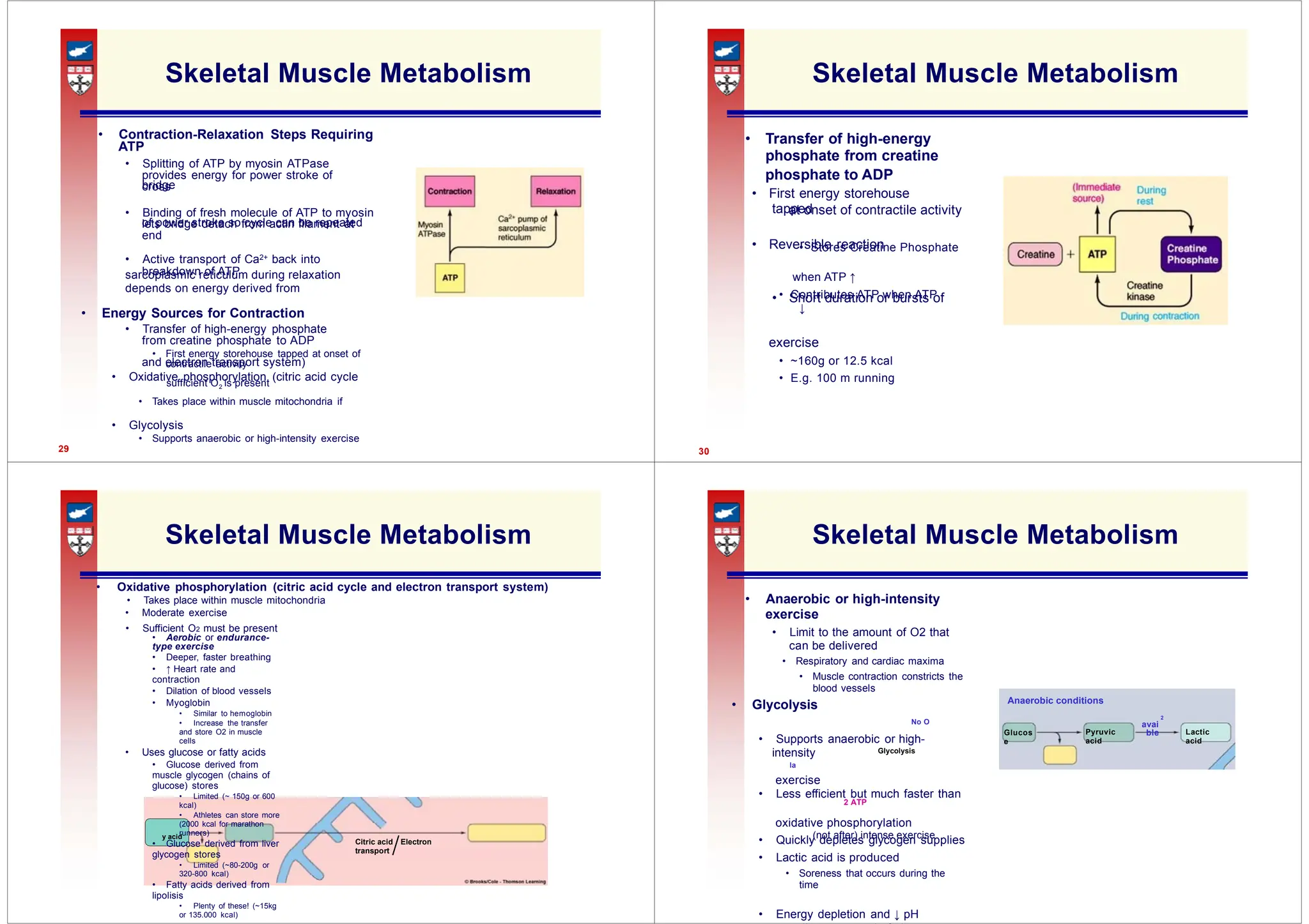
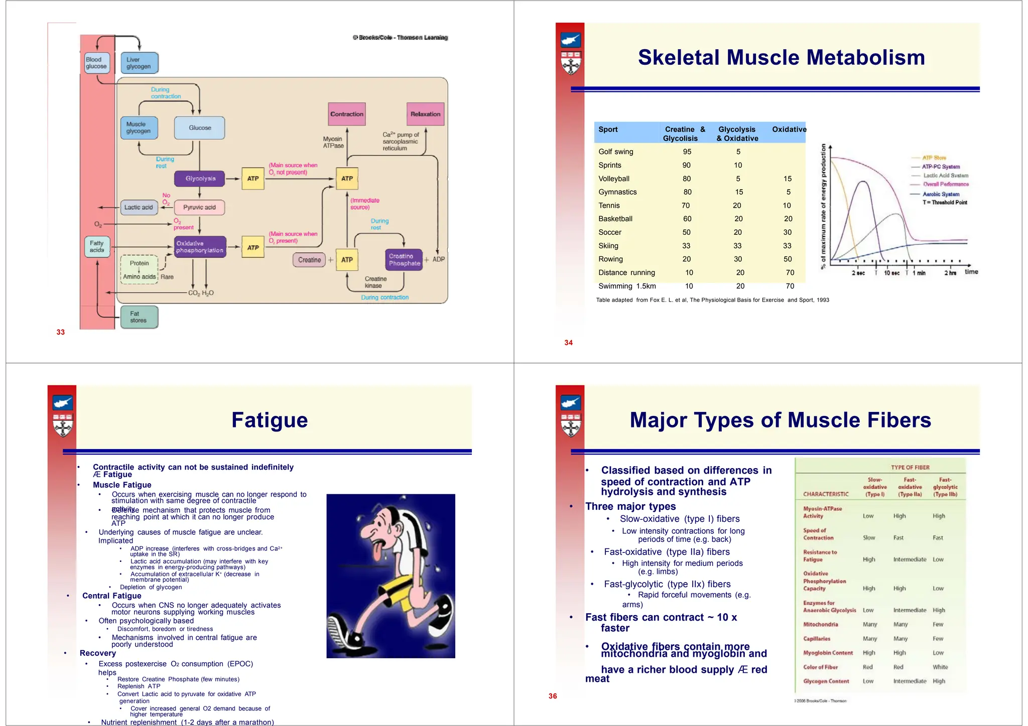
• Originate in spinal cord or brain stem and
• Stimulates muscle contraction
• Various regions of CNS exert control over
• Spinal cord, motor regions of cortex, basal
• Gradual degeneration of motor neurons
• Controlled muscle contraction allows muscle muscle
Skeletal Cardiac Smooth
external environment
Voluntary Involuntary
• Appears throughout the body
systems as
SNS - PYSIOLOGY
Somatic Nervous System
• Consists of axons of motor neurons
end on skeletal muscle
• Motor neuron releases neurotransmitter,
ACh
• Motor neurons = final common pathway
skeletal muscle activity
nuclei, cerebellum, and brain stem
• Pathologies
• Polio virus destroys the cell bodies of
motor neurons
• Amyotrophic Lateral Sclerosis (ALS)
• A.k.a. Lou Gehrig’s Disease
• Most common motor neuron disease
• Unknown cause
2
Somatic Nervous System
3
Muscle
• Comprises largest group of tissues in body
• Skeletal (30-40% BW), smooth and cardiac
(10% BW)
Striated Unstriated
• Purposeful movement of the whole body or parts
of the body
• Manipulation of external objects
• Propulsion of contents through various hollow
internal organs
• Emptying of contents of certain organs to
• Three types of muscle
muscle muscle muscle
• Skeletal muscle
• Make up muscular system
• Cardiac muscle
• Found only in the heart
• Smooth muscle
components of hollow
organs and tubes
muscle
muscle
Muscle
Connective
(a single
nerve terminal and muscle fiber
channels of the end plate
in motor neuron
calcium channel
molecules attaches and
• Ends end-plate potential and the
binds to receptor sites on motor end Action potential
in muscle fiber
contraction
movements) depolarize motor end Voltage-gated
depolarized end plate and
adjacent muscle cell membrane
brings
Acetycholinesterase
Acetylcholine
• Action potential is initiated and within muscle fiber
Chemically gated
Structure of Skeletal Muscle
• Muscle consists a number of
muscle fibers lying parallel
to Tendon
one another and held
together
by connective tissue
Muscle fiber
tissue
• Single skeletal muscle cell is
muscle cell)
known as a muscle fiber
• Multinucleated
• Large, elongated, and
cylindrically shaped
• Fibers usually extend
entire
length of muscle
Muscle fiber
Dark A band
Light I band
Myofibril
5
Neuromuscular Junction
• Axon terminal of motor
neuron forms
neuromuscular junction
with a single muscle cell
• Terminal button (of neuron)
• Motor End Plate (of muscle
cell)
6
Neuromuscular
Junction
• Signals are passed between
by means of
neurotransmitter
Action potential
ACh
propagation
Myelin sheath
• AP in motor neuron reaches
terminal Voltage-gated
Axon
• Voltage-gated Ca2+ channels open Vesicle of acetylcholine
terminal
• ACh is released by exocytosis
• ACh diffuses across the space and Terminal
button
plate of muscle cell membrane propagation
• Binding triggers opening of
cation channels in motor end
plate
• Na+ movements (larger than K+
plate, producing end-plate potential
Neuromuscular Junction
• Acetylcholinesterase
• On the chemically-
gated cation
• Inactivates ACh (as ACh
detaches from the receptors)
action potential
• Ensures prompt
termination of
8
• Myofibrils
• Contractile elements of muscle fiber of myofibril
displays alternating dark (the A
bands) and light bands (the I bands) Sarcomere
Thick filament
thin filaments
Z line
• Thin filaments – actin (protein) bridges
Myosin Actin
• Z lines connect thin filaments of two
Cross bridge
Portion
Myosin ATPase site
• Protein molecule consisting of
somewhat like a golf club Tail
each other
end
filament and globular heads
intervals Myosin molecule
thick and thin filaments
Thick filament Thin filament
critical to contractile process
Neuromuscular
Junction
• Neuromuscular junction is vulnerable to chemical agents and diseases
• Black widow spider venom
• Causes explosive release of ACh
• Prolonged depolarization keeps Na+ channels at inactive state
• Respiratory failure from diaphragm paralysis
• Botulism toxin
• From food infected with Clostridium Botulinum Æ Botulism
• Blocks release of ACh
• Respiratory failure from inability to contract diaphragm
• Curare
• Poisonous arrowheads
• Binds at ACh receptor sites but has no activity and is not degrated
• Organophosphates
• Pesticide and military nerve gases
• Prevent inactivation of Ach by inhibiting AChE
• Effect similar to Black widow spider venom
• Myasthenia gravis inactivates ACh receptor sites
• Autoimmune condition (Antibodies against ACh receptors)
• ACh is degraded before it can act.
• Antidote is neostigmine (inhibits AChE and prolongs ACh action)
9
Structure of Skeletal Muscle
Z line A band I band
Portion
• Viewed microscopically myofibril
M line H zone
giving appearance of striations Thin filament A band I band
• Regular arrangement of thick and
• Thick filaments – myosin (protein) Cross M line H zone
• Sarcomere
• Functional unit of skeletal muscle
• Found between two Z lines Thick filament Thin filament
adjoining sarcomeres
10
Thick filament
Thin filament
Structure of Skeletal Muscle
Z line A band I band
• Titin
• Giant, highly elastic protein of myofibril
• Largest protein in body
M line H zone
• Extends in both directions from Thick filament Sarcomere
along length of thick filament to Thin filament A band I
band
Z lines at opposite ends of
sarcomere
• Two important roles: Cross M line H
zone
• Helps stabilize position of thick bridges Z line
filaments in relation to thin
filaments
• Greatly augments muscle’s Myosin Actin
elasticity by acting like a spring
Cross bridge
11 Thick filament
Thin filament
Myosin
• Component of thick filament Myosin
molecule
• Several hundred of them
Actin binding site
two identical subunits shaped
Heads
• Tail ends are intertwined around
• Globular heads project out at one
• Tails oriented toward center of Thick
filament
protrude outward at regular
Cross bridge
• Heads form cross bridges between
• Cross bridge has two important sites
proteins
Tropomyosin
• attachment
• Thread-like molecules that lie cross bridge
Actin molecules
actin spiral
Actin helix Actin helix
+
leads to muscle contraction
• Made of three polypeptide units Tropomyosin Troponin
Tropomyosi
n
Cross-bridge binding
sites
Tropomyosi
n
Sarcomer
e
filaments from opposite sides of
together between thick filaments
A
band
same
I band
shorte
r
H
zone
shorte
r
sliding
process
Relaxed
Actin
• Primary structural component of
thin filaments
• Spherical in shape Binding site for
attachment
• Thin filament also has two other with myosin
proteins Actin molecules cross bridge
• Tropomyosin and troponin
• Each actin molecule has special
binding site for attachment with
myosin cross bridge
• Binding results in contraction of
+
muscle fiber
• Actin and
myosin are often
called
contractile
proteins.
Neither
Tropomyosin
Troponin
actually
contracts.
• Actin and myosin are not unique
to muscle cells, but are more
abundant and more highly
organized in muscle cells.
Thin filament
13
Tropomyosin and Troponin
• Often called regulatory
Binding site for
with myosin
end to end alongside groove of
• In this position, covers actin
sites blocking interaction that
• Troponin
• One binds to tropomyosin
• One binds to actin
• One can bind with Ca2+
Thin filament
14
Tropomyosin and Troponin
• Troponin
• When not bound to Ca2+,
troponin stabilizes tropomyosin Thin filament
in blocking position over actin’s
cross-bridge binding sites
Actin
• When Ca2+ binds to troponin,
tropomyosin moves away from
Cross-bridge binding site
blocking position
Actin binding site
M
y
o
s
i
n
c
r
o
s
s
b
r
i
d
g
e
Sliding Filament
Mechanism
• Thin filaments on each side of
sarcomere slide inward
• Over stationary thick filaments
• Toward center of A band
• They pull Z lines closer together
• Sarcomere shortens
• All sarcomeres throughout muscle
fiber’s length shorten
simultaneously
• Contraction is accomplished by thin
Z line H zone I band A band
Z line
each sarcomere sliding closer Relaxed
• Ca2+ plays a key role
• Increase in Ca2+ starts filament
width
Contracted
continues if ATP is
level in sarcoplasm
Energized Resting
attached until ATP
Binding
Detachment
Lateral
sac
• Foot proteins
• Cover the lateral sacs of
the
reticulu
m
recepto
r
• Span the gap between the SR
myofibril throughout its length reticulu
m
membrane
saclike regions – lateral sacs Latera
l
receptors on T tubules
receptors
portions of the muscle fiber
• Depolarization from AP opens
down into T-tubule
• Ca2+ release opens the
20
Power Stroke
• Activated cross bridge bends
toward center of thick filament,
“rowing” in thin filament to which
it is attached
• Sarcoplasmic reticulum releases
Ca2+ into sarcoplasm
• Myosin heads bind to
actin
• Myosin heads swivel toward
center of sarcomere (power
stroke)
• ADP released
• ATP binds to myosin head and
detaches it from actin
• Hydrolysis of ATP transfers
energy to myosin head and
reorients it
• Energy expended in the form of
ATP
17
Power Stroke
• Contraction
available and Ca2+
is high
• Myosin head remains
binds Æ rigor mortis
• Ca2+ released
• ATP quickly depleted
• Onset in a few hours
• Can last 1-2 days
• Ca2+ stores are in the
Bending (power stroke)
sarcoplasmic
reticulum
Rigor complex
18
Sarcoplasmic
Reticulum
• Sarcoplasmic Reticulum (SR)
• Modified endoplasmic
reticulum
• Consists of fine network of
interconnected compartments
that Surface
membrane of muscle fiber
surround each myofibril
Segments
of
• Not continuous but encircles sarcoplasmic
• Segments are wrapped around each
A band and each I band Myofibrils
• Ends of segments expand to form
(terminal cisternae)
sacs
• T tubules
Transverse
• Run perpendicularly from surface
of (T) tubule
muscle cell membrane into
central
Sarcoplasmic Reticulum
• Release of Ca2+
of sarcoplasmic
T tubule
sarcoplasmic reticulum
Dihydropyridine
Foot protein
and T tubules as well as SR
• Half interlock (“zipped”) with
Dihydropyridine (DHP)
• Dihydropyridine (DHP)
• Voltage sensors
Cytoplasm
Ca2+ channels of attached
foot
Ca2+
proteins
Excitation-Contraction Coupling
perio
d
bones
muscle internally into bundles
muscle to form tendons
• Muscle Contraction
from single AP
• Too short and too weak to be
useful
Sarcoplasmic Reticulum
• Relaxation - Reuptake of Ca2+
• AChE degrates ACh at the
endplate
• Electrical activity stops
• On-going activite of Ca2+-
ATPase pump returns the Ca2+
to the SR
• Trponin-tropomyosin complex
returns to blocking position
• No interaction between actin
and myosin
• Muscle fiber passively relaxes
21
Excitation-Contraction
Coupling
22
Excitation-Contraction Coupling
• Contractile activity Latent
• AP is very short (1-2 msec) Contraction Relaxation
• Contraction does not start until time time
enough Ca2+ is released
Muscle
twitch
• Latent period
• Contraction process requires Contractile
time to complete response
• Contraction time (~50 msec)
• Relaxation also requires time to
complete
• Relaxation time (~50 msec) Action
potential
Stimulation
23
Skeletal Muscle Mechanics
• Muscle consists of groups of muscle
fibers bundled together and attached to
• Connective tissue covering muscle divides
• Connective tissue extends beyond ends of
• Tendons attach muscle to bone
• Contractions of whole muscle can be of
varying strength
• Twitch – Contraction of single muscle fiber
• Brief, weak contraction
• Produced from single action potential
• Normally does not take place in body
• Two primary factors which can be adjusted to
accomplish gradation of whole-muscle
tension
• Number of muscle fibers contracting
within a muscle
• Tension developed by each contracting fiber
24
contraction
contraction
• Asynchronous recruitment of motor
Twitc
h
fatigu
e
twitc
h to move bones
force than actual weight of
Action
potentials
restimulated
before
relax between stimuli restimulated after
it
so rapidly that it does
not Biceps
Insertion
Motor Unit Recruitment
• Motor unit
• One motor neuron and the muscle fibers
it innervates
• Number of muscle fibers varies
among different motor units
• Number of muscle fibers per motor
unit and number of motor units per
muscle vary widely
• Muscles that produce precise, delicate
movements contain fewer fibers per
motor unit
• Muscles performing powerful, coarsely
Spinal cord
controlled movement have larger number
of fibers per motor unit
= Motor unit 1
units helps delay or prevent fatigue = Motor unit 2
• Muscle fibers which fatigue easily are = Motor unit
3
recruited later
• Can engage in endurance activities for a
long time but can only deliver full force for
brief periods of time
25
Factors Influencing Tension
• Factors influencing
extent to which tension
can be developed
• Varying from contraction to
• Frequency of stimulation
• Length of fiber at onset of
• Permanent or long term
adaptation
• Extent of fatigue
• Thickness of fiber
26
Frequency of
Stimulation
• Twitch summation
• Individual twitches are
summed Tetanus
• AP much sorter in time than
Stimulation contraction Æ Multiple APs Contractile activity
ceases or can be delivered Single summation
begins
• Results from sustained
elevation of cytosolic calcium
• Tetanus
• Occurs if muscle fiber is
stimulated so rapidly that it
does not have a chance to
Muscle fiber Muscle
fiber is Muscle fiber is
stimulated
h
a
s
c
o
Lever Systems
• Bones, muscles, and joints
interact to form lever systems
• Bones function as levers
• Joints function as fulcrums
• Skeletal muscles provide force
Fulcrum
• Muscles usually exert more
load!
• Advantages: higher speed,
more distance
o
f
b
i
c
e
p
s
Fulcrum
for lever
ATP
phosphate to ADP
bridge
at onset of contractile activity
of power stroke so cycle can be repeated
• Stores Creatine Phosphate
breakdown of ATP
• Short duration or bursts of
and electron transport system)
sufficient O2 is present
Anaerobic conditions
2
avai
ble
Pyruvic
acid
Lactic
acid
Glucos
e
(not after) intense exercise
y acid
Citric acid Electron
transport
Skeletal Muscle Metabolism
• Contraction-Relaxation Steps Requiring
• Splitting of ATP by myosin ATPase
provides energy for power stroke of
cross
• Binding of fresh molecule of ATP to myosin
lets bridge detach from actin filament at
end
• Active transport of Ca2+ back into
sarcoplasmic reticulum during relaxation
depends on energy derived from
• Energy Sources for Contraction
• Transfer of high-energy phosphate
from creatine phosphate to ADP
• First energy storehouse tapped at onset of
contractile activity
• Oxidative phosphorylation (citric acid cycle
• Takes place within muscle mitochondria if
• Glycolysis
• Supports anaerobic or high-intensity exercise
29
Skeletal Muscle Metabolism
• Transfer of high-energy
phosphate from creatine
• First energy storehouse
tapped
• Reversible reaction
when ATP ↑
• Contributes ATP when ATP
↓
exercise
• ~160g or 12.5 kcal
• E.g. 100 m running
30
Skeletal Muscle Metabolism
• Oxidative phosphorylation (citric acid cycle and electron transport system)
• Takes place within muscle mitochondria
• Moderate exercise
• Sufficient O2 must be present
• Aerobic or endurance-
type exercise
• Deeper, faster breathing
• ↑ Heart rate and
contraction
• Dilation of blood vessels
• Myoglobin
• Similar to hemoglobin
• Increase the transfer
and store O2 in muscle
cells
• Uses glucose or fatty acids
• Glucose derived from
muscle glycogen (chains of
glucose) stores
• Limited (~ 150g or 600
kcal)
• Athletes can store more
(2000 kcal for marathon
runners)
• Glucose derived from liver
glycogen stores
• Limited (~80-200g or
320-800 kcal)
• Fatty acids derived from
lipolisis
• Plenty of these! (~15kg
or 135.000 kcal)
Skeletal Muscle Metabolism
• Anaerobic or high-intensity
exercise
• Limit to the amount of O2 that
can be delivered
• Respiratory and cardiac maxima
• Muscle contraction constricts the
blood vessels
• Glycolysis
No O
• Supports anaerobic or high-
intensity Glycolysis
la
exercise
• Less efficient but much faster than
2 ATP
oxidative phosphorylation
• Quickly depletes glycogen supplies
• Lactic acid is produced
• Soreness that occurs during the
time
• Energy depletion and ↓ pH
Skeletal Muscle Metabolism
Æ Fatigue
hydrolysis and synthesis
stimulation with same degree of contractile
activity
• Slow-oxidative (type I) fibers
periods of time (e.g. back)
(e.g. limbs)
• Rapid forceful movements (e.g.
faster
mitochondria and myoglobin and
33
Skeletal Muscle Metabolism
Sport Creatine & Glycolysis Oxidative
Glycolisis & Oxidative
Golf swing 95 5
Sprints 90 10
Volleyball 80 5 15
Gymnastics 80 15 5
Tennis 70 20 10
Basketball 60 20 20
Soccer 50 20 30
Skiing 33 33 33
Rowing 20 30 50
Distance running 10 20 70
Swimming 1.5km 10 20 70
Table adapted from Fox E. L. et al, The Physiological Basis for Exercise and Sport, 1993
34
Fatigue
• Contractile activity can not be sustained indefinitely
• Muscle Fatigue
• Occurs when exercising muscle can no longer respond to
• Defense mechanism that protects muscle from
reaching point at which it can no longer produce
ATP
• Underlying causes of muscle fatigue are unclear.
Implicated
• ADP increase (interferes with cross-bridges and Ca2+
uptake in the SR)
• Lactic acid accumulation (may interfere with key
enzymes in energy-producing pathways)
• Accumulation of extracellular K+ (decrease in
membrane potential)
• Depletion of glycogen
• Central Fatigue
• Occurs when CNS no longer adequately activates
motor neurons supplying working muscles
• Often psychologically based
• Discomfort, boredom or tiredness
• Mechanisms involved in central fatigue are
poorly understood
• Recovery
• Excess postexercise O2 consumption (EPOC)
helps
• Restore Creatine Phosphate (few minutes)
• Replenish ATP
• Convert Lactic acid to pyruvate for oxidative ATP
generation
• Cover increased general O2 demand because of
higher temperature
• Nutrient replenishment (1-2 days after a marathon)
Major Types of Muscle Fibers
• Classified based on differences in
speed of contraction and ATP
• Three major types
• Low intensity contractions for long
• Fast-oxidative (type IIa) fibers
• High intensity for medium periods
• Fast-glycolytic (type IIx) fibers
arms)
• Fast fibers can contract ~ 10 x
• Oxidative fibers contain more
have a richer blood supply Æ red
meat
36
• Input to motor-neurons Primary
cortex
interneurons Interneurons
pathway
Efferent
pathway
• Responsible for fine voluntary
movement Response
central pattern generators
Muscle Adaptation & Repair
• Muscle has a high degree of plasticity
• Improvement of oxidative capacity
• From regular aerobic
exercise
• Capillaries and
mitochondria increase
• Hypertrophy
• From anaerobic high
intensity exercise
• Muscle fiber diameter increases (more
actin and myosin)
• Mainly fast-glycolytic fibers
• Testosterone and other steroids increase
the synthesis of actin and myosin
• Steroid abuse
• Fast muscle fibers are interconvertible
• Oxidative ↔ glycolytic
• But NOT fast ↔ slow
• Muscle atrophy
• Disuse atrophy (e.g. space exploration)
• Denervation atrophy (e.g. paralysis)
• Muscle has limited repair cababilities
• Satellite cells can create a few myoblasts
which fuse and create a few muscle fibers
37
Control of Motor Movement
Corticospinal tracts
• Input from afferent neurons motor
• Input from primary motor cortex
• Input from afferent neurons
• Usually through intervening
• Responsible for spinal reflexes
(e.g. withdrawal) Afferent
• Input from the primary motor
cortex
Muscles
• Corticospinal motor system
beginning from the motor cortex
Receptor
Stimulus
38
Control of Motor Movement
• Afferent sensory neuron
provide continues feedback
• Three levels of control and
coordination
• The Segmental Level
• The Projection Level
• The Precommand Level
39
Control of Motor Movement
• Three levels of control and
coordination
• The Segmental Level
• Spinal cord circuits including
(CPGs)
40
purposeful motor activity
specialized muscle fibers
Alpha motor
muscle fibers
connective tissue capsules
non-contractile central portion
endings of afferent
fibers
• Plays key role in stretch
muscle fibers
• Afferent
portion
)
* Efferent neurons to extrafusal fibers
are
44
of intrafusal fiber
of intrafusal
fiber
Control of Motor Movement
• Three levels of control and
coordination
• The Projection Level
• Premotor and Primary motor
cortex
• Plan and execute voluntary
movements
• Brain stem
•
Multineur
al
reflexes
• Regulation of involuntary
control of body posture
41
Control of Motor Movement
• Three levels of control and
coordination
• The Precommand Level
• Cerebellum
• Coordination of movement
• Maintenance of balance
• Control of eye movements
• Basal Nuclei
• Inhibit muscle tone
• Select and maintain
while suppressing unwanted
patterns of movement
• Monitor and coordinate slow
and sustained contractions
42
Muscle Receptors
• Receptors are necessary to
plan and control complicated
movement and balance
• The brain receives information
from all muscles and joints in
the body Æ proprioception
• Two types of muscle
receptors
• Muscle spindles
• Monitor muscle length and
tension
• Golgi tendon organs
• Monitor whole muscle
tension
43
Muscle Receptors
• Muscle Spindles
• Consist of collections of
Capsule
known as intrafusal fibers
• Lie within spindle-shaped neuron axon Intrafusal
(spindle)
parallel to extrafusal fibers Gamma motor
• Have contractile ends and a neuron axon Contractile end portions
• Each spindle has its own
Noncontractile
private nerve supply Secondary (flower-spray) central portion
reflex
• Efferent Primary (annulospiral)
• Gamma motor neurons* Extrafusal (“ordinary”) endings of afferent
fibers
• Primary (annulospiral)
endings (in the
central
• Secondary (flower-
spray) endings (at the
end segments)
and gamma motor
neurons
Extrafusa
l
muscle
fiber
cord muscl
e
stretch
tubes
• Not arranged in sarcomere pattern found
nucleus
within muscle
• Longer than those in skeletal
muscle
• Filaments of intermediate size
supports cell
shape
protein found in Z lines
Muscle Receptors
• Stretch Reflex
• Primary purpose
• Resist tendency for passive stretch of extensor muscles by gravitational
forces when person is standing upright
• Classic example is patellar tendon, or knee-jerk reflex
Extensor muscle of knee Muscle
(quadriceps femoris) spindle
Patellar tendon
Alpha motor
neuron
45
Muscle Receptors
• Coactivation of alpha
skeletal
• Spindle coactivation
during muscle
contraction Spinal
γ Intrafusal
• Spindle contracted to spindle fiber
reduce length α
• With no coactivation
• Slackened spindle
• Not sensitive to
• Adjustment to keep
muscle spindles
sensitive to stretch
46
Muscle Receptors
• Gogli Tendon Organs
• Provide necessary feedback for
overall muscle tension
• Integrates all factors which
influence tension
• Specialized nerve fibers
embedded in the tendons
• Stretch of tendons exerts force
on nerve endings
• Increase firing rate
• Part of this information reaches
conscious awareness
• We are aware of tension (but
not of length) of muscles
47
Smooth Muscle
• Found in walls of hollow organs and
• No striations
• Filaments do not form myofibrils
in skeletal muscle
• Spindle-shaped
cells with single
• Cells usually
arranged in sheets
• Cell has three
types of filaments
• Thick myosin
filaments
Relaxed
• Thin actin
filaments
• Contain
tropomyosin but
lack troponin
• Do not
Pacemaker smooth muscle cell Spontaneous action potential
nervous stimulation for contraction) Action potential spread
to nonpacemaker cell
as single unit
junctions
functional syncytium
Contraction is slow and energy-
• Gap junction smooth muscle cell
filaments Æ muscle
maintains
• Tension can be modified by Mitochondrio
n
neurotransmitt
er
• Ca2+ from the ECF (no
SR)
• Tone Axon
of
Pacemaker
potentials autonomi
c
absence of APs
system
branches
• Does not initiate APs but can
Smoot
h
cell
strength of contraction)
Smooth Muscle
• Two major types
• Multiunit smooth muscle
• Single-unit smooth muscle
• Multiunit Smooth Muscle
• Neurogenic (nerve initiated)
• Consists of discrete units that
function independently of one
another
• Units must be separately
stimulated by nerves to contract
• Found
• In walls of large blood vessels
• In large airways to lungs
• In muscle of eye that adjusts
lens for near or far vision
• In iris of eye
• At base of hair follicles
49
Smooth Muscle
• Single-unit Smooth Muscle
• Most smooth muscle
• Also called visceral smooth
muscleinduced by pacemaker potential
• Self-excitable (does not
require
• Fibers become excited and
contract
• Cells electrically linked by
gap
• Can also be described as a
Nonpacemaker
efficient
• Slow cross-bridge cycling
• Cross-bridges “latch-on” the thin
tension
• Well suited for forming walls of
distensible, hollow organs
50
Smooth Muscle
• Single-unit Smooth Muscle
Excitation
• Myogenic activity
(contraction initiated by the
muscle itself)
• Certain non-contractile cells can
initiate their own APs (self-
excitable)
• Small number distributed at key
locations
• Electrical activity of two types
• Pacemaker potentials
• Cells depolarize and initiate
APs spontaneously and
regularly
• Slow-wave potentials
• Cells depolarize and hyperpoplarize Slow-wave
potentials
rhythmically but not always initiate
APs
• Once initiated the action
potential spreads to the
contractile cells
Smooth Muscle
• Smooth muscle activity
• Gradation
• All muscle fibers are contracting
varying the intracellular Ca2+ Vesicle containing
Varicosity
• Many single-unit smooth muscle postganglionic
cells maintain a low level of neuron
tension (tone) even in the
Varicosities
• Effect of autonomic nervous
• Typically innervated by both
muscle
modify the activity (rate and Neurotransmitter
• Enhancement or
inhibition
• Smooth muscle cells interact with
more than one neurons
52
and smooth muscle
• Fibers are joined in branching
Smooth Muscle
• Smooth muscle activity
• Tension-Length relationship
• Increased tension when
stretched
• Can produce near-maximal
tension at lengths 2.5 x the
normal length
• When stretched, smooth
muscle has the ability to
relax
• These two properties are very
important for hollow organs
• Can accommodate varying
volumes while being able to
produce adequate contractile
force
53
Cardiac Muscle
• Found only in walls of the
heart
• Combines features of
skeletal
• Striated
• Cells are interconnected by gap
junctions
network
• Innervated by autonomic
nervous system
• You will learn more about
cardiac muscle in Cardiac
Physiology
54
THE END
55

somatic nervous system lecture lesson one

  • 1.
    • Originate inspinal cord or brain stem and • Stimulates muscle contraction • Various regions of CNS exert control over • Spinal cord, motor regions of cortex, basal • Gradual degeneration of motor neurons • Controlled muscle contraction allows muscle muscle Skeletal Cardiac Smooth external environment Voluntary Involuntary • Appears throughout the body systems as SNS - PYSIOLOGY Somatic Nervous System • Consists of axons of motor neurons end on skeletal muscle • Motor neuron releases neurotransmitter, ACh • Motor neurons = final common pathway skeletal muscle activity nuclei, cerebellum, and brain stem • Pathologies • Polio virus destroys the cell bodies of motor neurons • Amyotrophic Lateral Sclerosis (ALS) • A.k.a. Lou Gehrig’s Disease • Most common motor neuron disease • Unknown cause 2 Somatic Nervous System 3 Muscle • Comprises largest group of tissues in body • Skeletal (30-40% BW), smooth and cardiac (10% BW) Striated Unstriated • Purposeful movement of the whole body or parts of the body • Manipulation of external objects • Propulsion of contents through various hollow internal organs • Emptying of contents of certain organs to • Three types of muscle muscle muscle muscle • Skeletal muscle • Make up muscular system • Cardiac muscle • Found only in the heart • Smooth muscle components of hollow organs and tubes muscle muscle
  • 2.
    Muscle Connective (a single nerve terminaland muscle fiber channels of the end plate in motor neuron calcium channel molecules attaches and • Ends end-plate potential and the binds to receptor sites on motor end Action potential in muscle fiber contraction movements) depolarize motor end Voltage-gated depolarized end plate and adjacent muscle cell membrane brings Acetycholinesterase Acetylcholine • Action potential is initiated and within muscle fiber Chemically gated Structure of Skeletal Muscle • Muscle consists a number of muscle fibers lying parallel to Tendon one another and held together by connective tissue Muscle fiber tissue • Single skeletal muscle cell is muscle cell) known as a muscle fiber • Multinucleated • Large, elongated, and cylindrically shaped • Fibers usually extend entire length of muscle Muscle fiber Dark A band Light I band Myofibril 5 Neuromuscular Junction • Axon terminal of motor neuron forms neuromuscular junction with a single muscle cell • Terminal button (of neuron) • Motor End Plate (of muscle cell) 6 Neuromuscular Junction • Signals are passed between by means of neurotransmitter Action potential ACh propagation Myelin sheath • AP in motor neuron reaches terminal Voltage-gated Axon • Voltage-gated Ca2+ channels open Vesicle of acetylcholine terminal • ACh is released by exocytosis • ACh diffuses across the space and Terminal button plate of muscle cell membrane propagation • Binding triggers opening of cation channels in motor end plate • Na+ movements (larger than K+ plate, producing end-plate potential Neuromuscular Junction • Acetylcholinesterase • On the chemically- gated cation • Inactivates ACh (as ACh detaches from the receptors) action potential • Ensures prompt termination of 8
  • 3.
    • Myofibrils • Contractileelements of muscle fiber of myofibril displays alternating dark (the A bands) and light bands (the I bands) Sarcomere Thick filament thin filaments Z line • Thin filaments – actin (protein) bridges Myosin Actin • Z lines connect thin filaments of two Cross bridge Portion Myosin ATPase site • Protein molecule consisting of somewhat like a golf club Tail each other end filament and globular heads intervals Myosin molecule thick and thin filaments Thick filament Thin filament critical to contractile process Neuromuscular Junction • Neuromuscular junction is vulnerable to chemical agents and diseases • Black widow spider venom • Causes explosive release of ACh • Prolonged depolarization keeps Na+ channels at inactive state • Respiratory failure from diaphragm paralysis • Botulism toxin • From food infected with Clostridium Botulinum Æ Botulism • Blocks release of ACh • Respiratory failure from inability to contract diaphragm • Curare • Poisonous arrowheads • Binds at ACh receptor sites but has no activity and is not degrated • Organophosphates • Pesticide and military nerve gases • Prevent inactivation of Ach by inhibiting AChE • Effect similar to Black widow spider venom • Myasthenia gravis inactivates ACh receptor sites • Autoimmune condition (Antibodies against ACh receptors) • ACh is degraded before it can act. • Antidote is neostigmine (inhibits AChE and prolongs ACh action) 9 Structure of Skeletal Muscle Z line A band I band Portion • Viewed microscopically myofibril M line H zone giving appearance of striations Thin filament A band I band • Regular arrangement of thick and • Thick filaments – myosin (protein) Cross M line H zone • Sarcomere • Functional unit of skeletal muscle • Found between two Z lines Thick filament Thin filament adjoining sarcomeres 10 Thick filament Thin filament Structure of Skeletal Muscle Z line A band I band • Titin • Giant, highly elastic protein of myofibril • Largest protein in body M line H zone • Extends in both directions from Thick filament Sarcomere along length of thick filament to Thin filament A band I band Z lines at opposite ends of sarcomere • Two important roles: Cross M line H zone • Helps stabilize position of thick bridges Z line filaments in relation to thin filaments • Greatly augments muscle’s Myosin Actin elasticity by acting like a spring Cross bridge 11 Thick filament Thin filament Myosin • Component of thick filament Myosin molecule • Several hundred of them Actin binding site two identical subunits shaped Heads • Tail ends are intertwined around • Globular heads project out at one • Tails oriented toward center of Thick filament protrude outward at regular Cross bridge • Heads form cross bridges between • Cross bridge has two important sites
  • 4.
    proteins Tropomyosin • attachment • Thread-likemolecules that lie cross bridge Actin molecules actin spiral Actin helix Actin helix + leads to muscle contraction • Made of three polypeptide units Tropomyosin Troponin Tropomyosi n Cross-bridge binding sites Tropomyosi n Sarcomer e filaments from opposite sides of together between thick filaments A band same I band shorte r H zone shorte r sliding process Relaxed Actin • Primary structural component of thin filaments • Spherical in shape Binding site for attachment • Thin filament also has two other with myosin proteins Actin molecules cross bridge • Tropomyosin and troponin • Each actin molecule has special binding site for attachment with myosin cross bridge • Binding results in contraction of + muscle fiber • Actin and myosin are often called contractile proteins. Neither Tropomyosin Troponin actually contracts. • Actin and myosin are not unique to muscle cells, but are more abundant and more highly organized in muscle cells. Thin filament 13 Tropomyosin and Troponin • Often called regulatory Binding site for with myosin end to end alongside groove of • In this position, covers actin sites blocking interaction that • Troponin • One binds to tropomyosin • One binds to actin • One can bind with Ca2+ Thin filament 14 Tropomyosin and Troponin • Troponin • When not bound to Ca2+, troponin stabilizes tropomyosin Thin filament in blocking position over actin’s cross-bridge binding sites Actin • When Ca2+ binds to troponin, tropomyosin moves away from Cross-bridge binding site blocking position Actin binding site M y o s i n c r o s s b r i d g e Sliding Filament Mechanism • Thin filaments on each side of sarcomere slide inward • Over stationary thick filaments • Toward center of A band • They pull Z lines closer together • Sarcomere shortens • All sarcomeres throughout muscle fiber’s length shorten simultaneously • Contraction is accomplished by thin Z line H zone I band A band Z line each sarcomere sliding closer Relaxed • Ca2+ plays a key role • Increase in Ca2+ starts filament width Contracted
  • 5.
    continues if ATPis level in sarcoplasm Energized Resting attached until ATP Binding Detachment Lateral sac • Foot proteins • Cover the lateral sacs of the reticulu m recepto r • Span the gap between the SR myofibril throughout its length reticulu m membrane saclike regions – lateral sacs Latera l receptors on T tubules receptors portions of the muscle fiber • Depolarization from AP opens down into T-tubule • Ca2+ release opens the 20 Power Stroke • Activated cross bridge bends toward center of thick filament, “rowing” in thin filament to which it is attached • Sarcoplasmic reticulum releases Ca2+ into sarcoplasm • Myosin heads bind to actin • Myosin heads swivel toward center of sarcomere (power stroke) • ADP released • ATP binds to myosin head and detaches it from actin • Hydrolysis of ATP transfers energy to myosin head and reorients it • Energy expended in the form of ATP 17 Power Stroke • Contraction available and Ca2+ is high • Myosin head remains binds Æ rigor mortis • Ca2+ released • ATP quickly depleted • Onset in a few hours • Can last 1-2 days • Ca2+ stores are in the Bending (power stroke) sarcoplasmic reticulum Rigor complex 18 Sarcoplasmic Reticulum • Sarcoplasmic Reticulum (SR) • Modified endoplasmic reticulum • Consists of fine network of interconnected compartments that Surface membrane of muscle fiber surround each myofibril Segments of • Not continuous but encircles sarcoplasmic • Segments are wrapped around each A band and each I band Myofibrils • Ends of segments expand to form (terminal cisternae) sacs • T tubules Transverse • Run perpendicularly from surface of (T) tubule muscle cell membrane into central Sarcoplasmic Reticulum • Release of Ca2+ of sarcoplasmic T tubule sarcoplasmic reticulum Dihydropyridine Foot protein and T tubules as well as SR • Half interlock (“zipped”) with Dihydropyridine (DHP) • Dihydropyridine (DHP) • Voltage sensors Cytoplasm Ca2+ channels of attached foot Ca2+ proteins
  • 6.
    Excitation-Contraction Coupling perio d bones muscle internallyinto bundles muscle to form tendons • Muscle Contraction from single AP • Too short and too weak to be useful Sarcoplasmic Reticulum • Relaxation - Reuptake of Ca2+ • AChE degrates ACh at the endplate • Electrical activity stops • On-going activite of Ca2+- ATPase pump returns the Ca2+ to the SR • Trponin-tropomyosin complex returns to blocking position • No interaction between actin and myosin • Muscle fiber passively relaxes 21 Excitation-Contraction Coupling 22 Excitation-Contraction Coupling • Contractile activity Latent • AP is very short (1-2 msec) Contraction Relaxation • Contraction does not start until time time enough Ca2+ is released Muscle twitch • Latent period • Contraction process requires Contractile time to complete response • Contraction time (~50 msec) • Relaxation also requires time to complete • Relaxation time (~50 msec) Action potential Stimulation 23 Skeletal Muscle Mechanics • Muscle consists of groups of muscle fibers bundled together and attached to • Connective tissue covering muscle divides • Connective tissue extends beyond ends of • Tendons attach muscle to bone • Contractions of whole muscle can be of varying strength • Twitch – Contraction of single muscle fiber • Brief, weak contraction • Produced from single action potential • Normally does not take place in body • Two primary factors which can be adjusted to accomplish gradation of whole-muscle tension • Number of muscle fibers contracting within a muscle • Tension developed by each contracting fiber 24
  • 7.
    contraction contraction • Asynchronous recruitmentof motor Twitc h fatigu e twitc h to move bones force than actual weight of Action potentials restimulated before relax between stimuli restimulated after it so rapidly that it does not Biceps Insertion Motor Unit Recruitment • Motor unit • One motor neuron and the muscle fibers it innervates • Number of muscle fibers varies among different motor units • Number of muscle fibers per motor unit and number of motor units per muscle vary widely • Muscles that produce precise, delicate movements contain fewer fibers per motor unit • Muscles performing powerful, coarsely Spinal cord controlled movement have larger number of fibers per motor unit = Motor unit 1 units helps delay or prevent fatigue = Motor unit 2 • Muscle fibers which fatigue easily are = Motor unit 3 recruited later • Can engage in endurance activities for a long time but can only deliver full force for brief periods of time 25 Factors Influencing Tension • Factors influencing extent to which tension can be developed • Varying from contraction to • Frequency of stimulation • Length of fiber at onset of • Permanent or long term adaptation • Extent of fatigue • Thickness of fiber 26 Frequency of Stimulation • Twitch summation • Individual twitches are summed Tetanus • AP much sorter in time than Stimulation contraction Æ Multiple APs Contractile activity ceases or can be delivered Single summation begins • Results from sustained elevation of cytosolic calcium • Tetanus • Occurs if muscle fiber is stimulated so rapidly that it does not have a chance to Muscle fiber Muscle fiber is Muscle fiber is stimulated h a s c o Lever Systems • Bones, muscles, and joints interact to form lever systems • Bones function as levers • Joints function as fulcrums • Skeletal muscles provide force Fulcrum • Muscles usually exert more load! • Advantages: higher speed, more distance o f b i c e p s Fulcrum for lever
  • 8.
    ATP phosphate to ADP bridge atonset of contractile activity of power stroke so cycle can be repeated • Stores Creatine Phosphate breakdown of ATP • Short duration or bursts of and electron transport system) sufficient O2 is present Anaerobic conditions 2 avai ble Pyruvic acid Lactic acid Glucos e (not after) intense exercise y acid Citric acid Electron transport Skeletal Muscle Metabolism • Contraction-Relaxation Steps Requiring • Splitting of ATP by myosin ATPase provides energy for power stroke of cross • Binding of fresh molecule of ATP to myosin lets bridge detach from actin filament at end • Active transport of Ca2+ back into sarcoplasmic reticulum during relaxation depends on energy derived from • Energy Sources for Contraction • Transfer of high-energy phosphate from creatine phosphate to ADP • First energy storehouse tapped at onset of contractile activity • Oxidative phosphorylation (citric acid cycle • Takes place within muscle mitochondria if • Glycolysis • Supports anaerobic or high-intensity exercise 29 Skeletal Muscle Metabolism • Transfer of high-energy phosphate from creatine • First energy storehouse tapped • Reversible reaction when ATP ↑ • Contributes ATP when ATP ↓ exercise • ~160g or 12.5 kcal • E.g. 100 m running 30 Skeletal Muscle Metabolism • Oxidative phosphorylation (citric acid cycle and electron transport system) • Takes place within muscle mitochondria • Moderate exercise • Sufficient O2 must be present • Aerobic or endurance- type exercise • Deeper, faster breathing • ↑ Heart rate and contraction • Dilation of blood vessels • Myoglobin • Similar to hemoglobin • Increase the transfer and store O2 in muscle cells • Uses glucose or fatty acids • Glucose derived from muscle glycogen (chains of glucose) stores • Limited (~ 150g or 600 kcal) • Athletes can store more (2000 kcal for marathon runners) • Glucose derived from liver glycogen stores • Limited (~80-200g or 320-800 kcal) • Fatty acids derived from lipolisis • Plenty of these! (~15kg or 135.000 kcal) Skeletal Muscle Metabolism • Anaerobic or high-intensity exercise • Limit to the amount of O2 that can be delivered • Respiratory and cardiac maxima • Muscle contraction constricts the blood vessels • Glycolysis No O • Supports anaerobic or high- intensity Glycolysis la exercise • Less efficient but much faster than 2 ATP oxidative phosphorylation • Quickly depletes glycogen supplies • Lactic acid is produced • Soreness that occurs during the time • Energy depletion and ↓ pH
  • 9.
    Skeletal Muscle Metabolism ÆFatigue hydrolysis and synthesis stimulation with same degree of contractile activity • Slow-oxidative (type I) fibers periods of time (e.g. back) (e.g. limbs) • Rapid forceful movements (e.g. faster mitochondria and myoglobin and 33 Skeletal Muscle Metabolism Sport Creatine & Glycolysis Oxidative Glycolisis & Oxidative Golf swing 95 5 Sprints 90 10 Volleyball 80 5 15 Gymnastics 80 15 5 Tennis 70 20 10 Basketball 60 20 20 Soccer 50 20 30 Skiing 33 33 33 Rowing 20 30 50 Distance running 10 20 70 Swimming 1.5km 10 20 70 Table adapted from Fox E. L. et al, The Physiological Basis for Exercise and Sport, 1993 34 Fatigue • Contractile activity can not be sustained indefinitely • Muscle Fatigue • Occurs when exercising muscle can no longer respond to • Defense mechanism that protects muscle from reaching point at which it can no longer produce ATP • Underlying causes of muscle fatigue are unclear. Implicated • ADP increase (interferes with cross-bridges and Ca2+ uptake in the SR) • Lactic acid accumulation (may interfere with key enzymes in energy-producing pathways) • Accumulation of extracellular K+ (decrease in membrane potential) • Depletion of glycogen • Central Fatigue • Occurs when CNS no longer adequately activates motor neurons supplying working muscles • Often psychologically based • Discomfort, boredom or tiredness • Mechanisms involved in central fatigue are poorly understood • Recovery • Excess postexercise O2 consumption (EPOC) helps • Restore Creatine Phosphate (few minutes) • Replenish ATP • Convert Lactic acid to pyruvate for oxidative ATP generation • Cover increased general O2 demand because of higher temperature • Nutrient replenishment (1-2 days after a marathon) Major Types of Muscle Fibers • Classified based on differences in speed of contraction and ATP • Three major types • Low intensity contractions for long • Fast-oxidative (type IIa) fibers • High intensity for medium periods • Fast-glycolytic (type IIx) fibers arms) • Fast fibers can contract ~ 10 x • Oxidative fibers contain more have a richer blood supply Æ red meat 36
  • 10.
    • Input tomotor-neurons Primary cortex interneurons Interneurons pathway Efferent pathway • Responsible for fine voluntary movement Response central pattern generators Muscle Adaptation & Repair • Muscle has a high degree of plasticity • Improvement of oxidative capacity • From regular aerobic exercise • Capillaries and mitochondria increase • Hypertrophy • From anaerobic high intensity exercise • Muscle fiber diameter increases (more actin and myosin) • Mainly fast-glycolytic fibers • Testosterone and other steroids increase the synthesis of actin and myosin • Steroid abuse • Fast muscle fibers are interconvertible • Oxidative ↔ glycolytic • But NOT fast ↔ slow • Muscle atrophy • Disuse atrophy (e.g. space exploration) • Denervation atrophy (e.g. paralysis) • Muscle has limited repair cababilities • Satellite cells can create a few myoblasts which fuse and create a few muscle fibers 37 Control of Motor Movement Corticospinal tracts • Input from afferent neurons motor • Input from primary motor cortex • Input from afferent neurons • Usually through intervening • Responsible for spinal reflexes (e.g. withdrawal) Afferent • Input from the primary motor cortex Muscles • Corticospinal motor system beginning from the motor cortex Receptor Stimulus 38 Control of Motor Movement • Afferent sensory neuron provide continues feedback • Three levels of control and coordination • The Segmental Level • The Projection Level • The Precommand Level 39 Control of Motor Movement • Three levels of control and coordination • The Segmental Level • Spinal cord circuits including (CPGs) 40
  • 11.
    purposeful motor activity specializedmuscle fibers Alpha motor muscle fibers connective tissue capsules non-contractile central portion endings of afferent fibers • Plays key role in stretch muscle fibers • Afferent portion ) * Efferent neurons to extrafusal fibers are 44 of intrafusal fiber of intrafusal fiber Control of Motor Movement • Three levels of control and coordination • The Projection Level • Premotor and Primary motor cortex • Plan and execute voluntary movements • Brain stem • Multineur al reflexes • Regulation of involuntary control of body posture 41 Control of Motor Movement • Three levels of control and coordination • The Precommand Level • Cerebellum • Coordination of movement • Maintenance of balance • Control of eye movements • Basal Nuclei • Inhibit muscle tone • Select and maintain while suppressing unwanted patterns of movement • Monitor and coordinate slow and sustained contractions 42 Muscle Receptors • Receptors are necessary to plan and control complicated movement and balance • The brain receives information from all muscles and joints in the body Æ proprioception • Two types of muscle receptors • Muscle spindles • Monitor muscle length and tension • Golgi tendon organs • Monitor whole muscle tension 43 Muscle Receptors • Muscle Spindles • Consist of collections of Capsule known as intrafusal fibers • Lie within spindle-shaped neuron axon Intrafusal (spindle) parallel to extrafusal fibers Gamma motor • Have contractile ends and a neuron axon Contractile end portions • Each spindle has its own Noncontractile private nerve supply Secondary (flower-spray) central portion reflex • Efferent Primary (annulospiral) • Gamma motor neurons* Extrafusal (“ordinary”) endings of afferent fibers • Primary (annulospiral) endings (in the central • Secondary (flower- spray) endings (at the end segments)
  • 12.
    and gamma motor neurons Extrafusa l muscle fiber cordmuscl e stretch tubes • Not arranged in sarcomere pattern found nucleus within muscle • Longer than those in skeletal muscle • Filaments of intermediate size supports cell shape protein found in Z lines Muscle Receptors • Stretch Reflex • Primary purpose • Resist tendency for passive stretch of extensor muscles by gravitational forces when person is standing upright • Classic example is patellar tendon, or knee-jerk reflex Extensor muscle of knee Muscle (quadriceps femoris) spindle Patellar tendon Alpha motor neuron 45 Muscle Receptors • Coactivation of alpha skeletal • Spindle coactivation during muscle contraction Spinal γ Intrafusal • Spindle contracted to spindle fiber reduce length α • With no coactivation • Slackened spindle • Not sensitive to • Adjustment to keep muscle spindles sensitive to stretch 46 Muscle Receptors • Gogli Tendon Organs • Provide necessary feedback for overall muscle tension • Integrates all factors which influence tension • Specialized nerve fibers embedded in the tendons • Stretch of tendons exerts force on nerve endings • Increase firing rate • Part of this information reaches conscious awareness • We are aware of tension (but not of length) of muscles 47 Smooth Muscle • Found in walls of hollow organs and • No striations • Filaments do not form myofibrils in skeletal muscle • Spindle-shaped cells with single • Cells usually arranged in sheets • Cell has three types of filaments • Thick myosin filaments Relaxed • Thin actin filaments • Contain tropomyosin but lack troponin • Do not
  • 13.
    Pacemaker smooth musclecell Spontaneous action potential nervous stimulation for contraction) Action potential spread to nonpacemaker cell as single unit junctions functional syncytium Contraction is slow and energy- • Gap junction smooth muscle cell filaments Æ muscle maintains • Tension can be modified by Mitochondrio n neurotransmitt er • Ca2+ from the ECF (no SR) • Tone Axon of Pacemaker potentials autonomi c absence of APs system branches • Does not initiate APs but can Smoot h cell strength of contraction) Smooth Muscle • Two major types • Multiunit smooth muscle • Single-unit smooth muscle • Multiunit Smooth Muscle • Neurogenic (nerve initiated) • Consists of discrete units that function independently of one another • Units must be separately stimulated by nerves to contract • Found • In walls of large blood vessels • In large airways to lungs • In muscle of eye that adjusts lens for near or far vision • In iris of eye • At base of hair follicles 49 Smooth Muscle • Single-unit Smooth Muscle • Most smooth muscle • Also called visceral smooth muscleinduced by pacemaker potential • Self-excitable (does not require • Fibers become excited and contract • Cells electrically linked by gap • Can also be described as a Nonpacemaker efficient • Slow cross-bridge cycling • Cross-bridges “latch-on” the thin tension • Well suited for forming walls of distensible, hollow organs 50 Smooth Muscle • Single-unit Smooth Muscle Excitation • Myogenic activity (contraction initiated by the muscle itself) • Certain non-contractile cells can initiate their own APs (self- excitable) • Small number distributed at key locations • Electrical activity of two types • Pacemaker potentials • Cells depolarize and initiate APs spontaneously and regularly • Slow-wave potentials • Cells depolarize and hyperpoplarize Slow-wave potentials rhythmically but not always initiate APs • Once initiated the action potential spreads to the contractile cells Smooth Muscle • Smooth muscle activity • Gradation • All muscle fibers are contracting varying the intracellular Ca2+ Vesicle containing Varicosity • Many single-unit smooth muscle postganglionic cells maintain a low level of neuron tension (tone) even in the Varicosities • Effect of autonomic nervous • Typically innervated by both muscle modify the activity (rate and Neurotransmitter • Enhancement or inhibition • Smooth muscle cells interact with more than one neurons 52
  • 14.
    and smooth muscle •Fibers are joined in branching Smooth Muscle • Smooth muscle activity • Tension-Length relationship • Increased tension when stretched • Can produce near-maximal tension at lengths 2.5 x the normal length • When stretched, smooth muscle has the ability to relax • These two properties are very important for hollow organs • Can accommodate varying volumes while being able to produce adequate contractile force 53 Cardiac Muscle • Found only in walls of the heart • Combines features of skeletal • Striated • Cells are interconnected by gap junctions network • Innervated by autonomic nervous system • You will learn more about cardiac muscle in Cardiac Physiology 54 THE END 55