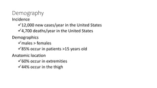
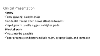
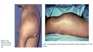
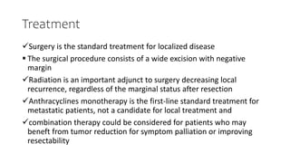
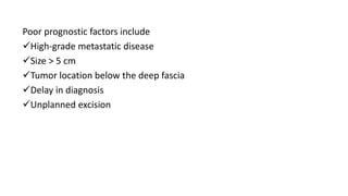
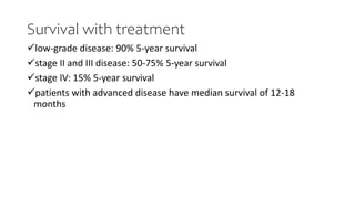
Soft tissue sarcoma
DrSharew Delelegn
Orthopedics&TraumaSurgery Resident
AdamaHospitaMedicalCollege
July-2024
Outline
Introduction
Etiology
Classification
Clinical presentation
Diagnosis & staging
Treatment
Summary
Introduction
Soft-tissue sarcomas (STS) represent a cohort of rare and
heterogeneous tumors that account for 1% of all adult malignancies.
Mesenchymal in origin
The condition usually presents in patients > 15 years old with a slow-
growing, painless soft tissue mass.
Demography
Incidence
12,000 new cases/year in the United States
4,700 deaths/year in the United States
Demographics
males > females
85% occur in patients >15 years old
Anatomic location
60% occur in extremities
44% occur in the thigh
Etiology
Genetics
classic translocations
t(X;18); SYT-SSX fusion synovial sarcoma
t(2:13)rhabdomyosarcoma (alveolar)
t(12;16)(q13:p11)myxoid liposarcoma
Associated conditions
neurofibromatosis type-1malignant peripheral nerve sheath tumor (MPNST)
Stuart-Treves syndrome  angiosarcoma
chronic lymphedema  angiosarcoma
Radiation exposure
Chemical exposure
Chronic inflammation
Previous trauma
Idiopathic
Classification of soft tissue sarcoma
Classification of STS is based on histology, with over 50 types
recognized, all STS have the similar presentation, imaging, differential
diagnosis, and treatment.
Histologic findings and molecular signatures are used to distinguish
individual types.
WHO Classification of STS
Clinical Presentation
History
slow growing, painless mass
 incidental trauma often draws attention to mass
rapid growth usually suggests a higher grade
Physical exam
mass may be palpable
poor prognostic indicators include >5cm, deep to fascia, and immobile
Most sarcomas have a greater tendency to metastasise via blood
vessels to lungs rather than to lymph nodes.
Only 5% of soft tissue sarcomas metastasize to lymph nodes
soft tissue sarcomas that metastasize to lymph nodes (RACES to the
lymph nodes)
 Rhabdomyosarcoma,
 Angiosarcoma,
 Clear cell sarcoma,
 Epithelioid sarcoma,
 Synovial sarcoma
Imaging
Radiographs
 may show soft tissue shadow or mineralization (most commonly with
synovial sarcoma)
CT chest
 obtain to assess for metastatic disease
 an exception is that a CT chest/abdomen/pelvis is indicated for myxoid
liposarcoma
MRI with contrast
 T1: low signal intensity (isointense with muscle)
 T2: high signal intensity
 IV gadolinium: peripheral enhancing zone and non-enhancing necrotic
center
French Federation of cancer centers sarcoma group
AJCC 8th edition
Treatment
Surgery is the standard treatment for localized disease
 The surgical procedure consists of a wide excision with negative
margin
Radiation is an important adjunct to surgery decreasing local
recurrence, regardless of the marginal status after resection
Anthracyclines monotherapy is the first-line standard treatment for
metastatic patients, not a candidate for local treatment and
combination therapy could be considered for patients who may
beneft from tumor reduction for symptom palliation or improving
resectability
Poor prognostic factors include
High-grade metastatic disease
Size > 5 cm
Tumor location below the deep fascia
Delay in diagnosis
Unplanned excision
Survival with treatment
low-grade disease: 90% 5-year survival
stage II and III disease: 50-75% 5-year survival
stage IV: 15% 5-year survival
patients with advanced disease have median survival of 12-18
months
Reference
Upto date
Orthobullet
Basics of Oncology (Frederi (Z-Library).pdf
The 2020 WHO Classification of Soft Tissue Tumours: news and
perspectives DOI:10.32074/1591-951X-213
SEOM Clinical Guideline of management of soft-tissue sarcoma
(2020) https://doi.org/10.1007/s12094-020-02534-0
Soft tissue sarcomas in adults and children: A comparison , Walter
Lawrence, Jr., MD
THANK YOU !

Soft tissue sarcoma ,Dr Sharew Delelegn

  • 1.
    Soft tissue sarcoma DrSharewDelelegn Orthopedics&TraumaSurgery Resident AdamaHospitaMedicalCollege July-2024
  • 2.
  • 3.
    Introduction Soft-tissue sarcomas (STS)represent a cohort of rare and heterogeneous tumors that account for 1% of all adult malignancies. Mesenchymal in origin The condition usually presents in patients > 15 years old with a slow- growing, painless soft tissue mass.
  • 4.
    Demography Incidence 12,000 new cases/yearin the United States 4,700 deaths/year in the United States Demographics males > females 85% occur in patients >15 years old Anatomic location 60% occur in extremities 44% occur in the thigh
  • 6.
    Etiology Genetics classic translocations t(X;18); SYT-SSXfusion synovial sarcoma t(2:13)rhabdomyosarcoma (alveolar) t(12;16)(q13:p11)myxoid liposarcoma Associated conditions neurofibromatosis type-1malignant peripheral nerve sheath tumor (MPNST) Stuart-Treves syndrome  angiosarcoma chronic lymphedema  angiosarcoma Radiation exposure Chemical exposure Chronic inflammation Previous trauma Idiopathic
  • 7.
    Classification of softtissue sarcoma Classification of STS is based on histology, with over 50 types recognized, all STS have the similar presentation, imaging, differential diagnosis, and treatment. Histologic findings and molecular signatures are used to distinguish individual types.
  • 8.
  • 11.
    Clinical Presentation History slow growing,painless mass  incidental trauma often draws attention to mass rapid growth usually suggests a higher grade Physical exam mass may be palpable poor prognostic indicators include >5cm, deep to fascia, and immobile
  • 14.
    Most sarcomas havea greater tendency to metastasise via blood vessels to lungs rather than to lymph nodes. Only 5% of soft tissue sarcomas metastasize to lymph nodes soft tissue sarcomas that metastasize to lymph nodes (RACES to the lymph nodes)  Rhabdomyosarcoma,  Angiosarcoma,  Clear cell sarcoma,  Epithelioid sarcoma,  Synovial sarcoma
  • 15.
    Imaging Radiographs  may showsoft tissue shadow or mineralization (most commonly with synovial sarcoma) CT chest  obtain to assess for metastatic disease  an exception is that a CT chest/abdomen/pelvis is indicated for myxoid liposarcoma MRI with contrast  T1: low signal intensity (isointense with muscle)  T2: high signal intensity  IV gadolinium: peripheral enhancing zone and non-enhancing necrotic center
  • 17.
    French Federation ofcancer centers sarcoma group
  • 18.
  • 19.
    Treatment Surgery is thestandard treatment for localized disease  The surgical procedure consists of a wide excision with negative margin Radiation is an important adjunct to surgery decreasing local recurrence, regardless of the marginal status after resection Anthracyclines monotherapy is the first-line standard treatment for metastatic patients, not a candidate for local treatment and combination therapy could be considered for patients who may beneft from tumor reduction for symptom palliation or improving resectability
  • 21.
    Poor prognostic factorsinclude High-grade metastatic disease Size > 5 cm Tumor location below the deep fascia Delay in diagnosis Unplanned excision
  • 22.
    Survival with treatment low-gradedisease: 90% 5-year survival stage II and III disease: 50-75% 5-year survival stage IV: 15% 5-year survival patients with advanced disease have median survival of 12-18 months
  • 23.
    Reference Upto date Orthobullet Basics ofOncology (Frederi (Z-Library).pdf The 2020 WHO Classification of Soft Tissue Tumours: news and perspectives DOI:10.32074/1591-951X-213 SEOM Clinical Guideline of management of soft-tissue sarcoma (2020) https://doi.org/10.1007/s12094-020-02534-0 Soft tissue sarcomas in adults and children: A comparison , Walter Lawrence, Jr., MD
  • 24.