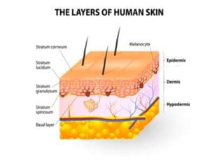
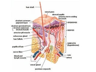
This document discusses transdermal drug delivery systems. It defines transdermal delivery as delivering drugs through the skin into systemic circulation at predetermined rates over prolonged periods. The key advantages are avoiding gastrointestinal degradation and first-pass metabolism, providing controlled drug levels, and increasing patient compliance. The document covers skin anatomy, permeation pathways, factors influencing permeation like drug properties, and technologies used to develop transdermal patches.

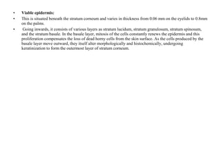
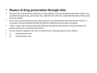

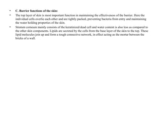
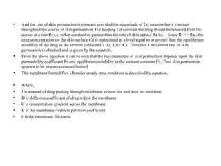
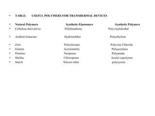
![• Various methods for preparation of transdermal drug delivery systems :
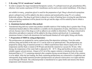
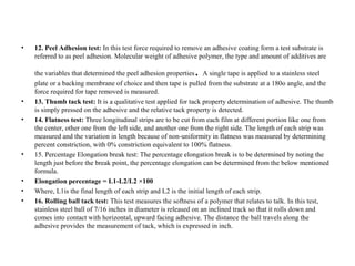
• 1. Asymmetric TPX membrane method:
• A prototype patch can be fabricated by a heat sealable polyester film (type 1009, 3m) with a concave of
1cm diameter used as the backing membrane. Drug sample is dispensed into the concave membrane,
covered by a TPX {poly (4-methyl-1-pentene)} asymmetric membrane, and sealed by adhesive.
• 1.1 Asymmetric TPX membrane preparation
• These are fabricated by using the dry/wet inversion process. TPX is dissolved in a mixture of solvent
(cyclohexane) and nonsolvent additives at 60°c to form a polymer solution. The polymer solution is kept at
40°C for 24 hrs and cast on a glass plate to a pre-determined thickness with a gardener knife. After that the
casting film is evaporated at 50°C for 30 sec, then the glass plate is to be immersed immediately in
coagulation bath [maintained the temperature at 25°C]. After 10 minutes of immersion, the membrane can
be removed, air dry in a circulation oven at 50°C for 12 hrs].](https://image.slidesharecdn.com/shubhtdds-190404141435/85/Shubh-tdds-54-320.jpg)
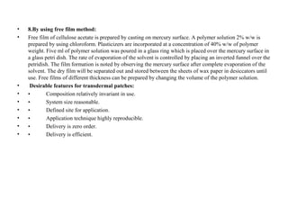
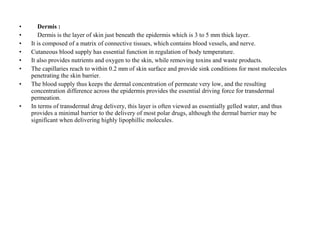
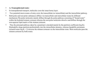
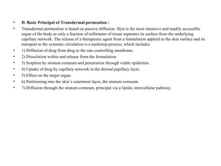
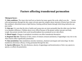
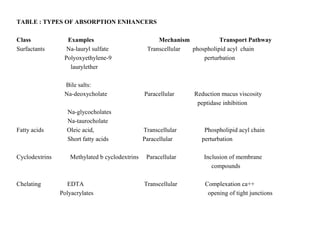
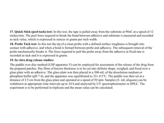
![• Evaluation parameters :
• Interaction studies: Excipients are integral components of almost all pharmaceutical dosage forms. The stability of a
formulation amongst other factors depends on the compatibility of the drug with the excipients. The drug and the
excipients must be compatible with one another to produce a product that is stable, thus it is mandatory to detect any
possible physical or chemical interaction as it can affect the bioavailability and stability of the drug. If the excipients
are new and have not been used in formulations containing the active substance, the compatibility studies play an
important role in formulation development. Interaction studies are commonly carried out in Thermal analysis, FT-IR,
UV and chromatographic techniques by comparing their physicochemical characters such as assay, melting
endotherms, characteristic wave numbers, absorption maxima etc.,
• 2. Thickness of the patch: The thickness of the drug loaded patch is measured in different points by using a digital
micrometer and determines the average thickness and standard deviation for the same to ensure the thickness of the
prepared patch.
• 3. Weight uniformity: The prepared patches are to be dried at 60°c for 4hrs before testing. A specified area of patch
is to be cut in different parts of the patch and weigh in digital balance. The average weight and standard deviation
values are to be calculated from the individual weights.
• 4. Folding endurance: A strip of specific are is to be cut evenly and repeatedly folded at the same place till it broke.
The number of times the film could be folded at the same place without breaking gave the value of the folding
endurance.
• 5. Percentage Moisture content: The prepared films are to be weighed individually and to be kept in a desiccator
containing fused calcium chloride at room temperature for 24 hrs. After 24 hrs the films are to be reweighed and
determine the percentage moisture content from the below mentioned formula.
• Percentage moisture content = [Initial weight- Final weight/ Final weight] ×100.](https://image.slidesharecdn.com/shubhtdds-190404141435/85/Shubh-tdds-59-320.jpg)
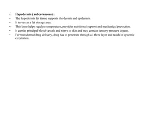
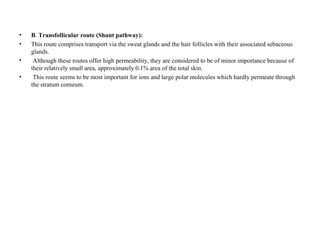
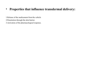
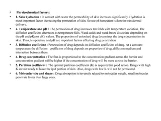
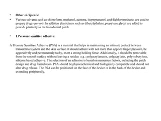
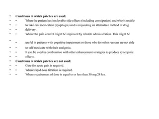
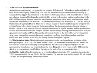
![• 6. Percentage Moisture uptake: The weighed films are to be kept in a desiccator at room
temperature for 24 hrs containing saturated solution of potassium chloride in order to maintain
84% RH. After 24 hrs the films are to be reweighed and determine the percentage moisture
uptake from the below mentioned formula.
• Percentage moisture uptake = [Final weight- Initial weight/ initial weight] ×100.
• 7. Water vapour permeability (WVP) evaluation: Water vapour permeability can be determined
with foam dressing method the air forced oven is replaced by a natural air circulation oven. The
WVP can be determined by the following formula
• WVP=W/A
• Where, WVP is expressed in gm/m2 per 24hrs, W is the amount of vapour permeated through
the patch expressed in gm/24hrs and A is the surface area of the exposure samples expressed in
m2
• 8. Drug content: A specified area of patch is to be dissolved in a suitable solvent in specific
volume. Then the solution is to be filtered through a filter medium and analyse the drug contain
with the suitable method (UV or HPLC technique). Each value represents average of three
different samples.](https://image.slidesharecdn.com/shubhtdds-190404141435/85/Shubh-tdds-60-320.jpg)


![How Big Brands are Taking Your Traffic in Alberta [Data Inside].pptx](https://cdn.slidesharecdn.com/ss_thumbnails/howbigbrandsaretakingyourtrafficinalbertadatainside-260123180142-42d276f3-thumbnail.jpg?width=640&height=640&fit=bounds)