

![[Final] Purification Of B-Gal Formal Report](https://cdn.slidesharecdn.com/ss_thumbnails/3fffdfc3-0490-430c-8d52-b8b026281187-150402132241-conversion-gate01-thumbnail.jpg?width=640&height=640&fit=bounds)


![[13386905 nova biotechnologica et chimica] phage endolysin a way to unders...](https://cdn.slidesharecdn.com/ss_thumbnails/13386905-novabiotechnologicaetchimicaphageendolysinawaytounderstandabindingfunctionofc-terminaldomai-210118083506-thumbnail.jpg?width=640&height=640&fit=bounds)


Sgm Poster Final
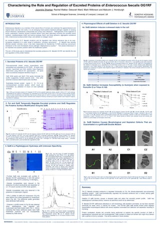
- 1. Characterising the Roleand Regulation of Excreted Proteins of EnterococcusfaecalisOG1RFJayendra Shankar, Rachel Walker, Deborah Ward, Mark Wilkinson and Malcolm J. HorsburghSchool of Biological Sciences, University of Liverpool, Liverpool, UKDOROTHY HODGKINSPOSTGRADUATE AWARDS4. Physiological Effects of salBDeletion in E. faecalis OG1RFINTRODUCTIONEnterococcusfaecalisis is a member of the natural floraof humans and colonises the gastrointestinal and vaginal tracts, and the oral cavity. It is an emerging Gram-positive bacterial pathogen capable of causing wound infections, bacteraemia, endocarditis and urinary tract infections1. Pathogenesis of this organism is poorly understood, however several virulence factors have been described including the excreted toxins cytolysin, metalloprotease (GelE) and serine protease (SprE) and three adhesins: aggregation substance (AS), enterococcal surface protein (Esp), and adhesin to collagen (Ace)2. An increased study of E. faecalis virulence and its regulation has clinical relevance due to its ever-increasing resistance to a wide range of antibiotics, including vancomycin3 . E. faecaliswas recently afforded greater notoriety, since it has been responsible for transferring Tn1546 conferring vancomycin resistance to the more formidable clinical pathogen Staphylococcus aureus4 . This places enterococci in an important and dynamic position within the healthcare system. The aim of this study was to characterise the excreted proteome of E. faecalisOG1RF and identify the role and regulation of this protein fraction.4a. SalB deletion induces a stressed state in the cellpH4pH7pH4pH7ABFig 5A: Comparison of excreted proteins of E. faecalis OG1RF (Cy3 labelled blue-green spots, 80 μg) and its isogenic mutant OG1RF salB (Cy5 labelled red spots, 80 μg) by 2D DIGE after 8 h of growth in BHI at 37°C. Cy2 labelled purple spots represent a pooled internal control (40 μg + 40 μg). Fig 5B: 500 μg of secreted proteins of E. faecalis OG1RF salB at 8 h precipitated by the method of Chaussee et. al., separated by 2D SDS-PAGE across a pH 4-7 IPG strip. Arrows indicate spots differentially expressed in the mutant relative to the wild-type (Fig 1). Black arrows indicate proteins to which putative identification was assigned by comparing its peptide mass fingerprint against the MASCOT database. Red arrows indicate proteins for which no identity was assigned.1. Secreted Proteins of E. faecalis OG1RFPost-exponential phase culture supernatants were precipitated and separated by 2D PAGE. 59 spots were excised,trypsinised and their peptide mass fingerprint determined using MALDI-ToF spectrometry. 44 spots were successfully assigned identities (Figure 1).GelE (9/44 spots) and SprE (7/44 spots) dominate the profile, suggesting multiple species exist. Few other potential virulence determinants were identified.Sulphatase domain protein (spots 11, 12, 13) is an uncharacterised excreted protein, which could aid colonisation via utilisation of mucin on host epithelial surfacesTable 1: Stress related proteins expressed by OG1RF salB4b. SalB Deletion Increases Susceptibility to Autolysis when exposed to Penicillin G or Triton X-100.pH4pH7Fig 1: 500 μg of excreted proteins of E. faecalisOG1RF at 8h of growth (stationary phase, growth in BHI at 37°C with shaking) precipitated by the method of Chaussee et. al.5separated by 2D SDS PAGE across a pH 4-7 IPG strip. Arrows indicate excised spots on which identification was identified. Red arrows indicate spots for which no putative identification was assigned. Black arrows indicate spots to which putative identities were assigned following comparing its tryptic peptide mass fingerprint against the MASCOT database.AB2. Fsr and GelE Temporally Regulate Excreted proteins and GelE Regulates the Putative Surface Modification Enzyme SalB.ABFig 6: Autolysis assays for E. faecalis OG1RF () and its isogenic mutants OG1RF gelE (), OG1RF fsrB (), OG1RF salB (▲), and double mutants OG1RF gelE/salB () and OG1RF fsrB/salB () conducted under 0.1mgml-1 Penicillin G (A) or 0.1% (v/v) Triton X-100 (B) induced stress. Exponentially growing cells were harvested, washed in PBS and resuspended in PBS containing 0.1mgml-1 Penicillin G or 0.1% v/v Triton X 100. Optical density at 600nm was collected hourly and turbidity was plotted as a percentage survival graph versus time.*4c. SalB Deletion Causes Morphological and Septation Defects That are Exacerbated in a gelE/salBDouble Mutant.Fig 2 A: Growth of E. faecalis OG1RF and the isogenic mutants OG1RF fsrB and OG1RF gelE over 8 hours cultured in BHI at 37°C with shaking (150 rpm). Error bars represent SEM from a triplicate experiment. Fig 2B: Protein profiles of E. faecalis OG1RF and the isogenic mutants OG1RF fsrB and gelE collected over growth and separated by SDS-PAGE (11%). 3h, 5h and 8h correspond to time points on Fig 2A indicated by arrows. ON = overnight; M = Bio-Rad broad range marker. * indicates SalB, a secreted protein, post-translationally regulated by GelE.3. SalB is a PeptidoglycanHydrolase with Unknown Specificity.A1A2C1C2Fig 3: 5μg, 1μg and 0.5μg of purified His-tagged SalB separated by SDS PAGE (11%,) (A)stained with coommassie brilliant blue. A corresponding 11% gel supplemented with purified peptidoglycan from E. faecalis OG1RF (B) was treated overnight in renaturing buffer (0.1% v/v Triton X-100, 10 mM MgCl2, 25 mM Tris-HCl pH 7.5) and stained with 0.1% w/v Methylene blue in 0.01% w/v KOH. After staining, the gel was washed in distilled water repeatedly until zones of clearing were observed. Zones of clearing indicate peptidoglycan hydrolytic activity of SalB. M = Promega prestained marker.ABB1B2D1D2Purified SalB was incubated with purified E. faecalis OG1RF peptidoglycan in 200 mM sodium phosphate buffer (pH 6) containing 10 mM MgCl2.