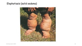
Session 11: Blood Borne
Helminths –
WUCHERELIA
BANCROFTI
Learning Objectives
By the end of this session, students are expected to be able to:
• Define Wucherelia bancrofti
• Describe characteristics of Wucherelia bancrofti
• Describe life cycle of Wucherelia bancrofti
• Explain medical importance of Wucherelia bancrofti
Saturday, July 13, 2024 2
Overview of Wuchereria Bancrofti
• Wuchereria bancrofti is the blood borne helminth that
belongs to the superfamily Filarioidea of the phylum
nematoda.
• Filarioidea group include spilurate filliform nematodes
adapted in inhabiting the deeper tissues like the circulatory,
lymphatic, and connective tissue layers.
• Wuchereria bancrofti is responsible for causing bancroftian
filariasis.
Saturday, July 13, 2024 3
Habitats
• Found in tropics and subtropics, chiefly in Asia and Africa
• Found coiled in lymphatic glands or lying in lymphatic vessels
• Microfilaria: Occur in lymphatic vessels and in the
peripheral blood, normally at night
• Infective larvae: Are found in the gut and muscles of
certain species of mosquitoes which have taken up infected
blood, later infective larvae are present in their mouth
parts
Saturday, July 13, 2024 4
Characteristics of Wucherelia Bancrofti
• Thread like white worms found in the lymphatic vessels and
glands.
• It contains short, thick proximal and a whip like distal
portion ending in a hook shape
• The egg lies in the upper uterus enclosed in a chorionic
membrane.
• The chorionic membrane becomes a sheath for the living
embryos after the egg hatch into a microfilaria.
• Wuchereria bancrofti are viviparous (reproduce by giving
birth to young living microfilaria).
• Female adult measures 80-100 x 0.25 mm and the male
measures 40 x 0.1 mm.
Saturday, July 13, 2024 5
• Adults of both sexes lie tightly coiled in the nodular
dilatations of the lymphatic vessels and sinuses of the lymph
nodes while the microfilariae live in the blood and lymph.
• The length of life span in man is considered to be 5 years.
• Microfilaria circulate at night (nocturnal periodicity), when
their mosquito vector is most likely to bite.
• Decreased peripheral temperature may attract more
microfilariae to circulate to peripheral blood.
Saturday, July 13, 2024 6
Figure 1: Morphological Feature of W. Bancrofti
Microfilaria
Source: Beye & Lawless, 2004
Saturday, July 13, 2024 7
NB:
Microfilariae:
• Measures 225 – 300 x 10 µm
• Sheathed unstained or stained pinkish – mauve
• Few large curves
• Nuclei round and separate
• Tail pointed and no nuclei in the tip
• Periodic: Are nocturnal and diurnal
Saturday, July 13, 2024 8
Mode of Transmission
• Man is the only known definitive host
• Filariasis is spread from infected persons to uninfected
persons through mosquitoes bites
• Mosquitos involved include
- Culex spp
- Mansonia spp
- Aedes spp
- Anopheles spp
• In Africa, the vectors are mainly Culex spp in urban areas
and mainly Anopheles spp in rural areas
Saturday, July 13, 2024 9
Life cycle
• Infective larvae are transmitted by infected biting
arthropods during a blood meal.
• After penetrating the skin through the bite would, the
larvae pass into lymphatic vessels and nodes where they grow
to maturity. This takes about a year.
• The adult worms tend to inhabit the varices of the
lymphatic vessels of the lower limbs, the groin glands and
epididymis in the male, and the labial glands in the female
leading to filariasis.
• The adult females release large numbers of very small
sheathed worm larvae (microfilariae), which circulate in an
infected person's bloodstream.
Saturday, July 13, 2024 10
 When the person is bitten by a mosquito, the mosquito can
ingest the microfilariae.
• In the mosquito the microfilariae shed the sheath and
migrate into muscles of the mosquito where they develop
from L1 to L3 stages of development. This takes about 6 to
20 days.
• After 6 to 20 days of development, the larvae force their
way out of the muscles, causing considerable damage and
migrate to the proboscis.
• During the blood meal, the developed larvae (L3) emerge
from the proboscis into the skin of man and the cycle starts
over.
Saturday, July 13, 2024 11
Saturday, July 13, 2024 12
Medical Importance of Wucherelia Bancrofti
Disease Caused by Wuchereria bancrofti
• Bancroftian filariasis which presents in the form of
- Lymphangitis
- Hydrocele
- Elephantiasis
Saturday, July 13, 2024 13
Elephatiasis (solid oedema)
Saturday, July 13, 2024 14
Swelling of the lymphnode .
Saturday, July 13, 2024 15
Diagnosis
• Specimen Collection
- Blood to be drawn at night
• Routine technique
Microscopy
- Wet preparation to see the live microfilaria
- Giemsa stain of thick blood film to identify
microfilariae
• Other techniques
- Polymerase chain reaction (PCR)
- Rapid diagnostic test
Saturday, July 13, 2024 16
Drugs of Choice
• Ivermectin
• Diethylcarbamazine
Saturday, July 13, 2024 17
Key Points
• Wuchereria bancrofti are helminths which are adapted in
inhabiting the deeper tissues like the circulatory, lymphatic,
and connective tissue layers.
• Wuchereria bancrofti is the causative agent for bancroftian
filariasis in man and man is the only definitive host.
• The infective stage is filariform larvae.
• Vectors of microfilariae include Culex, Anopheles spp, Aedes
spp, and Mansoni mosquitoes.
• Laboratory diagnosis of Wuchereria bancrofti infection is
based on microscopic examination of fresh blood to identify
microfilariae.
Saturday, July 13, 2024 18
Evaluation
• What are the characteristics of W. Bancrofti?
• What are the vectors involved in the transmission of W.
bancrofti?
• What are the drugs of choice for the treatment of W.
bancrofti infection?
Saturday, July 13, 2024 19
References
• Cook, G. (2000). Manson’s Tropical Diseases (22nd ed.). London:
WB Saunders Company Ltd.
• Harwood, R.F. &James, M.T. (1979). Entomology in Human and
Animal Health (7th ed.). Pulman: Washington state University
• Levinson, W. (2004). Medical Microbiology and Immunology (8th
ed.). New York: McGraw Hill Medical Publishing & Davson.
• Mike, S. (2004). Medical Entomology for Students. London:
Oxford University Press.
• Monica, C. (1987). Medical Laboratory Manual for Tropical
Countries. Volume 1 (2nd ed.). Oxford: ELBS Butterworth,
Heinemann Ltd.
• Monica, C. (1998). District Laboratory Practice in Tropical
Countries. Part 1. Tropical Health Technology. Noida India: Gapson
Papers Ltd.
Saturday, July 13, 2024 20

Session11. Wucherelia Bancroft.Presentation

  • 1.
    Session 11: BloodBorne Helminths – WUCHERELIA BANCROFTI
  • 2.
    Learning Objectives By theend of this session, students are expected to be able to: • Define Wucherelia bancrofti • Describe characteristics of Wucherelia bancrofti • Describe life cycle of Wucherelia bancrofti • Explain medical importance of Wucherelia bancrofti Saturday, July 13, 2024 2
  • 3.
    Overview of WuchereriaBancrofti • Wuchereria bancrofti is the blood borne helminth that belongs to the superfamily Filarioidea of the phylum nematoda. • Filarioidea group include spilurate filliform nematodes adapted in inhabiting the deeper tissues like the circulatory, lymphatic, and connective tissue layers. • Wuchereria bancrofti is responsible for causing bancroftian filariasis. Saturday, July 13, 2024 3
  • 4.
    Habitats • Found intropics and subtropics, chiefly in Asia and Africa • Found coiled in lymphatic glands or lying in lymphatic vessels • Microfilaria: Occur in lymphatic vessels and in the peripheral blood, normally at night • Infective larvae: Are found in the gut and muscles of certain species of mosquitoes which have taken up infected blood, later infective larvae are present in their mouth parts Saturday, July 13, 2024 4
  • 5.
    Characteristics of WuchereliaBancrofti • Thread like white worms found in the lymphatic vessels and glands. • It contains short, thick proximal and a whip like distal portion ending in a hook shape • The egg lies in the upper uterus enclosed in a chorionic membrane. • The chorionic membrane becomes a sheath for the living embryos after the egg hatch into a microfilaria. • Wuchereria bancrofti are viviparous (reproduce by giving birth to young living microfilaria). • Female adult measures 80-100 x 0.25 mm and the male measures 40 x 0.1 mm. Saturday, July 13, 2024 5
  • 6.
    • Adults ofboth sexes lie tightly coiled in the nodular dilatations of the lymphatic vessels and sinuses of the lymph nodes while the microfilariae live in the blood and lymph. • The length of life span in man is considered to be 5 years. • Microfilaria circulate at night (nocturnal periodicity), when their mosquito vector is most likely to bite. • Decreased peripheral temperature may attract more microfilariae to circulate to peripheral blood. Saturday, July 13, 2024 6
  • 7.
    Figure 1: MorphologicalFeature of W. Bancrofti Microfilaria Source: Beye & Lawless, 2004 Saturday, July 13, 2024 7
  • 8.
    NB: Microfilariae: • Measures 225– 300 x 10 µm • Sheathed unstained or stained pinkish – mauve • Few large curves • Nuclei round and separate • Tail pointed and no nuclei in the tip • Periodic: Are nocturnal and diurnal Saturday, July 13, 2024 8
  • 9.
    Mode of Transmission •Man is the only known definitive host • Filariasis is spread from infected persons to uninfected persons through mosquitoes bites • Mosquitos involved include - Culex spp - Mansonia spp - Aedes spp - Anopheles spp • In Africa, the vectors are mainly Culex spp in urban areas and mainly Anopheles spp in rural areas Saturday, July 13, 2024 9
  • 10.
    Life cycle • Infectivelarvae are transmitted by infected biting arthropods during a blood meal. • After penetrating the skin through the bite would, the larvae pass into lymphatic vessels and nodes where they grow to maturity. This takes about a year. • The adult worms tend to inhabit the varices of the lymphatic vessels of the lower limbs, the groin glands and epididymis in the male, and the labial glands in the female leading to filariasis. • The adult females release large numbers of very small sheathed worm larvae (microfilariae), which circulate in an infected person's bloodstream. Saturday, July 13, 2024 10
  • 11.
     When theperson is bitten by a mosquito, the mosquito can ingest the microfilariae. • In the mosquito the microfilariae shed the sheath and migrate into muscles of the mosquito where they develop from L1 to L3 stages of development. This takes about 6 to 20 days. • After 6 to 20 days of development, the larvae force their way out of the muscles, causing considerable damage and migrate to the proboscis. • During the blood meal, the developed larvae (L3) emerge from the proboscis into the skin of man and the cycle starts over. Saturday, July 13, 2024 11
  • 12.
  • 13.
    Medical Importance ofWucherelia Bancrofti Disease Caused by Wuchereria bancrofti • Bancroftian filariasis which presents in the form of - Lymphangitis - Hydrocele - Elephantiasis Saturday, July 13, 2024 13
  • 14.
  • 15.
    Swelling of thelymphnode . Saturday, July 13, 2024 15
  • 16.
    Diagnosis • Specimen Collection -Blood to be drawn at night • Routine technique Microscopy - Wet preparation to see the live microfilaria - Giemsa stain of thick blood film to identify microfilariae • Other techniques - Polymerase chain reaction (PCR) - Rapid diagnostic test Saturday, July 13, 2024 16
  • 17.
    Drugs of Choice •Ivermectin • Diethylcarbamazine Saturday, July 13, 2024 17
  • 18.
    Key Points • Wuchereriabancrofti are helminths which are adapted in inhabiting the deeper tissues like the circulatory, lymphatic, and connective tissue layers. • Wuchereria bancrofti is the causative agent for bancroftian filariasis in man and man is the only definitive host. • The infective stage is filariform larvae. • Vectors of microfilariae include Culex, Anopheles spp, Aedes spp, and Mansoni mosquitoes. • Laboratory diagnosis of Wuchereria bancrofti infection is based on microscopic examination of fresh blood to identify microfilariae. Saturday, July 13, 2024 18
  • 19.
    Evaluation • What arethe characteristics of W. Bancrofti? • What are the vectors involved in the transmission of W. bancrofti? • What are the drugs of choice for the treatment of W. bancrofti infection? Saturday, July 13, 2024 19
  • 20.
    References • Cook, G.(2000). Manson’s Tropical Diseases (22nd ed.). London: WB Saunders Company Ltd. • Harwood, R.F. &James, M.T. (1979). Entomology in Human and Animal Health (7th ed.). Pulman: Washington state University • Levinson, W. (2004). Medical Microbiology and Immunology (8th ed.). New York: McGraw Hill Medical Publishing & Davson. • Mike, S. (2004). Medical Entomology for Students. London: Oxford University Press. • Monica, C. (1987). Medical Laboratory Manual for Tropical Countries. Volume 1 (2nd ed.). Oxford: ELBS Butterworth, Heinemann Ltd. • Monica, C. (1998). District Laboratory Practice in Tropical Countries. Part 1. Tropical Health Technology. Noida India: Gapson Papers Ltd. Saturday, July 13, 2024 20