
Recommended
Recommended
More Related Content
What's hot
What's hot (20)
Similar to Sequential Ipsilateral Avulsion of the Anterior Inferior Iliac Spine and the Anterior Superior Iliac Spine in an Adolescent Patient
Similar to Sequential Ipsilateral Avulsion of the Anterior Inferior Iliac Spine and the Anterior Superior Iliac Spine in an Adolescent Patient (20)
Recently uploaded
Recently uploaded (20)
Sequential Ipsilateral Avulsion of the Anterior Inferior Iliac Spine and the Anterior Superior Iliac Spine in an Adolescent Patient
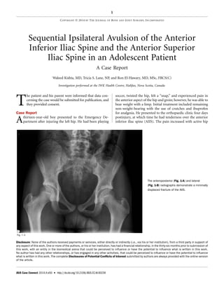
- 1. Sequential Ipsilateral Avulsion of the Anterior Inferior Iliac Spine and the Anterior Superior Iliac Spine in an Adolescent Patient A Case Report Waleed Kishta, MD, Tricia S. Lane, NP, and Ron El-Hawary, MD, MSc, FRCS(C) Investigation performed at the IWK Health Centre, Halifax, Nova Scotia, Canada T he patient and his parent were informed that data con- cerning the case would be submitted for publication, and they provided consent. Case Report Athirteen-year-old boy presented to the Emergency De- partment after injuring the left hip. He had been playing soccer, twisted the hip, felt a ‘‘snap,’’ and experienced pain in the anterior aspect of the hip and groin; however, he was able to bear weight with a limp. Initial treatment included remaining non-weight-bearing with the use of crutches and ibuprofen for analgesia. He presented to the orthopaedic clinic four days postinjury, at which time he had tenderness over the anterior inferior iliac spine (AIIS). The pain increased with active hip Fig. 1-A The anteroposterior (Fig. 1-A) and lateral (Fig. 1-B) radiographs demonstrate a minimally displaced fracture of the AIIS. Disclosure: None of the authors received payments or services, either directly or indirectly (i.e., via his or her institution), from a third party in support of any aspect of this work. One or more of the authors, or his or her institution, has had a financial relationship, in the thirty-six months prior to submission of this work, with an entity in the biomedical arena that could be perceived to influence or have the potential to influence what is written in this work. No author has had any other relationships, or has engaged in any other activities, that could be perceived to influence or have the potential to influence what is written in this work. The complete Disclosures of Potential Conflicts of Interest submitted by authors are always provided with the online version of the article. 1 COPYRIGHT Ó 2014 BY THE JOURNAL OF BONE AND JOINT SURGERY, INCORPORATED JBJS Case Connect 2014;4:e50 d http://dx.doi.org/10.2106/JBJS.CC.M.00239
- 2. flexion, active knee extension, and passive knee flexion. He did not have swelling or deformity at the level of the pelvis. The remainder of the hip and knee examinations was normal. The gait was observed to be antalgic on the left side. Other than weakness in ipsilateral knee extension, the bilateral motor and sensory neurological evaluation from L2 to S1 was normal. Anteroposterior and lateral radiographs of the pelvis revealed a minimally displaced avulsion fracture of the left AIIS (Figs. 1-A and 1-B). The patient underwent six weeks of treatment that con- sisted of oral analgesia and rest, as well as avoidance of contact sports, running, and jumping. After that period of treatment, he was pain-free, and there was no swelling, tenderness, or deformity through the hip, thigh, or knee. Specifically, there was no tenderness over the AIIS. He had no pain with resisted hip flexion or knee extension. Gait examination was grossly normal. Anteroposterior and lateral radiographs revealed a healing fracture of the AIIS. He returned back to sports on a gradual basis and was fully functional ten weeks after the injury. Twenty-one months later, at the age of fifteen, the patient experienced another left pelvic injury. He felt a ‘‘pop’’ while he was lunging during gym class. He was seen in the Emergency Department and was treated with crutches and with oral an- algesia until he was assessed by the orthopaedic team one week postinjury. On examination, he had an antalgic gait on the left side. He had weakness in hip flexion (grade 1 of 5) and knee extension (grade 2 of 5). Other than this ipsilateral weakness, the remainder of the bilateral lower-extremity neurological evaluation was grossly normal. Anteroposterior and lateral radiographs of the pelvis demonstrated an avulsion fracture of the left anterior superior iliac spine (ASIS) (Figs. 2-A and 2-B). Treatment was nonoperative. The patient remained Fig. 1-B Fig. 2-A The anteroposterior (Fig. 2-A) and lateral (Fig. 2-B) radiographs demonstrate that the AIIS fracture had healed; however, a minimally displaced fracture of the ASIS was evident. 2 JBJS CASE CONNECTOR VOLU ME 4 d NU MBE R 2 d JUNE 25, 2014 SEQUENTIA L AVULSION OF THE ANTERIOR INFERIOR ILIAC SPINE AND THE ANTERIOR SUP ERIOR ILIAC SPINE
- 3. non-weight-bearing with the use of crutches, was treated with oral analgesia, and avoided sporting activity for six weeks. After that period of treatment, he was able to walk without crutches and had full range of motion of the hip. He had no pain with passive hip extension or knee flexion or with re- sisted hip flexion or knee extension. He then returned back to sports on a gradual basis as tolerated, and he was able to return to full activity ten weeks after the second injury. Blood tests, including complete blood count, metabolic screening, and thyroid-stimulating hormone screening, were performed to rule out any underlying medical abnormality. The labo- ratory tests were all normal, with the exception of a slightly low phosphorus level. The last clinic visit was approximately fifteen months after the second injury, at which time the patient had no pain and was comfortably performing all activities. On examination, he walked without a limp and had full range of movement of the hip and the knee. Radiographic examination revealed that the each fracture had healed in an acceptable position and with satisfactory alignment (Fig. 3). Discussion Avulsion fractures of the pelvis are common injuries in the adolescent population and generally occur during ath- letic activities1 . Isolated avulsions of either the AIIS or the ASIS are the most common locations1-3 . Although bilateral simultaneous avulsion fractures and ipsilateral avulsion fractures of the AIIS and ischial tuberosity have been reported by Rossi and Dragoni1 , and simultaneous avulsion of the AIIS and ASIS with and without hip dislocation also has been re- ported4,5 , to the best of our knowledge, this is the first report of sequential ipsilateral avulsion fractures of the AIIS and the ASIS. Fig. 2-B Fig. 3 Thirty-six months postinjury to the AIIS and fifteen months postinjury to the ASIS, the anteroposterior radiograph demonstrates that both fractures had healed in an acceptable position. 3 JBJS CASE CONNECTOR VOLU ME 4 d NU MBE R 2 d JUNE 25, 2014 SEQUENTIA L AVULSION OF THE ANTERIOR INFERIOR ILIAC SPINE AND THE ANTERIOR SUP ERIOR ILIAC SPINE
- 4. The AIIS ossifies around fourteen years of age and fuses about four years later; the ASIS ossifies at fifteen years of age and fuses with the ilium about four years later. The sartorius muscle and the tensor fasciae latae muscle both originate from the ASIS, while the direct head of the rectus femoris muscle originates from the AIIS. Each muscle spans two joints, which may produce substantial loads during muscle contraction6 . These sudden and substantial muscle contractions may be too powerful for the immature cartilaginous apophysis and may result in avulsion fractures in the skeletally immature po- pulation. Most injuries occur in boys because of their delayed physeal closure; the mean age for these fractures is 13.8 years1,2,7 . Apophyseal avulsion fractures of the pelvis in adolescents are common during sports such as soccer and gymnastics. Etiologic data support that these injuries are the result of indirect trauma secondary to sudden, violent concentric or eccentric muscle contraction1 . The distribution related to the type of athletic activity undertaken reveals that soccer and tennis are the most common sports in which participants may sustain AIIS lesions, while soccer and gymnastics are the most common sports that produce ASIS lesions. When the total number of lesions in each sport was compared with the overall number of radiographic examinations performed on partici- pants in that sport, soccer and gymnastics had the highest prevalence of these injuries1 . Injuries to the ASIS and AIIS in soccer generally result from a superiorly directed movement of the lower extremity while ‘‘kicking the air’’ or from a strike at the goal with maximum flexion of the hip and extension of the knee. Despite injuring the pelvis while playing soccer, our pa- tient was not in the process of swinging the lower extremity in order to kick the soccer ball. The injury occurred in the stance phase and was likely similar to the mechanism of injury that can occur during the push-off for a sprint. At the beginning of a sprint, there is simultaneous hip extension, knee flexion, and ankle plantar flexion. The ensuing eccentric muscle activation can stress both the AIIS and the ASIS. The second injury from lunging occurred during the swing phase of the lunge and may have been the result of the sudden, forceful concentric con- traction of the anterior thigh muscles in order to flex the hip and extend the knee. Another predisposing factor contributing to these in- juries is the repetitive stresses that are produced while inten- sively training for these sports. In the skeletally immature, these forces may weaken the epiphyseal plate so that it may not be able to withstand the powerful forces produced by muscles that are hypertrophied by training. The contribution of physeal maturity to pelvic fracture patterns is evident as these patterns of injury change with skeletal maturity8 . As a result, adolescents with open triradiate cartilage are prone not only to avulsion fractures, but to iliac wing and pubic rami fractures as well. In contrast, adults are more likely to sustain high-energy injuries to the acetabulum and the pelvic ring8 . The diagnosis of acute AIIS or ASIS avulsion is usually made clinically and then confirmed radiographically. The history is often consistent with an acute pain that was pre- cipitated by a certain maneuver during a sporting activity. The pain is often severe, sharp, and localized to the buttocks for ischial tuberosity injuries and to the anterior aspect of the pelvis for ASIS and AIIS injuries. The pain is aggravated by stretching or by actively contracting the involved mus- cle(s). This hip and lower-extremity pain is generally ac- companied by restricted range of motion, and the area over the avulsed fragment can be swollen and tender9 . In a review of twenty cases of pelvic avulsion fractures, Fernbach and Wilkinson found that AIIS avulsions were less painful than ASIS avulsions10 . Chronic injuries secondary to repetitive loading at the tendinous origin often are more difficult to diagnose. Radio- graphs of the pelvis may demonstrate both lytic and sclerotic lesions at the site of injury1,7 . Computed tomography and magnetic resonance imaging may be necessary to confirm the diagnosis of an avulsion injury9 . Nontraumatic avulsions of the pelvis may be related to an underlying weakness of the sur- rounding tissues (e.g., with a malignancy)11 . The literature has suggested that symptomatic care with early weight-bearing for the majority of ASIS and AIIS injuries generally results in full recovery without any sporting limita- tions by two months postinjury12-14 . The most commonly re- ported complication related to nonoperative treatment of pelvic avulsion injuries has been exostosis formation15,16 . In- dications for open reduction and internal fixation of a pelvic avulsion injury have included substantial fragment displace- ment of more than 3 cm and fracture nonunion; open reduc- tion and internal fixation has also been indicated in athletes, who may require a shorter convalescent period17-19 . Com- plications related to open reduction and internal fixation of the AIIS or ASIS have included injury to the lateral femoral cutaneous nerve with resultant meralgia paresthetica, intra- articular screw penetration, and substantial blood loss14 . Be- cause of the low potential for complications, complete recovery and return to athletic activities have been reported for most cases, regardless of whether they were treated operatively or nonoperatively2,7,14,18 . Sequential ipsilateral avulsion fractures of the AIIS and ASIS are an uncommon occurrence. In our patient, the se- quential ipsilateral avulsion fractures of the AIIS and the ASIS had typical features on history, physical examination, radio- graphic imaging, and prognosis; the results of the nonoperative treatment were the same as would be expected with either of these injuries in isolation. n Waleed Kishta, MD Tricia S. Lane, NP Ron El-Hawary, MD, MSc, FRCS(C) IWK Health Centre, 5850 University Avenue, P.O. Box 9700, Halifax, NS, B3K 6R8, Canada. E-mail address for R. El-Hawary: ron.el-hawary@iwk.nshealth.ca 4 JBJS CASE CONNECTOR VOLU ME 4 d NU MBE R 2 d JUNE 25, 2014 SEQUENTIA L AVULSION OF THE ANTERIOR INFERIOR ILIAC SPINE AND THE ANTERIOR SUP ERIOR ILIAC SPINE
- 5. References 1. Rossi F, Dragoni S. Acute avulsion fractures of the pelvis in adolescent com- petitive athletes: prevalence, location and sports distribution of 203 cases col- lected. Skeletal Radiol. 2001 Mar;30(3):127-31. 2. Fernbach SK, Wilkinson RH. Avulsion injuries of the pelvis and proximal femur. AJR Am J Roentgenol. 1981 Sep;137(3):581-4. 3. Rosenberg N, Noiman M, Edelson G. Avulsion fractures of the anterior superior iliac spine in adolescents. J Orthop Trauma. 1996;10(6):440-3. 4. Oldenburg FP, Smith MV, Thompson GH. Simultaneous ipsilateral avulsion of the anterior superior and anterior inferior iliac spines in an adolescent. J Pediatr Orthop. 2009 Jan-Feb;29(1):29-30. 5. Meyer NJ, Schwab JP, Orton D. Traumatic unilateral avulsion of the anterior superior and inferior iliac spines with anterior dislocation of the hip: a case report. J Orthop Trauma. 2001 Feb;15(2):137-40. 6. Ogden JA. Skeletal injury in the child. 2nd ed. Philadelphia: WB Saunders; 2000. 7. Sundar M, Carty H. Avulsion fractures of the pelvis in children: a report of 32 fractures and their outcome. Skeletal Radiol. 1994 Feb;23(2):85-90. 8. Silber JS, Flynn JM. Changing patterns of pediatric pelvic fractures with skeletal maturation: implications for classification and management. J Pediatr Orthop. 2002 Jan-Feb;22(1):22-6. 9. Stevens MA, El-Khoury GY, Kathol MH, Brandser EA, Chow S. Imaging features of avulsion injuries. Radiographics. 1999 May-Jun;19(3):655-72. 10. Fernbach SK, Wilkinson RH. Avulsion injuries of the pelvis and proximal femur. AJR Am J Roentgenol. 1981 Sep;137(3):581-4. 11. Bui-Mansfield LT, Chew FS, Lenchik L, Kline MJ, Boles CA. Nontraumatic avul- sions of the pelvis. AJR Am J Roentgenol. 2002 Feb;178(2):423-7. 12. Gomez JE. Bilateral anterior inferior iliac spine avulsion fractures. Med Sci Sports Exerc. 1996 Feb;28(2):161-4. 13. Fernbach SK, Wilkinson RH. Avulsion injuries of the pelvis and proximal femur. AJR Am J Roentgenol. 1981 Sep;137(3):581-4. 14. Saluan PM, Weiker GG. Avulsion of the anterior inferior iliac spine. Orthopedics. 1997 Jun;20(6):558-9. 15. Irving MH. Exostosis formation after traumatic avulsion of the anterior inferior iliac spine: report of two cases. J Bone Joint Surg Br. 1964 Nov;46: 720-2. 16. Watts HG. Fractures of the pelvis in children. Orthop Clin North Am. 1976 Jul;7(3):615-24. 17. Aksoy B, Ozt¨urk K, Ensenyel CZ, Kara AN. Avulsion of the iliac crest apophysis. Int J Sports Med. 1998 Jan;19(1):76-8. 18. Smith J, Mears DC. Children. In: Mears DC, Rubash HE, editors. Pelvic and acetabular fractures. Thorofare, NJ: Slac. 1986; p. 497-501. 19. Veselko M, Smrkolj V. Avulsion of the anterior-superior iliac spine in athletes: case reports. J Trauma. 1994 Mar;36(3):444-6. 5 JBJS CASE CONNECTOR VOLU ME 4 d NU MBE R 2 d JUNE 25, 2014 SEQUENTIA L AVULSION OF THE ANTERIOR INFERIOR ILIAC SPINE AND THE ANTERIOR SUP ERIOR ILIAC SPINE