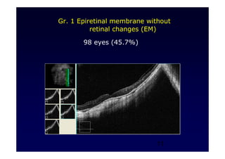
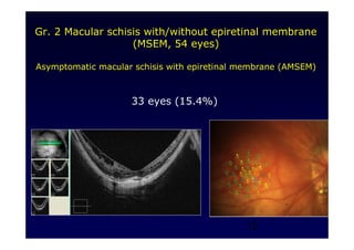
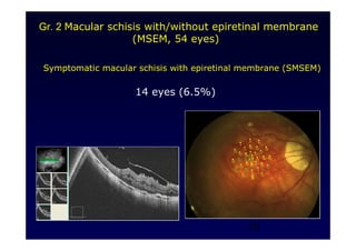
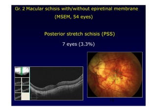
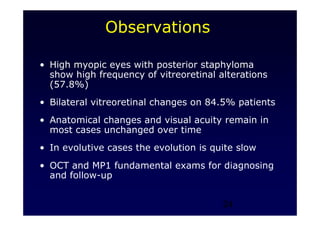
1
The author has no potential
conflict of interest to disclose
2
Modificazioni Vitreo-Retiniche negli
Stafilomi nel Miope Elevato ed Eventuali
Indicazioni Chirurgiche.
Guido Ripandelli
Tommaso Rossi*
Fabio Scarinci
Mario Stirpe
IRCCS – Fondazione Bietti, Roma
*Ospedale Oftalmico, Roma
3
Macular Vitreo-Retinal Interface
Abnormalities in Highly Myopic Eyes with
Posterior Staphyloma:
5-Years Follow-Up.
Guido Ripandelli, Tommaso Rossi, * Fabio Scarinci,
Cecilia Scassa, Vincenzo Parisi, Mario Stirpe.
Retina 2012;32:1531-8.
4
• Purpose: to review prevalence, long-term
progression and prognosis of VR interface
modifications in highly myopic eyes with
posterior staphyloma
When surgery is necessary?
Which is the best timing for surgery?
• Study design: retrospective, single
Institution series
5
Patients’ characteristics
• December 1999 - December 2004
• 408 phakic eyes / 204 subjects
• 108 Males / 96 Females
• Age 22-58 years
• Myopia 18-30 D and PS (Axial lenght 30-34
mm)
6
Inclusion Criteria
• AL > 30 mm
• Presence of posterior staphyloma
• Availability of OCT, MP1 at baseline, at
latest examination, (before and after
surgery)
7
Exclusion Criteria
Patients with incomplete charts, lost at follow-
up, with significant media opacities, with
previous ocular surgery
8
Characteristics of patients
with VR changes
• Prevalence of posterior VR changes: 236/408 (57.8%) eyes
• Minimum follow-up: 5 years
• Lost to follow-up: 22/236 eyes (9.3%)
• Included in the study: 214 eyes (116 subjects)
– Bilateral VR changes 98/116 (84.4%)
– 54 males /62 females
– Age 24-59 years
– Myopia 19-30 D (29.5-34 mm)
– 209 eyes: visual acuity 20/50-20/20
– 5 eyes: C F – 20/100
9
Testing
• Visual acuity (VA)
• Biomicroscopy
• Optical Coherence Tomography (OCT)
• MP1 Microperimetry
• A, B-scan ultrasound
10
Group 1 Epiretinal membrane without retinal changes (EM) : 98 eyes
Group 2 Macular schisis with/without epiretinal membrane (MSEM): 54 eyes
Asymptomatic macular schisis with EM (AMSEM, 33 eyes)
Symptomatic macular schisis with EM (SMSEM, 14 eyes)
Posterior stretch schisis (PSS, 7 eyes)
Group 3 Partial thickness macular hole (PTMH): 24 eyes
PTHM with retinal schisis (15 eyes)
PTHM without retinal schisis (9 eyes)
Group 4 Full thickness macular hole (FTMH): 18 eyes
Asymptomatic full thickness macular hole (AFTMH, 9 eyes)
Symptomatic full thickness macular hole (SFTMH, 9 eyes)
Group 5 Posterior retinal detachment (PRD): 20 eyes
PRD with macula on (PRD M-on, 11 eyes)
PRD with macula off (PRD M-off, 4 eyes)
PRD with macular hole (PRDMH, 5 eyes)
11
Gr. 1 Epiretinal membrane without
retinal changes (EM)
98 eyes (45.7%)
12
Gr. 2 Macular schisis with/without epiretinal membrane
(MSEM, 54 eyes)
Asymptomatic macular schisis with epiretinal membrane (AMSEM)
33 eyes (15.4%)
13
Symptomatic macular schisis with epiretinal membrane (SMSEM)
14 eyes (6.5%)
Gr. 2 Macular schisis with/without epiretinal membrane
(MSEM, 54 eyes)
14
Posterior stretch schisis (PSS)
7 eyes (3.3%)
Gr. 2 Macular schisis with/without epiretinal membrane
(MSEM, 54 eyes)
15
Gr. 3 Partial thickness macular hole ( PTMH, 24 eyes)
PTMH with macular schisis
15 eyes (7%)
16
Gr. 3 Partial thickness macular hole ( PTMH, 24 eyes)
PTMH without macular schisis
9 eyes (4.2%)
17
Asymptomatic full thickness macular hole (AFTMH)
9 eyes (4.2%)
Gr. 4 Full thickness macular hole ( FTMH, 18 eyes)
18
Symptomatic full thickness macular hole (SFTMH)
9 eyes (4.2%)
Gr. 4 Full thickness macular hole ( FTMH, 18 eyes)
19
PRD in the staphyloma area with macula on (PRD M-on)
11 eyes (5.1%)
Gr. 5 Posterior retinal detachment (PRD, 20 eyes)
20
PRD in the staphyloma area with macula off (PRD M-off)
4 eyes (1.8%)
Gr. 5 Posterior retinal detachment (PRD, 20 eyes)
21
PRD with macular hole (PRDMH)
5 eyes (2.3%)
Gr. 5 Posterior retinal detachment (PRD, 20 eyes)
22
Evolution to pathologies requiring surgery: 24/205 (11.7%)
(9 PRD eyes underwent surgery upon presentation)
Gr. 1 Epiretinal membrane without retinal changes (EM) : 2/98 (2V)
Gr. 2 Asymptomatic macular schisis (AMSEM): 6/33 (3V, 3V+PB)
Symptomatic macular schisis (SMSEM): 7/14 (4V, 3V+PB)
Posterior stretch schisis (PSS): 0/7 -
Gr. 3 PTHM with macular schisis (PTMH-MS): 5/15 (3V, 2V+PB)
PTMH without macular schisis (PTMH-w/o MS): 0/9 -
Gr. 4 Asymptomatic macular hole (AFTMH): 2/9 (2V)
Symptomatic macular hole (SFTMH): 1/9 (1V)
Gr. 5 PRD in staphyloma area / macula ON (PRD M-on): 1/11 (1V)
PRD in staphyloma area/ macula off (PRD M-off): 4/4 (2V, 2V+PB)
PRD with macular hole (PRD-MH): 5/5 (2V, 3V+PB)
V= vitrectomy, PB= posterior buckle
23
Observations
High myopia with posterior staphyloma
can be associated with a broad
spectrum of vitreoretinal alterations
and represents an elusive nosological
entity, whose natural history,
progression, therapeutic options and
surgical timing remain uncertain.
24
Observations
• High myopic eyes with posterior staphyloma
show high frequency of vitreoretinal alterations
(57.8%)
• Bilateral vitreoretinal changes on 84.5% patients
• Anatomical changes and visual acuity remain in
most cases unchanged over time
• In evolutive cases the evolution is quite slow
• OCT and MP1 fundamental exams for diagnosing
and follow-up
25
Observations
• Microperimetry provides important data for a
timely surgical decision
• Foveal sensitivity decrease and worsening of
fixation stability are significantly associated to
the need for surgery.
26
Observations
• The decrease of sensitivity explored by MP1 is
a sign that may precede the objective
worsening of visual acuity
VA 20/25 VA 20/25
27
•Surgical removal of ER tissue represents the first
attempt to resolve the pathology
Observations
28
• The reduction of posterior staphyloma by means of
posterior buckle is a method to use to repair the
unsuccessfull cases
Observations
29
Observations
Vitreoretinal interface alterations in
highly myopic eyes have not been
univocally classified yet and the
concept itself of disease progression
and surgical indication are largely
debatable and deserve
standardization.
30
Observations
Foveal sensitivity and fixation patterns
are associated to a variable risk of
progression and visual prognosis differs
significantly among groups. We believe
this variability represents underlying
separate pathologies that warrant
diversified treatment.

Ripandelli the author has no potential

  • 1.
    1 The author hasno potential conflict of interest to disclose
  • 2.
    2 Modificazioni Vitreo-Retiniche negli Stafilominel Miope Elevato ed Eventuali Indicazioni Chirurgiche. Guido Ripandelli Tommaso Rossi* Fabio Scarinci Mario Stirpe IRCCS – Fondazione Bietti, Roma *Ospedale Oftalmico, Roma
  • 3.
    3 Macular Vitreo-Retinal Interface Abnormalitiesin Highly Myopic Eyes with Posterior Staphyloma: 5-Years Follow-Up. Guido Ripandelli, Tommaso Rossi, * Fabio Scarinci, Cecilia Scassa, Vincenzo Parisi, Mario Stirpe. Retina 2012;32:1531-8.
  • 4.
    4 • Purpose: toreview prevalence, long-term progression and prognosis of VR interface modifications in highly myopic eyes with posterior staphyloma When surgery is necessary? Which is the best timing for surgery? • Study design: retrospective, single Institution series
  • 5.
    5 Patients’ characteristics • December1999 - December 2004 • 408 phakic eyes / 204 subjects • 108 Males / 96 Females • Age 22-58 years • Myopia 18-30 D and PS (Axial lenght 30-34 mm)
  • 6.
    6 Inclusion Criteria • AL> 30 mm • Presence of posterior staphyloma • Availability of OCT, MP1 at baseline, at latest examination, (before and after surgery)
  • 7.
    7 Exclusion Criteria Patients withincomplete charts, lost at follow- up, with significant media opacities, with previous ocular surgery
  • 8.
    8 Characteristics of patients withVR changes • Prevalence of posterior VR changes: 236/408 (57.8%) eyes • Minimum follow-up: 5 years • Lost to follow-up: 22/236 eyes (9.3%) • Included in the study: 214 eyes (116 subjects) – Bilateral VR changes 98/116 (84.4%) – 54 males /62 females – Age 24-59 years – Myopia 19-30 D (29.5-34 mm) – 209 eyes: visual acuity 20/50-20/20 – 5 eyes: C F – 20/100
  • 9.
    9 Testing • Visual acuity(VA) • Biomicroscopy • Optical Coherence Tomography (OCT) • MP1 Microperimetry • A, B-scan ultrasound
  • 10.
    10 Group 1 Epiretinalmembrane without retinal changes (EM) : 98 eyes Group 2 Macular schisis with/without epiretinal membrane (MSEM): 54 eyes Asymptomatic macular schisis with EM (AMSEM, 33 eyes) Symptomatic macular schisis with EM (SMSEM, 14 eyes) Posterior stretch schisis (PSS, 7 eyes) Group 3 Partial thickness macular hole (PTMH): 24 eyes PTHM with retinal schisis (15 eyes) PTHM without retinal schisis (9 eyes) Group 4 Full thickness macular hole (FTMH): 18 eyes Asymptomatic full thickness macular hole (AFTMH, 9 eyes) Symptomatic full thickness macular hole (SFTMH, 9 eyes) Group 5 Posterior retinal detachment (PRD): 20 eyes PRD with macula on (PRD M-on, 11 eyes) PRD with macula off (PRD M-off, 4 eyes) PRD with macular hole (PRDMH, 5 eyes)
  • 11.
    11 Gr. 1 Epiretinalmembrane without retinal changes (EM) 98 eyes (45.7%)
  • 12.
    12 Gr. 2 Macularschisis with/without epiretinal membrane (MSEM, 54 eyes) Asymptomatic macular schisis with epiretinal membrane (AMSEM) 33 eyes (15.4%)
  • 13.
    13 Symptomatic macular schisiswith epiretinal membrane (SMSEM) 14 eyes (6.5%) Gr. 2 Macular schisis with/without epiretinal membrane (MSEM, 54 eyes)
  • 14.
    14 Posterior stretch schisis(PSS) 7 eyes (3.3%) Gr. 2 Macular schisis with/without epiretinal membrane (MSEM, 54 eyes)
  • 15.
    15 Gr. 3 Partialthickness macular hole ( PTMH, 24 eyes) PTMH with macular schisis 15 eyes (7%)
  • 16.
    16 Gr. 3 Partialthickness macular hole ( PTMH, 24 eyes) PTMH without macular schisis 9 eyes (4.2%)
  • 17.
    17 Asymptomatic full thicknessmacular hole (AFTMH) 9 eyes (4.2%) Gr. 4 Full thickness macular hole ( FTMH, 18 eyes)
  • 18.
    18 Symptomatic full thicknessmacular hole (SFTMH) 9 eyes (4.2%) Gr. 4 Full thickness macular hole ( FTMH, 18 eyes)
  • 19.
    19 PRD in thestaphyloma area with macula on (PRD M-on) 11 eyes (5.1%) Gr. 5 Posterior retinal detachment (PRD, 20 eyes)
  • 20.
    20 PRD in thestaphyloma area with macula off (PRD M-off) 4 eyes (1.8%) Gr. 5 Posterior retinal detachment (PRD, 20 eyes)
  • 21.
    21 PRD with macularhole (PRDMH) 5 eyes (2.3%) Gr. 5 Posterior retinal detachment (PRD, 20 eyes)
  • 22.
    22 Evolution to pathologiesrequiring surgery: 24/205 (11.7%) (9 PRD eyes underwent surgery upon presentation) Gr. 1 Epiretinal membrane without retinal changes (EM) : 2/98 (2V) Gr. 2 Asymptomatic macular schisis (AMSEM): 6/33 (3V, 3V+PB) Symptomatic macular schisis (SMSEM): 7/14 (4V, 3V+PB) Posterior stretch schisis (PSS): 0/7 - Gr. 3 PTHM with macular schisis (PTMH-MS): 5/15 (3V, 2V+PB) PTMH without macular schisis (PTMH-w/o MS): 0/9 - Gr. 4 Asymptomatic macular hole (AFTMH): 2/9 (2V) Symptomatic macular hole (SFTMH): 1/9 (1V) Gr. 5 PRD in staphyloma area / macula ON (PRD M-on): 1/11 (1V) PRD in staphyloma area/ macula off (PRD M-off): 4/4 (2V, 2V+PB) PRD with macular hole (PRD-MH): 5/5 (2V, 3V+PB) V= vitrectomy, PB= posterior buckle
  • 23.
    23 Observations High myopia withposterior staphyloma can be associated with a broad spectrum of vitreoretinal alterations and represents an elusive nosological entity, whose natural history, progression, therapeutic options and surgical timing remain uncertain.
  • 24.
    24 Observations • High myopiceyes with posterior staphyloma show high frequency of vitreoretinal alterations (57.8%) • Bilateral vitreoretinal changes on 84.5% patients • Anatomical changes and visual acuity remain in most cases unchanged over time • In evolutive cases the evolution is quite slow • OCT and MP1 fundamental exams for diagnosing and follow-up
  • 25.
    25 Observations • Microperimetry providesimportant data for a timely surgical decision • Foveal sensitivity decrease and worsening of fixation stability are significantly associated to the need for surgery.
  • 26.
    26 Observations • The decreaseof sensitivity explored by MP1 is a sign that may precede the objective worsening of visual acuity VA 20/25 VA 20/25
  • 27.
    27 •Surgical removal ofER tissue represents the first attempt to resolve the pathology Observations
  • 28.
    28 • The reductionof posterior staphyloma by means of posterior buckle is a method to use to repair the unsuccessfull cases Observations
  • 29.
    29 Observations Vitreoretinal interface alterationsin highly myopic eyes have not been univocally classified yet and the concept itself of disease progression and surgical indication are largely debatable and deserve standardization.
  • 30.
    30 Observations Foveal sensitivity andfixation patterns are associated to a variable risk of progression and visual prognosis differs significantly among groups. We believe this variability represents underlying separate pathologies that warrant diversified treatment.