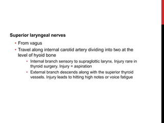
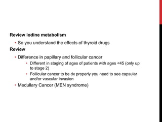
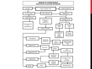
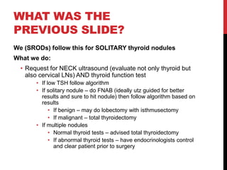
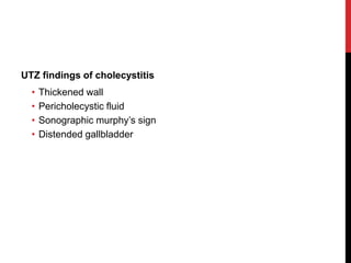
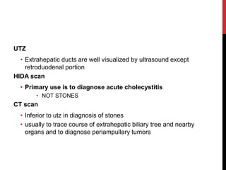
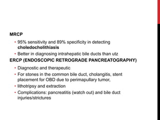
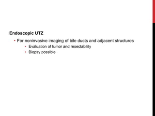
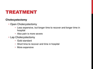
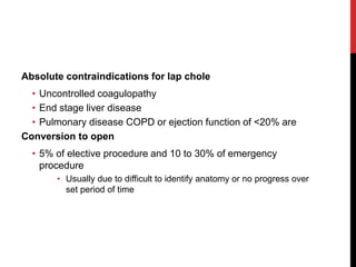
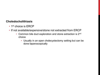
This document provides tips and information for revalida exams. It discusses general study tips like pacing oneself and taking breaks. It also covers specific topics that may come up like breast diseases, thyroid surgery, and gallbladder conditions. For breast exams, it outlines diseases and treatments like lumpectomy. For thyroidectomy, it discusses anatomy like the recurrent laryngeal nerve. Gallbladder topics include cholecystectomy, cholelithiasis, and imaging tests for stones. The document emphasizes knowing content well while also resting the brain between study sessions.
The document discusses the pelvis and cephalopelvic disproportion (CPD). It defines the false pelvis and true pelvis, and lists the normal diameters of the pelvic inlet, cavity, and outlet. It describes a contracted pelvis as having one or more diameters reduced by 1 cm or more below normal. Causes of a contracted pelvis include developmental factors, trauma, infections, tumors, and metabolic bone diseases. Degrees of CPD range from minor to extreme. Management depends on the degree of disproportion and may include a trial of vaginal delivery, cesarean section, or craniotomy. Complications can affect both mother and fetus.
This document provides an overview of the physiotherapy assessment of cardiac conditions. It discusses the importance of obtaining an accurate subjective and objective assessment in order to develop an appropriate treatment plan. The assessment includes gathering a patient history, performing objective measurements, inspecting the patient, palpating pulses and edema, auscultating heart sounds, and evaluating disability. Signs and symptoms associated with common cardiac conditions are reviewed. The grading of murmurs and scales to assess functions are also outlined.
This document provides an overview of how to perform an abdominal examination, including the key steps and techniques. It begins with an anatomical review of the organs located in each abdominal quadrant. It then details how to properly position the patient and prepare for the exam. The core components of the abdominal exam are described in the recommended order of inspection, auscultation, percussion, and palpation. Specific techniques for examining organs like the liver, spleen, and kidneys are outlined. The document concludes with descriptions of how to assess for possible conditions like ascites, acute abdomen, and acute pancreatitis based on exam findings.
OBSTETRIC AND GYNAECILOGICAL PHYSICAL EXAMINATION.pptxmucunguziamos495
This document provides guidance on performing a physical examination. It begins by outlining examination of general appearance and vital signs. It then provides detailed instructions on examination of specific body systems including hands, fingers, pulse, face, eyes, mouth, neck, lymph nodes, breasts, abdomen, and obstetric assessment. The physical examination is thorough, with emphasis on inspection, palpation, and assessing relevant physical signs.
This document provides information on clinically examining the breast. It begins with the external and internal anatomy of the breast, including lobes, lobules and ducts. It describes the arterial and venous blood supply and lymphatic drainage sites. The summary discusses taking a history regarding lumps, pain, discharge, risk factors and more. The physical exam methods are outlined, including inspection for symmetry, masses and skin/nipple changes, and palpation techniques to feel the breasts and axillary lymph nodes. Self exam instructions are provided to help with early cancer detection. Other clinical findings like gynecomastia and Mondor's disease are also mentioned.
This document provides details from a presentation on ovarian carcinoma including:
1) An introduction defining ovarian tumors and their classification.
2) A WHO classification of ovarian tumors into 9 categories.
3) Details on the clinical signs, symptoms, differentiation and complications of benign vs malignant ovarian tumors.
The document provides information on clinical examination of the breast. It describes the anatomy of the breast including shape, size, base, areola, and nipple. It outlines the process of breast examination including inspection for symmetry, masses, skin changes and palpation of the breasts and axilla. Characteristics of benign and malignant breast lumps are compared. The document also discusses breast self-examination and conditions like gynecomastia and Mondor's disease.
This document provides information about performing a physical examination of the abdomen. It describes the anatomy of the abdominal wall and contents. The peritoneum lines the abdominal cavity. Structures within the cavity include solid organs like the liver and hollow organs like the stomach. The document outlines the steps of inspecting, auscultating, and palpating the abdomen and defines normal and abnormal findings for each step.
Ca breast, diagnosis, clinical examination and diagnostic workup Satyajeet Rath
This document provides an overview of the clinical presentation, examination, and diagnostic workup for breast cancer. It discusses collecting a history including symptoms like breast lumps or nipple discharge. Physical examination involves inspecting and palpating the breasts and axillary lymph nodes. Diagnostic workup includes imaging like mammography or ultrasound, as well as pathology studies to confirm a diagnosis and determine tumor characteristics. Staging further involves tests like bone scans or CT scans to identify distant metastases. A thorough evaluation is important for developing a treatment plan tailored to each patient's specific cancer.
This document presents the case of a 40-year-old male farmer who presented with a large swelling on the front of his neck and a smaller swelling on his right collarbone. Examination and investigations revealed a large multinodular goiter in his left thyroid lobe and a cystic mass on his right collarbone. Biopsy of the thyroid swelling suggested nodular colloid goiter or follicular neoplasm. CT imaging showed the thyroid mass compressing his trachea and a metastatic bone lesion in his right collarbone and rib, suggesting follicular thyroid carcinoma with skeletal metastasis. He was scheduled for a total thyroidectomy.
This document provides advice and tips for the FRCR 2A exam, including:
- Make prayers and prepare well in advance by sleeping well the day before.
- Carefully read the question and scan the scenario to find key details.
- Take breaks between questions to take deep breaths.
- Stick closely to the answer choices provided rather than outside knowledge.
- Remain logical and imagine applying the scenario to real life.
- The exam is long so bring water and food for breaks.
This document provides information on breast examination including history taking, examination techniques, common findings, diagnoses, and recommendations. It discusses systematically examining the breasts through inspection and palpation using various patterns to check for lumps or abnormalities. Recommendations include performing monthly breast self-exams after age 20 and regular clinical exams and mammograms starting at age 40.
The document provides guidance on performing a physical examination, beginning with an overview of the general survey, vital signs, and pain assessment. It describes how to observe the patient's general appearance, measure vital signs like blood pressure and temperature, and evaluate pain. Key steps include observing the patient's state of health, dress, posture, and skin while ensuring accurate measurement of vital signs and thorough pain assessment using techniques like COLDERR. The document aims to guide healthcare practitioners in conducting a complete initial physical evaluation of the patient.
This document summarizes guidelines for antenatal care including the number of recommended appointments based on pregnancy history, risk factors requiring obstetrician-led care, vitamins and supplements recommended during pregnancy, testing for Rhesus D status, monitoring fetal growth, and how to perform an obstetric examination. Key points include recommendations for 10 appointments with a midwife for first pregnancies and 7 for subsequent ones, higher dose folic acid and vitamin D for those at high risk, testing all women for Rhesus D status, monitoring fetal size with fundal height measurements and ultrasounds if high risk, and steps for examining the mother and fetus during an obstetric exam.
Sources for EDiR are same as FRCR 2A especially Crack the core and 2 b (all cases books especially if updated) plus very very important: the mock exams in their website. Its importance: EDiR certifies that a candidate has passed a high quality examination (in English) in general radiology. EDiR is definitely an added value to the candidates’ CV, differentiating them from other contenders when applying for a job or fellowship
Note that EDiR does not replace any national board certificate .(COPIED from website of EDIR) Consist of three parts in one day 1. Multiple response questions (MRQs): Computer-based examination (90 minutes). FRCR2 A BOOKS WILL HELP YOU IN THIS STEP PLUS THE MOST IMPORTANT CRACK THE CORE, take care it is not Single best answer, it is mutiple response questions (may be more than 1 answer is correct)
2. Short cases (SCs): Computer-based examination (90 minutes).
3. Clinically Oriented Reasoning Evaluation (CORE): Practical-oriented cases, computer-based examination (90 minutes). FRCR 2B BOOKS WILL HELP in 2 and 3 my advice if you are fresh and good prepared توكل على الله and go on if not wait
Abnormal labour process and management for nursing studentsbrownmunde108
This document discusses abnormal labor including cephalo pelvic disproportion (CPD) and contracted pelvis. It defines CPD as a disparity between the fetal head size and the mother's pelvis size. CPD is assessed using the Muller-Kerr method where the head is pushed into the pelvis and overlapping is observed. Contracted pelvis refers to a pelvis with reduced diameters and is classified based on type and degree of contraction. Management of CPD and contracted pelvis may include trial of labor, induction, or cesarean section depending on the severity. Complications of abnormal labor can arise during each stage of labor.
This document provides an overview of pediatric and geriatric assessment. For pediatrics, it describes taking a thorough history, modifying examination techniques, and identifying common assessment findings for different developmental levels. For geriatrics, it identifies common age-related changes to body systems and geriatric syndromes. Key aspects of assessment for both populations include positive communication, patience, and encouraging participation.
Sonographic anatomy of abdomen and pelvic organmanishyadav513
This document reviews the normal sonographic anatomy of abdominal and pelvic organs. It describes the sonographic appearance and measurements of organs like the liver, gallbladder, biliary tree, pancreas, kidneys, spleen, urinary bladder, prostate, uterus, ovaries, abdominal aorta, inferior vena cava, and appendix. Key points about echogenicity, size, vascular structures and changes through the menstrual cycle are provided. Diagrams supplement the written descriptions to illustrate sonographic views and anatomy.
This document provides guidance on performing a newborn examination. It begins by classifying newborns by gestational age and birth weight. It then describes how to assess vital signs, growth measurements, and the different body systems. Key parts of the examination are classified including the skin, head, eyes, chest, heart, abdomen, genitals and nervous system. Important reflexes are outlined to assess neurological development. The document emphasizes the importance of estimating gestational age and recognizing normal and abnormal findings during the newborn examination.
The document summarizes the anatomy and function of the spleen. It describes the spleen's location in the upper left quadrant of the abdominal cavity. It has three borders, two surfaces, and two poles. The spleen filters blood and removes old red blood cells, microbes, and debris. It is supplied by the splenic artery and drained by the splenic vein. Microscopically, it contains white pulp with lymphocytes and red pulp that filters the blood and removes particulate matter.
general Examination in paediatric medicine Sujit Balodiya
This 22 month old boy presents with pallor. His mother notes no other symptoms but was concerned due to comments from relatives. On exam, he appears pale but is otherwise healthy and active. Lab work shows microcytic anemia. The cause is likely iron deficiency due to a diet high in milk and low in iron-rich foods. Counseling is provided on an iron-rich diet.
Cell Therapy Expansion and Challenges in Autoimmune DiseaseHealth Advances
There is increasing confidence that cell therapies will soon play a role in the treatment of autoimmune disorders, but the extent of this impact remains to be seen. Early readouts on autologous CAR-Ts in lupus are encouraging, but manufacturing and cost limitations are likely to restrict access to highly refractory patients. Allogeneic CAR-Ts have the potential to broaden access to earlier lines of treatment due to their inherent cost benefits, however they will need to demonstrate comparable or improved efficacy to established modalities.
In addition to infrastructure and capacity constraints, CAR-Ts face a very different risk-benefit dynamic in autoimmune compared to oncology, highlighting the need for tolerable therapies with low adverse event risk. CAR-NK and Treg-based therapies are also being developed in certain autoimmune disorders and may demonstrate favorable safety profiles. Several novel non-cell therapies such as bispecific antibodies, nanobodies, and RNAi drugs, may also offer future alternative competitive solutions with variable value propositions.
Widespread adoption of cell therapies will not only require strong efficacy and safety data, but also adapted pricing and access strategies. At oncology-based price points, CAR-Ts are unlikely to achieve broad market access in autoimmune disorders, with eligible patient populations that are potentially orders of magnitude greater than the number of currently addressable cancer patients. Developers have made strides towards reducing cell therapy COGS while improving manufacturing efficiency, but payors will inevitably restrict access until more sustainable pricing is achieved.
Despite these headwinds, industry leaders and investors remain confident that cell therapies are poised to address significant unmet need in patients suffering from autoimmune disorders. However, the extent of this impact on the treatment landscape remains to be seen, as the industry rapidly approaches an inflection point.
Does Over-Masturbation Contribute to Chronic Prostatitis.pptxwalterHu5
In some case, your chronic prostatitis may be related to over-masturbation. Generally, natural medicine Diuretic and Anti-inflammatory Pill can help mee get a cure.
Osteoporosis - Definition , Evaluation and Management .pdfJim Jacob Roy
Osteoporosis is an increasing cause of morbidity among the elderly.
In this document , a brief outline of osteoporosis is given , including the risk factors of osteoporosis fractures , the indications for testing bone mineral density and the management of osteoporosis
share - Lions, tigers, AI and health misinformation, oh my!.pptxTina Purnat
• Pitfalls and pivots needed to use AI effectively in public health
• Evidence-based strategies to address health misinformation effectively
• Building trust with communities online and offline
• Equipping health professionals to address questions, concerns and health misinformation
• Assessing risk and mitigating harm from adverse health narratives in communities, health workforce and health system
These lecture slides, by Dr Sidra Arshad, offer a quick overview of the physiological basis of a normal electrocardiogram.
Learning objectives:
1. Define an electrocardiogram (ECG) and electrocardiography
2. Describe how dipoles generated by the heart produce the waveforms of the ECG
3. Describe the components of a normal electrocardiogram of a typical bipolar lead (limb II)
4. Differentiate between intervals and segments
5. Enlist some common indications for obtaining an ECG
6. Describe the flow of current around the heart during the cardiac cycle
7. Discuss the placement and polarity of the leads of electrocardiograph
8. Describe the normal electrocardiograms recorded from the limb leads and explain the physiological basis of the different records that are obtained
9. Define mean electrical vector (axis) of the heart and give the normal range
10. Define the mean QRS vector
11. Describe the axes of leads (hexagonal reference system)
12. Comprehend the vectorial analysis of the normal ECG
13. Determine the mean electrical axis of the ventricular QRS and appreciate the mean axis deviation
14. Explain the concepts of current of injury, J point, and their significance
Study Resources:
1. Chapter 11, Guyton and Hall Textbook of Medical Physiology, 14th edition
2. Chapter 9, Human Physiology - From Cells to Systems, Lauralee Sherwood, 9th edition
3. Chapter 29, Ganong’s Review of Medical Physiology, 26th edition
4. Electrocardiogram, StatPearls - https://www.ncbi.nlm.nih.gov/books/NBK549803/
5. ECG in Medical Practice by ABM Abdullah, 4th edition
6. Chapter 3, Cardiology Explained, https://www.ncbi.nlm.nih.gov/books/NBK2214/
7. ECG Basics, http://www.nataliescasebook.com/tag/e-c-g-basics
One health condition that is becoming more common day by day is diabetes.
According to research conducted by the National Family Health Survey of India, diabetic cases show a projection which might increase to 10.4% by 2030.
Histololgy of Female Reproductive System.pptxAyeshaZaid1
Dive into an in-depth exploration of the histological structure of female reproductive system with this comprehensive lecture. Presented by Dr. Ayesha Irfan, Assistant Professor of Anatomy, this presentation covers the Gross anatomy and functional histology of the female reproductive organs. Ideal for students, educators, and anyone interested in medical science, this lecture provides clear explanations, detailed diagrams, and valuable insights into female reproductive system. Enhance your knowledge and understanding of this essential aspect of human biology.
Clinic ^%[+27633867063*Abortion Pills For Sale In Tembisa Central19various
Clinic ^%[+27633867063*Abortion Pills For Sale In Tembisa Central Clinic ^%[+27633867063*Abortion Pills For Sale In Tembisa CentralClinic ^%[+27633867063*Abortion Pills For Sale In Tembisa CentralClinic ^%[+27633867063*Abortion Pills For Sale In Tembisa CentralClinic ^%[+27633867063*Abortion Pills For Sale In Tembisa Central
Local Advanced Lung Cancer: Artificial Intelligence, Synergetics, Complex Sys...Oleg Kshivets
Overall life span (LS) was 1671.7±1721.6 days and cumulative 5YS reached 62.4%, 10 years – 50.4%, 20 years – 44.6%. 94 LCP lived more than 5 years without cancer (LS=2958.6±1723.6 days), 22 – more than 10 years (LS=5571±1841.8 days). 67 LCP died because of LC (LS=471.9±344 days). AT significantly improved 5YS (68% vs. 53.7%) (P=0.028 by log-rank test). Cox modeling displayed that 5YS of LCP significantly depended on: N0-N12, T3-4, blood cell circuit, cell ratio factors (ratio between cancer cells-CC and blood cells subpopulations), LC cell dynamics, recalcification time, heparin tolerance, prothrombin index, protein, AT, procedure type (P=0.000-0.031). Neural networks, genetic algorithm selection and bootstrap simulation revealed relationships between 5YS and N0-12 (rank=1), thrombocytes/CC (rank=2), segmented neutrophils/CC (3), eosinophils/CC (4), erythrocytes/CC (5), healthy cells/CC (6), lymphocytes/CC (7), stick neutrophils/CC (8), leucocytes/CC (9), monocytes/CC (10). Correct prediction of 5YS was 100% by neural networks computing (error=0.000; area under ROC curve=1.0).
Integrating Ayurveda into Parkinson’s Management: A Holistic ApproachAyurveda ForAll
Explore the benefits of combining Ayurveda with conventional Parkinson's treatments. Learn how a holistic approach can manage symptoms, enhance well-being, and balance body energies. Discover the steps to safely integrate Ayurvedic practices into your Parkinson’s care plan, including expert guidance on diet, herbal remedies, and lifestyle modifications.
3. 1. Study well
2. No one can ever “be prepared”
3. Pace yourself
4. Take a break
5. Know your panel
6. Pray
4. STUDY
You’ve been doing it for 4-5 years. Don’t stop because it
won’t stop.
Enjoy studying. Imagine how many people you’ll be helping
from just reading a book – or a couple.
5. NEVER PREPARED
No one was and will ever be prepared for interviews, Q&A, or
this
So don’t be. Don’t think about it.
Focus on you’re goal. I wrote down my goal and hung it by
my desk.
Motivation will produce hard work.
6. PACE
People have different paces. Some do it fast. Some need to
read a book more than once.
I read fast. But I read more than once. I scan as 1st read, read
and memorize 2nd, and analyze 3rd. But I schedule my
studying based on my pace.
Set a date for each topic/subject/specialty. If you don’t finish,
move on. Better to read more than one subject than just to
finish one.
7. HAVE A KITKAT
Just like exercising, you need to rest some muscles while
training the others
Rest your brain, watch a movie, swim, eat, go to events, etc.
– it helps more than you know
But everything in moderation
Also there are no cheat days. Once you start cheating, it will
only escalate. Please, don’t
8. KNOW YOUR PANEL
Know more or less how they think
Know how they were in previous revalidas
They are not there to fail you. There are there to test how well
you can handle REAL patients ALONE
9. PRAY
Let your gentle spirit be known to all men. The Lord is near.
Be anxious for nothing, but in everything by prayer and
supplication with thanksgiving let your requests be made
known to God. And the peace of God, which surpasses all
comprehension, will guard your hearts and your minds in
Christ Jesus.
• Philippians 4:5-7
10. SECRETS
Panel
• They can’t ask under their specialty unless you “force/push”
them to.
• If you say something erroneous, they’ll say something about
it. So be conscious
Chairperson
• He/she has the power (sometimes) to change the votes of the
tribe. So know the chair well.
11. On cases
• Your residents help
• In the OPD, we give (or at least try until we almost die) the
cases that are obvious.
• In the wards, we are asked every day to pass a list of cases
that will be included as ward cases. We still give the basic
ones. We also try limiting admissions during revalida season
• Visit the wards and opd before your day. Tell the residents in
the opd/ward of your impending success
12. MANDATOR
Y ICE
CAUTION: THE NEXT FEW SLIDES WERE MADE
AS ADD ON READING. NOT MEANT FOR “I WON’T
READ THE BOOK ANYMORE BECAUSE I HAVE
THIS” SORT OF CRAP.
14. SURGERY
Most common cases given
1. Breast
2. Thyroid
3. Skin lesions (sebaceous cyst, lipoma, etc)
• I wont be discussing skin lesions anymore. Get it from your
opd lecture ppts
4. Gallbladder
15. TO BE PRESENTED AS
1. History
2. PE
3. Assessment
4. Diagnostics
5. Management
• Conservative
• Surgical/Invasive
(This is how you’re really supposed to present anyway)
16. BREAST
15 to 20 lobes lobules
Cooper’s ligament – fibrous band providing support
Female mature breast from second or third rib to the
inframammary fold 6th to 7th rib
Deep surface lies above pectoralis, serratus anterior, and
external oblique msucle
17. Axillary tail of spence is part of the breast
Upper outer quadrant greatest volume of tissue than other
quadrants
During pregnancy and lactation, breasts become larger and
increase in volume and density, in contrast during sensence
18. Blood supply
• Perforating branches of internal mammary artery
• Lateral branches of posterior intercostal a.
• Branches from the axillary a.
• Highest thoracic, lateral thoracic, pectoral branch of
thoracoacromial a.
• Veins follow arterial course
• BATSON’S vertebral venous plexus extent from skull to
sacrum which may provide route for distant mets to bones
and cns
19. Lymph nodes
• assigned levels according to their anatomic relationship to the
pectoralis minor muscle
• Level I: lateral or lower border of pec minor
• Level II: central and interpectoral groups
• Level III: medial or upper border
Review on effect of estrogen and progesterone on the breast
20. BENIGN BREAST
DISEASES
Cysts
• volume of a typical cyst is 5 to 10 mL, but it may be 75 mL or
more. If the fluid that is aspirated is not bloodstained, then the
cyst is aspirated to dryness. When cystic fluid is bloodstained,
fluid can be sent for cytologic examination.
Abscess
• Usually by staph aureus
• Tender, red, sometimes with fever, with purulent discharge
sometimes
• Treat with incision and drainage and antibiotics
21. Fibroadenoma
• Varies in size, usually less than 3 cm (giant fibroadenoma
size is >3cm)
• Rubbery, smooth, rounded, regular, movable, mostly
nontender, in the young (less than 25), mostly solitary
• Maybe observed, unless the patient wants it to be removed
• If found in older people, core needle, or mammogram may be
indicated to rule out breast cancer
• Treatment: excision
22. Fibrocystic change
• Multiple lesions, variable in size, mostly regular non tender
cystic lesions involving both breasts during or close to
menstruation.
• Enlarges during menstruation, also may become painful
during this time
• Treatment: observe, if with large lesion may excise
• If found in higher ages, may need imaging and biopsy
23. Phyllodes Tumors
• Mostly large in a small amount of time, movable, irregularly
shaped
• Classified as benign, borderline, malignant
• But only less than 5% are truly malignant (spreads to lungs if
it is)
• Leaf like appearance in histopath
• Treat: wide excision, if almost or whole breast, do
mastectomy
• 1cm margin for wide excision
24. REVIEW:
FOR BREAST CANCER
Modified Radical Mastectomy (what do you preserve?)
• Thoracodorsal nerve
• Long thoracic nerve
• Borders
• Lat dorsi laterally
• Clavical superiorly
• Inframmamary area inferiorly
• Sternum medially
• Axillary lymph node dissection up to level I and II unless you
have palpable “diseased” lymph node at level III
For breast conservation therapy
• lumpectomy/wide excision/qudratectomy + sentinel lymph
node/ axillary lymph node dissection + radiation therapy
25.
26. THYROID
Weighs 20g but varies based on body weight and iodine
intake
Adjacent to thyroid cartilage connected by isthmus just
inferior to cricoid cartilage
Strap muscles (sternohyoid, sternothyroid, omohyoid)
located anteriorly
Covered by loose fascia formed from deep cervical fascia
condensed posterior into the berry’s ligament near cricoid
cartilage
27. Blood supply
• Superior thyroid from the external carotid artery
• Divered to anterior and posterior apices of thyroid
• Inferior thyroid from the thyrocervical trunk
• Enter thyroid at midpoint
• Descends with recurrent laryngeal nerve so identify before
ligate
• Thyroidea ima from aorta or inominate
• Enters isthmus
28. Recurrent Laryngeal Nerve
• Left RLN from vagus
• Crosses aortic arch, loops at ligamentum arteriosum, ascends
medially in the tracheoesophageal groove
• Right RLN from vagus
• Crosses right subclavian artery
• passes posterior to the artery
• Innervate all intrinsic muscles except cricothyroid muscle
• injury to one = paralysis of ipsilateral vocal cord = lie
paramedian or abducted position
• Paramedian – weak but normal voice
• Abducted – hoarse voice or ineffective cough
• Injury to two may lead to airway obstruction
29. Superior laryngeal nerves
• From vagus
• Travel along internal carotid artery dividing into two at the
level of hyoid bone
• Internal branch sensory to supraglottic larynx. Injury rare in
thyroid surgery. Injury = aspiration
• External branch descends along with the superior thyroid
vessels. Injury leads to hitting high notes or voice fatigue
30. Review iodine metabolism
• So you understand the effects of thyroid drugs
Review
• Difference in papillary and follicular cancer
• Different in staging of ages of patients with ages <45 (only up
to stage 2)
• Follicular cancer to be dx properly you need to see capsular
and/or vascular invasion
• Medullary Cancer (MEN syndrome)
31.
32. WHAT WAS THE
PREVIOUS SLIDE?
We (SRODs) follow this for SOLITARY thyroid nodules
What we do:
• Request for NECK ultrasound (evaluate not only thyroid but
also cervical LNs) AND thyroid function test
• If low TSH follow algorithm
• If solitary nodule – do FNAB (ideally utz guided for better
results and sure to hit nodule) then follow algorithm based on
results
• If benign – may do lobectomy with isthmusectomy
• If malignant – total thyroidectomy
• If multiple nodules
• Normal thyroid tests – advised total thyroidectomy
• If abnormal thyroid tests – have endocrinologists control
and clear patient prior to surgery
33.
34. GALLBLADDER
7-10cm long
30 to 50cc capacity but can carry up to 300cc
• Liver can produce up to 1L of bile per day
Cystic artery comes from right hepatic artery 90% of the time
• Triangle of calot = location of artery = cystic duct, common
hepatic duct, liver margine
• Good to know: lymph node of calot overlies insertion of cystic
artery into the gallbladder
Wall thickened at 4mm
35. Bile ducts
• Left and right hepatic common hepatic common hepatic
+ cystic duct = common bile duct pass through sphincter of
Oddi through ampulla of Vater together with main pancreatic
duct second part of duodenum
Bile duct diameter = 5 to 10mm but filipino standards bile
duct is already dilated by >7mm
Ducts of luschka – ducts draining directly from liver to body
of gallbladder
• If not identified might cause bile leak post cholecystectomy
36. Primary bile salts = cholate and chenodeoxycholate
• Conjugated with taurine and glycine
Secondary bile salts = deoxycholate and lithocholate – 20%
of the bile salts not absorbed in the terminal ileum that are
dehydroxylated.
• Absorbed in the colon and recycled.
95% reabsorbed via enterohepatic circulation
5% excreted in stool
37. IMAGING
UTZ
• Initial investigation for patients suspected of biliary tree
diseases
• Dependent on technical skills and experience
• Will show stones within the gallbladder WITH >90%
sensitivity and specificity
• Stones – echoes with posterior acoustic shadowing
• Polyp – may be revealed as echoes but NO acoustic
shadowing
39. UTZ
• Extrahepatic ducts are well visualized by ultrasound except
retroduodenal portion
HIDA scan
• Primary use is to diagnose acute cholecystitis
• NOT STONES
CT scan
• Inferior to utz in diagnosis of stones
• usually to trace course of extrahepatic biliary tree and nearby
organs and to diagnose periampullary tumors
40. MRCP
• 95% sensitivity and 89% specificity in detecting
choledocholithiasis
• Better in diagnosing intrahepatic bile ducts than utz
ERCP (ENDOSCOPIC RETROGRADE PANCREATOGRAPHY)
• Diagnostic and therapeutic
• For stones in the common bile duct, cholangitis, stent
placement for OBD due to perimapullary tumor,
• lithotripsy and extraction
• Complications: pancreatitis (watch out) and bile duct
injuries/strictures
41. Endoscopic UTZ
• For noninvasive imaging of bile ducts and adjacent structures
• Evaluation of tumor and resectability
• Biopsy possible
42. GALLSTONE DISEASE
3% of asymptomatic individuals become symptomatic per
year
• Prophylactic cholecystectomy not indicated unless:
1. Elderly patients with diabetes
2. Isolated from medical care for extended periods of time
3. Populations at increased risk of gallbladder ca
Porcelain gallbladder – absolute indicated for
cholecystectomy
43. CHOLESTEROL
STONES
Pure cholesterol stones <10% of gallstones
Single, smooth surface
Most have variable amounts of bile pigments and calcium but
>70% are cholesterol by weight
Ursodeoxycholic acid can be used to “melt” this kind of
stone
44. PIGMENT STONES
<20% cholesterol, dark because of calcium bilirubinate
Black stones
• Supersaturation of minerals
• Secondary to hemolytic disorders mostly
Brown stones
• Usually secondary to infection caused by bile stasis
45. CHOLECYSTITIS
Acute
• Pain ruq and epigastric more than 24hrs
• Murphy’s sign (pathognomonic)
• May radiate to shoulder/back
Chronic
• Recurrent colicky ruq and epigastric pain
• May not present with murphy’s sign
Read on Mirrizzi’s syndrome
46. TREATMENT
Cholecystectomy
• Open Cholecystectomy
• Less expensive, but longer time to recover and longer time in
hospital
• Also pain is more severe
• Lap Cholecystectomy
• Gold standard
• Short time to recover and time in hospital
• More expensive
47. Absolute contraindications for lap chole
• Uncontrolled coagulopathy
• End stage liver disease
• Pulmonary disease COPD or ejection function of <20% are
Conversion to open
• 5% of elective procedure and 10 to 30% of emergency
procedure
• Usually due to difficult to identify anatomy or no progress over
set period of time
48. Choledocholithiasis
• 1st choice is ERCP
• If not available/expensive/stone not extracted from ERCP
• Common bile duct exploration and stone extraction is 2nd
choice
• Usually in an open cholecystectomy setting but can be
done laparoscopically
49. Read on
• Courvoisier gallbladder
• in the presence of an enlarged gallbladder which is nontender
and accompanied with mild jaundice, the cause is unlikely to
be gallstones
50. THAT’S MY PART
Disclaimer: most data came from schwartz some from other
materials I used as a resident. Listen to your review lectures
in the coming days, they’ll contain more.
If there are any info wrong, contact me/correct me.
Reproduce with caution.
Commonly given in revalida:
Fibroadenoma
Breast abscess – if no fibroadenoma
Phyllodes tumor – because it is really obvious
Fibrocystic change – please ask menstruation changes
Remember:
Gold standard for biopsy is excision since you get whole mass
But we do core needle biopsy because it is the less invasive procedure that provides us with the closest histopathologic reading to a excision biopsy (if done right, it is equal with an incision biopsy but without the risk of seeding)
In core needle: you can see invasion should there be cancer
In fine needle: you only see cells not the invasion
Consultants may say otherwise, based on experience, but we follow things based on guidelines/books to back us up
Open chole and lap chole same goal – remove gallbladder