SlideShare a Scribd company logo
1 of 153
RESPIRATORY SYSTEM
Objectives:
1. To recognize, recall, and
explain the functions of the
respiratory system
2. To assess related pathology/
diseases of respiratory system
3. To learn accompanying
diagnostic and therapeutic
interventions
RESPIRATORY SYSTEM
(GAS EXCHANGE SYSTEM)
WHY DO PEOPLE BREATH?
HOW DOES BREATHING HAPPENS
Neurochemical Control
Respiratory center: lateral medulla oblongata
Group of Respiratory Neurons
a. Dorsal – autonomic rhythm
b. Ventral – active when there is a demand for
increased ventilatory effort
c. Pneumotaxic and apneustic – modulate an
established rhythm
 Pneumotaxic – affects inspiratory effort by
limiting the volume of air inspired
 Apneustic – prevents excessive inflation of
the lungs
HERING-BREUER INFLATION REFLEX-inhibitory
impulses sent to inspiratory neurons in the
brainstem to prevent lung overdistention
FUCTIONS OF RESPIRATION:
DEFReSS
1. Deliver O2 to blood for transport
to tissues/ cells
2. Excrete waste products of cellular
metab_
3. Filter, cleanse, warm, humidify air
to the lungs
4. Regulate Blood Ph_
5. Sound for speech, singing
6. Stimulus for Olfaction
_
EXTERNAL RESPIRATION- gas
exchange between air in ALVEOLI
and blood PULMONARY
CAPILLARIES
INTERNAL RESPIRATION- gas
exchange between TISSUE CELLS
and blood SYSTEMIC CAPILLARIES
UPPER
RESPIRATORY
TRACT (N P L)
LOWER
RESPIRATORY
TRACT (T B L)
FUNCTIONS:
Upper portion- filters, moistens, warms air
 NOSE
 PHANRYNX divisions:
- NOL
- laryngopharynx opens to esophagus and larynx
- Phonation
- TONSILS: Adenoids, Palatine, Lingual
 LARYNX (Voice box)
- composed of 9 cartilages in box-like formation
- thyroid cartilage (Adam’s Apple)
- epiglottis
- cricoid
- vocal cords: TRUE and FALSE
FUNCTIONS:
Lower Portion – gas exchange and regulation of pH,
PO2, PaCO2
 TRACHEA
- smooth muscle, C-shaped cartilage
- 10-11 cm (4 ½ inches)
 BRONCHIAL TREE
- bronchi: Left and Right
- right: more vertical and larger than the left
- bronchioles
- terminal bronchioles
 LUNGS
- Lobes: Left and Right
- mediastinum
- carina
- bronchi
- bronchioles
- alveoli
- surfactant
- Pleura: Visceral, Parietal
- Pleurisy
- Pleural Effusion
Defense Mechanisms
1.Mucociliary system
- Production of mucus and cilia action
2. Secretory immunity
- Production of antibody in mucosal
secretions that initiates immune
response
- Macrophage
3. Surfactant – keeps alveoli open
4. Irritant reflex
- Reflex brochospasm, followed by
coughing
INSPIRATION
1.Respiratory Muscle Contract
2.Thorax increases in size
3.Lungs increases in size
4.Diaphragm flattens
5.Negative pressure in the
pleural space_
6.Gas rushes from higher
atmospheric pressure to
negative intrapulmonic
pressure
Inspiration is complete
EXPIRATION
relax
decreases
decreases
Elevates
Positive pressure in
the pleural space
high pressure in the
lung to lower
pressure in the
atmosphere
Expiration is
complete
PHYSIOLOGY OF RESPIRATION
GAS EXCHANGE
Ventilation
- External respiration
- Air flow
Perfusion
- Internal respiration
- Blood flow
TERMS
Tidal Volume (V1)
- Movement of air in and out of the lungs with
each respiration
- 500-700ml
Hyperventilation
- Excess 02 causing C02 to fall below normal
- 38-42 mmHg
Compliance- volume of air lung is able to accept
vs pressure from properties that inhibit
adequate lung expansion
The greater the pressure needed to provide a
given volume of air to overcome inhibiting
properties, the lower is the lung compliance.
Regulation of Breathing
Factors:
Lung volume and capacity
Lung compliance (ability for
expansion)
Resistance to airflow
Lung Compliance
 Alteration may occur from the lung
or chest wall from thoracic
deformity, muscle spasm and
abdominal distention_
Resistance
 Changes may occur in lung tissues,
chest wall or airways
 Opposition to airflow
 Refers to homeostasis of the
hydrogen ion concentration in body
fluids in the ECF
 Acid – substance that donates
hydrogen ions
 Base – substance that accepts
hydrogen ions
Regulation of Acid-Base Balance
1. Buffer System-
-control hydrogen ion concentration in the
blood and kidney tubules
Bicarbonate-carbonic acid system
(Carbonate System) – body’s primary
buffer system
 Bicarbonate helps stabilize pH by
combining reversibly with hydrogen ions
◦ Carbonic Acid (H2CO3)-dissolved
C02 in water
◦ Sodium Bicarbonate (NaHCO3)-
electrolyte, alkalinizing agent
Production of Bicarbonate
 Increase
◦H2CO3=CO2 + H2O (breakdown
of carbonic acid)
 Decrease
◦H2CO3= H+ HCO3 (formation of
carbonic acid)
 ARTERIAL BLOOD GAS
– a diagnostic laboratory studies that
measures the amount of dissolved O2
and CO2 in arterial blood and indicate
the acid-base state.
Blood pH– 7.35 to 7.45
PaCO2 – 35 to 45 mmHg
PaO2 – 80 to 100 mmHg
HCO3 – 22 to 26 meq/L
O2 Sat – 96-100%
Signs and Symptoms of Acid-Base
Imbalance
 Metabolic acidosis – kussmaul,
headache, hyperkalemia)
 Metabolic alkalosis – dizziness,
confusion, tetany, convulsion
 Respiratory alkalosis –
lightheadedness, convulsions, tetany
 Respiratory acidosis – hyperpnea,
headache, drowsiness, coma,
confusion
Equipment:
 Sterile tuberculin syringe
 Heparin
 Rubber stopper
 Ice
 Kidney basin
Procedure:
 Draw blood into heparinized syringe (90°
angle)
 Apply pressure to the puncture site for 5 to
10 mins
 Sterile technique should be observed
 SITE: right radial artery
Normal Values:
Blood pH – 7.35 to 7.45
PaCO2 – 35 to 45 mmHg
PaO2 – 80 to 100 mmHg
HCO3 – 22 to 26 meq/L
O2 Sat – 96-100%
HOW TO ANALYZE ABG
1. Check the pH
 pH < 7.35 ( acidosis)
 pH = 7.40 (normal)
 pH > 7.45 (alkalosis)
2. Determine primary cause of
disturbance
Acidosis
 If PCO2 > 40 – respiratory
 If HCO3 < 24 – metabolic
Alkalosis
 If PCO2 < 40 – respiratory
 If HCO3 > 24 – metabolic
 ROME
3. Determine the degree of compensation
Fully compensated
-pH is normal
Partially compensated
-PCO2 & HCO3 are abnormal, pH
abnormal
Uncompensated
-pH is abnormal, either of PCO2 or
HCO3 is normal
 Health History
 Presenting problem
◦ Nose/nasal sinuses: symptoms may
include colds, discharge, epistaxis, sinus
problems (swelling, pain)
◦ Throat: symptoms may include sore throat,
hoarseness, difficulty swallowing, strep
throat
◦ Lungs: symptoms may include
 Cough: note duration; frequency; type (dry,
hacking, bubbly, barky, hoarse, congested);
 sputum (productive vs nonproductive);
 circumstances related to cough (time of day,
positions, talking, anxiety)
 treatment
 Health History
 Presenting problem
 Dyspnea: note onset, severity, duration,
efforts to treat,
 if accompanied by cough or diaphoresis
 time of day when it most likely occurs
 interference with ADL
 whether precipitated by any specific
activities,
 whether accompanied by cyanosis.
 Wheezing
 Chest pain
 Hemoptysis
 PAIN
 Health History
 Life-style:
 smoking
 occupation
 geographical location
 type and frequency of exercise/recreation
◦ Nutrition/diet:
◦ Past medical history:
 immunizations (yearly immunizations for
colds/flu
 frequency and results of tuberculin skin
testing)
 allergies (foods, drugs, contact or inhalant
allergens
 precipitating factors, specific treatment,
desensitization)
Types of secretions
 Sputum – an aggregation of secretions from
the tracheobronchial tree, mouth, pharynx,
nose and sinuses
 Phlegm – refers to secretion of the
tracheobronchial tree and lungs. Normal is
100 ml/24 hours
Physical Assessment
 Inspection (DOB, RR & Cyanosis)
 Palpation- masses, respiratory
excursion, fremitus
 Percussion – resonance (normal
lung)
 Auscultation
 Physical Examination
 Inspect for configuration of the
chest (kyphosis, scoliosis, barrel
chest) and cyanosis.
 Determine rate and pattern of
breathing (normal rate 12-
18/minute); note tachypnea,
hyperventilation, or labored
breathing pattern.
 Palpate skin, subcutaneous
structures, and muscles for texture,
temperature, and degree of
development.
FACTORS AFFECTING THE
APPERANCE OF CYANOSIS
1. Pigmentation and thickness
Cyanosis must be examined carefully
Very thin skin, unpigmented and whose
capillaries are superficial and
numerous (e.g. tip of the tongue,
buccal mucosa, cutaneous surfaces of
lips, tips of fingers and toes, nail
beds, earlobes and tips of the nose)
must be observed
Some areas are highly vascular (e.g.
heels of the newborns)
Mucous membranes (for dark skin
patients)
 Anemia – may not appear cyanotic
 Polycythemia Vera – cyanotic with
lesser degree of dissaturation than
the normal individuals
Breath Sounds & Patterns
 Normal Breath Sounds
◦ Vesicular – heard over most of the lungs
◦ Bronchovesicular – main stem bronchi
◦ Bronchial / Tubular – trachea
 Abnormal Breath Sounds
 Rales – discrete, non-continuous
sounds produce by moisture in the
tracheobronchial tree. Heard best on
inspiration
Fine Crackles - COPD, CHF,
Pneumonia
Coarse Crackles – Pneumonia,
pulmonary edema, bronchitis and
atelectasis
Rhonchi & Wheezes-passage of
air in narrowed airways
(inflammation, tumor,
spastic). Prominent on
expiration
◦Asthma, bronchitis, tumor,
bronchiolar spasm, foreign
body obstruction
Friction rubs
-cracking, grating sounds
originating in an inflamed
pleura
-predominant in inspiration &
expiration
◦Pleurisy, TB, pulmonary
infarction, lung abscess
Stridor – crowing. harsh, high
pitched sounds usually associated
with an obstruction in the upper
trachea or vocal cords Predominantly
in inspiration
◦Croup, foreign body obstruction,
large airway obstruction
Breathing Pattern
Eupnea
Tachypnea
Bradypnea
Apnea
Cheyne-stokes – periodic breathing
characteristic by periods of slowly
waxing and wanning respirations
separated by period of apnea
Hyperpnea – increase rate and depth
of breathing
DIAGNOSTIC ASSESSMENTS
 Radiographic Studies – used for
screening and diagnostic purposes
 Chest X-ray – identify pathologic
changes in the lungs
◦ Also used to determine
placement of catheters and
tubes
Possible Findings
Infiltrates
Solid masses (tumor)
Areas of necrosis (TB)
Excessive air trapping (Emphysema)
Abnormal accumulation of air or fluid
in the pleural space (pleural effusion
or pneumothorax)
Gross abnormalities
◦Tomography (Plamigraphy,
Laminography or Sectional Radiograph)
◦ techniques used to demonstrate
intrathoracic lesions (such as
cavities, cysts or calcification) that
are observed by overlying structures
◦Fluoroscopy – observation of the
motion of the pulmonary and cardiac
structures
◦Pulmonary Angiography – rapid
injection of the radiopaque dye into
the pulmonary circulation. Useful in
the determination of the site of
pulmonary embolism
◦Bronchography – refer to roent-
geographic visualization of the size,
shape, patency and number of bronchi
◦Lung scan (Pulmonary
Scintiphotography) – use of scanning
device that records and outline of the
pulmonary radioactivity following
injection or inhalation of radiopaque
particles
◦Sinus X-ray – obtains information
about the size and shape of the
sinuses
 Examination by Direct Visualization
 Rhinoscopy – examination of the
interior of the nasal cavities
 Laryngoscopy
b.1 Indirect – instructed to stick out
the tongue and produce a sounds
as in “ah” or “e-e-e”
b.2 Direct – preparation
◦ Bronchoscopy – visualization of the
larger bronchi of the
tracheobronchial tree wherein a
bronchoscope is inserted through
the mouth
Prep: NPO 6-8 hours, dentures
removal, oral hygiene
Post Procedure:
◦NPO until cough, swallowing and gag
reflex returns. Talking should be
discouraged (complication: laryngeal
edema /hemorrhage)
◦ Bronchofiberoscopy (flexible fiber
optic bronchoscopy –FFB) – direct
visualization using fiber optic
bronchoscope which are flexible
tube that transmit light and a clear
image around corners
- Used to diagnose tumors,
granulomatous lesions, to find
hemorrhage sites, to evaluate
trauma or nerve paralysis, obtain
biopsy
◦Media Tinoscopy – a small incision is
made in the supersternal notch where
the media tinoscope is inserted usually
performed under general anesthesia
-Can identify carcinoma,sarcoidosis,
histoplasmosis
-Used for staging lung tumors
◦ Transillumination – method of
examining the frontal and the
maxillary sinuses by directing a
beam
◦ Lung biopsy – aspiration of secretion
of a needle
2 Approaches
 Transtracheobronchial – performed during
bronchoscopy with the aid of a fluoroscope
 Transthoracic – includes percutaneous
needle lung biopsy or aspiration and the
open thorachotomy technique (open lung
biopsy)
 Pleural biopsy – specimen may be obtained
from percutaneous needle lung biopsy
Laboratory Studies
◦ Hematological
◦ Cytological Studies – sputum,
tracheobronchial secretions and
pleural fluid to detect the presence
of CA cells (e.g. PAP Smear)
◦ Bacteriological – secretions from
nasopharynx, chest and pleural
cavities
◦ Sputum Studies – goblet cells
normally produce 100 ml of mucus
per day
d.1 Culture of sputum
d.2 Sensitivity studies
Sputum Color Pathology
•Mucoid •Tracheobronchitis,
asthma
•Yellow or green •Bacterial infection
•Rust or blood tinge •Pneumonia,
pulmonary
infarction, TB
Sputum Color Pathology
•Black •Black lung disease
•Pink •Pulmonary edema
d.3 Cytologic Exam (CA)
 5 to 10 ml of sputum is sufficient; c/s
must be done prior to antimicrobial
therapy. Occasionally all sputum collected
over a period of 24-72 hours is needed and
can be refrigerated in cases of delay
e. Thoracentesis – aspiration of fluids or air
from the pleural space. May be diagnostic or
therapeutic purposes.
SITE: 2nd or 3rd ICS MCL for air, 7th or 8th
ICS PAL for fluid
POSITION: Over bed table; seated in bed
with the affected hand raised over the head
NURSING RESPONSIBILITIES:
 Inform patient to remain immobile
 Pressure sensation during procedure
 No anticipated discomforts after the
procedure
 Place patient on unaffected side
approximately 1 hour to permit the pleural
site to seal itself and prevent fluid seepage
from cough or gravitational force
f. Skin Test for TB – PPD (Purified Protein
Derivatives)
f. 1 Mantoux Test – read after 42-
72 hours
Interpretation:
 10 mm – positive (either infected or
indicative of presence of antibodies)
 5-9 mm – doubtful
 <5mm – negative
ASSESSMENT OF PULMONARY
FUNCTION
 Pulmonary Function Test
◦ to assess lung functions
◦ to rule-out non-organic types of
dyspnea such as that cause by
psychoneurotic disorders
◦ to differentiate diseases of the lungs
(obstruction, restriction or both)
Lung Capacities – sum of two or more
primary non-overlapping lung volume
a.1 Vital Capacity (VC) – quantity of air
that a person can expel by a forcible
expiration after the deepest
inspiration possible;
average: 4000-4800 ml for an adult man
a.2. Normal Capacity (NC)
average: 2900-3000 ml
a.3 Total Lung Capacity (TLC) –
total amount of air in the lungs after
maximal expiration
average: 5400 to 5800 ml
a.4 Inspiratory Capacity (IC) – maximal
amount of air that can be inspired after
a normal expiration
a.5 Functional Residual Capacity (FRC)
amount of air left in the lungs after
normal expiration
Lung Volumes
b.1 Tidal Volume (TV) – volume of air inspired
and expired with a normal breath. Average
is 500 ml.
b.2 Inspiratory Reserve Volume (IRV) – maximal
volume that can be inspired from the end of
normal inspiration. 1800-2000 ml
b.3 Expiratory Reserve Volumes (ERV) –
maximal volume that can be exhaled by force
expiration after a normal expiration. About
1400 ml
b.4 Residual Volume (RV) – volume of air left in
the lung after maximal expiration. Approx.
1200 ml or “Dead Space” (does not contribute
to gas exchange)
b.5 Minute Volume (MV) – volume inspired and
expired in one minute of normal breathing.
About 1000 ml
 Spirometry – the means by which the lung
capacity volumes and flow rate are measured.
NURSING DIAGNOSIS
 Ineffective Airway Clearance
 Impaired Gas Exchange
 Decrease Cardiac Output
 Ineffective Tissue Perfusion
OTHER NURSING DIAGNOSIS
 Deficient knowledge
 Activity intolerance
 Disturbed Sleep Pattern
 Imbalanced Nutrition
 Acute pain
 Anxiety
Nursing Management:
 Assess and monitor
◦ Fluid and electrolyte status
◦ Respiratory status
 Nursing activities
◦ Administer oxygen as prescribed
◦ Maintain direct pressure over arterial
puncture site for 5 to 10 minutes after
drawing the sample
◦ Avoid O2 contamination of arterial specimen
◦ Report result as soon as possible
PLANNING
1. Health Promotion
 Adequate ventilation
 Prevent inhalation of dust and fumes
discourage over use of inhalers, sprays and
nose drops
2. Restoration and Maintenance
◦ Coughing techniques – 5x q1-2 hours
◦ Suctioning
◦ Reducing metabolic demands
 Rest
 Decreasing effort of breathing
 Maintaining nutrition and hydration
 Maintaining elimination
3. Preventing and controlling infection
 Medical asepsis
 Prophylaxis (e.g. vaccines)
 Medications
 Oxygen therapy
Purposes:
 Improved tissue oxygenation
 Decrease work of breathing in
dyspneic clients
 Decrease work of the heart in
clients with cardiac disease
 Hypoxemia
 Hypoxia
Types
 Hypoxic hypoxia – results from decrease in
the diffusion of O2 from the lungs into
the arterial blood (e.g. decrease O2 in
inspired air (high altitude): lung problems
– Atelectasis, pneumonia and pulmonary
edema
 Anemic hypoxia – occurs in conditions where
there is insufficient hgb as in anemic
problems and CO poisoning
 Ischemic hypoxia – results from decrease
tissue perfusion (e.g. MI, CHF, hypovolemic
shock, vascular diseases and thrombosis
 Histotoxic Hypoxia / Cellular Hypoxia –
conditions in which cells have an increase
demand for O2 (e.g. hypermetabolism)
Assessment for the Need for Oxygen
 Increase HR
 Dyspnea, rapid, shallow breathing
 cardiac dysrhythmias
 Drowsiness
 Headache
 disorientation, excitement,
apprehension
 flaring nostrils
 yawning
 restlessness
 cyanosis
 ICS and sternal retractions
GOALS OF O2 THERAPY
 Psychological ad physical comfort
◦ Information regarding purposes
◦ Comfort measures such as hygiene (skin. Oral, nasal
at least q 2 hours
◦ Proper positioning and repositioning
 Promoting safety
 Knowledge dissemination regarding O2
properties such as odorless, colorless,
tasteless and heavier than air
 O2 supports combustion (no smoking rule,
electrical equipment should be far from O2
proximity
 Drying effect of O2
 Maintaining adequate oxygen
Mode of Supply
 Low-flow System – system that delivers
O2 at a rate less than the inspiratory rate
requirement
Types
 Nasal Cannula (Nasal Prong) – O2 flow 1-
6 lpm which delivers 21-24%. Mouth
breathing is discouraged
 Face mask – O2 flow rates from 35-60 %
between 5-10 lpm. Inspired air should be
equal or higher than minute ventilation
of the client. The following may be used:
◦ Partial re breathing mask (50 to 70%)
◦ Face tents or hoods
◦ Incubators (22 – 40%)
◦ Humidifying tent – provide mists to transport O2 to
terminal alveoli
◦ O2 tent – suitable for administration of moderately
high concentration of O2. (croup disease – 21 to
30%)
CANNULA VENTURI MASK
SIMPLE O2 MASK RESERVOIR MASK
 High-flow – provides the total volume of
inspired gas for the client; that is the
client only breathes the gas that is being
supplied
TYPES:
 Non-rebreathing mask – concentration
is high as 95%. Useful therapy for MI
 Venturi Mask – deliver a precise,
fixed concentration of O2 ranging
from 24-50%
◦ Humidifier is not required to 30% below
◦ Used in COPD because increase
concentration of O2 might depress
ventilation. It prevents abrupt changes on
PaO2 and PCO2 and is thereby an
effective method to control the amount of
inspired air.
Other ways
 Tracheostomy
 Portable O2
 Hyperbaric oxygenation – delivering a 100%
O2 in an environment of increase
atmospheric pressure. Used to treat carbon
monoxide poisoning, air embolism, acute
cyanide poisoning
 Incentive Spirometry - The client will blow
air out of the lungs and then to inhale
deeply through a mouth piece attached to a
device that measures the client’s maximum
inspiration.
VOLUMETRIC INCENTIVE SPIROMETER
Aerosol Therapy
 To add moisture to oxygen delivery systems
 To hydrate thick sputum and prevent mucus
plugging
 To administer various drugs in the airway
◦ NSS
◦ Detergents – e.g. propyl glycol or glycerine
to decrease viscosity of secretions by
reducing surface tension
AEROSOL MASK
 Mucolytics – e.g. acetylcysteine change
the physical characteristics of bronchial
secretions and increase mobilization
 Others – antibiotics, steroids,
vasoconstricrtors, bronchodilators
Devices
 Nebulizers
 Humidifiers
MANUAL RESUSCITATOR-BIG VALVE MASK
 IPPB – refers to pressure greater than the
atmospheric pressure at the airway opening during
the inspiration. The client’s inspiratory effort
triggers the ventilator which pushes air to the
lungs
 Pressure assisted
 Client controlled
 Prescribed QID
adults – 10 to 15 minutes
children – 10 to 15 minutes
 Observe for signs of
 Hyperventilation:
◦ Headache, chest pain, tingling of the fingers and
toes, numbness, vertigo, syncope
 Gastric distention
 Dangers of worsening pneumothorax
 Possible air trapping in clients with obstructive
diseases
 Artificial airway
Functions:
 Ensure open airway
 Facilitate administration of high
concentration of oxygen and
humidification
 Facilitate mechanical ventilation
TYPES:
 Oropharyngeal airway – prevents the tongue
from falling back and blocking the airway
 Endotracheal intubation – inserted through
the mouth (orotracheal) or nostril
(nasotracheal)
ENDOTRACHEAL TUBES
ORAL AIRWAYS NASAL TRUMPHETS
 Tracheostomy – inflated with at least 1-3 cc
of air to seal off. A slight air leak should
always be left to decrease pressure in the
trachea and prevent ischemic necrosis thus
preventing the development of tracheo-
esophageal fistulas.
Signs to Determine Adequate Amount of
Air
 Aphonia because air flow in the vocal
cords
 Absence of an audible escape of air from
the nose, mouth and tracheostomy tube
when occluded
TRACHEOSTOMY CARE
3 MAIN PRINCIPLES
1. maintain patent airway – suction
10-15 minutes during the first 24
hours
Sign of mental occlusion
 changes in vital signs
 changes in mental attitude
2. Prevent infection
 Remove inner cannula and cleanse it as
often as necessary
 Dressing should be change and sites must
be inspected for inflammation
3. Prevent drying and crusting of the
mucosa
 Provide adequate hydration
 Installation of at least 2-3 cc of
NSS
 Ventilation Therapy (Mechanical
Ventilation) – use of mechanical device
Purposes:
 To maintain adequate alveolar
ventilation
 To provide pulmonary system the
mechanical power to maintain
physiologic ventilation
 To manipulate ventilatory patterns and
airway pressures to improve efficiency of
ventilation
 To decrease myocardial workload by
diminishing the work of breathing
Indications:
1. Impaired ventilation
 Chronic airway obstruction
 Restrictive defects
 Neuromuscular defects
 Respiratory center damage or
depression
2. Impaired gas exchange and diffusion
 ARDS
 Atelectasis
 Pneumonia
 Tumor
3. Respiratory Failure as evidenced by ABG
 Continuous decrease in oxygenation
 An increase in arterial carbon dioxide
 Persistence of acidosis
Types:
POSITIVE-PRESSURE VENTILATORS
 Pressure cycled
– permits air to flow into the client’s lung until a
predetermined pressure is reached (e.g. Bird
and Bennet, PR-1, PR-2)
- The major limitation is that the volume of air
can vary as the patient’s airway resistance or
compliance change
 Volume cycled
◦ delivers a predetermined volume of gas into the
patient’s lung with each breath and exhalation
occurs passively
◦ Intended for long term use for patient with
primary pulmonary disease
◦ Volume may range up to 2000 cc or more (e.g.
Bennet MA-1, Engstron, Ohio and Emerson
Critical Care Ventilators)
 Time cycled ventilators
◦Terminates or control inspiration after
preset time
◦Volume of air the patient receives is
regulated by the length of inspiration and
flow rate of the air
◦Used in newborns and infants
Accessory Attachments:
 Intermittent Mandatory Ventilation (IMV)
– a method for providing both spontaneous
breathing and breathing by machine. This
is also used for weaning.
 Continuous Positive Airway Pressure
(CPAP) - a non-mechanical means of
ventilation.
◦ It provides continuous positive airway
pressure by maintaining pressure above zero
at the end of expiratory phase thereby
preventing alveolar collapse
◦ An endotracheal tube or face mask may be
used to delivered measured concentration of
O2.
 Positive End-Expiratory Pressure (PEEP)
– a method of maintaining a pressure
higher than the atmospheric pressure in
the lungs at the end of expiration
◦ - CPAP differs from PEEP in that in the
client in CPAP is breathing spontaneously and
may not be on ventilator whereby in PEEP, it
is controlled by a ventilator.
PEEP VALVE
Ventilator Setting
 FiO2 – fraction of inspired oxygen; the
concentration of O2 delivered (21 – 100%)
 Tidal Volume – the amount of air the machine has
been set to deliver with each ventilator breath
◦ Pt’s wt. in kilograms multiplied by 10-15 cc
 Respiratory rate – no. of positive pressure per
minute
 Sigh – additional volumes of air delivered several
times each hour. Average is 10 to 15 per hour
 Respirator mode
Potential Complications:
 ET displacement or trache extubation
 Infections
 Barotrauma – leads to rupture of over distended alveolus
 Central nervous system disturbances due to decrease
cerebral venous return
 Psychological trauma – fear, anxiety, inability to
communicate
 Oxygen toxicity – high alveolar oxygen tensions can impair
respiratory function
 Hemodynamic alterations caused by
◦ Restriction of left ventricular filling
◦ Limitation of expansion and filling of the
ventricles during systole
◦ Limitation of the intrathoracic pump
 Musculoskeletal complications
◦ Tissue necrosis on mouth (ET), trache stoma,
infection and pressure sores
CARE OF MECHANICALLY
VENTILATED PATIENTS
 Basic airway care
 Prevention of respiratory complications
◦ Humidification, USN, suctioning, turning, deep
breathing, CPT
 Prevention of infection
◦ Aseptic technique
◦ Clean possible sources of contamination
◦ Use sterile supplies
◦ Daily dressing
◦ Nutrition and hygiene
 Prevention of Oxygen toxicity
◦ Limit the use of 100% O2 to brief periods
◦ Decrease FiO2 to lowest possible level to maintain
PaO2 at 60 to 90 mmHg
◦ Up to 70% O2 may be used safely for 24 hours
◦ After 2 days, an FiO2 about $0% is potentially
toxic
◦ Prolonged use of FiO2 below 40% rarely causes O2
toxicity
◦ Up to 50% O2 may be used safely for 2 days
 Prevention of other complications
◦ Bedsore formation
Areas of Concerns:
 Nutrition
 Fluid and electrolyte balance
 Muscle tone and skin integrity
 Communication, emotional and psychological support
 Environmental factors
TROUBLE SHOOTING
VENTILATOR PROBLEMS
CAUSES INTERVENTIONS
Increased secretions Suction the patient
Kinked ventilator tubing
or ET tube
Unkink the tubing
Assess & check the cm
markings on the ET at
the lips or nares
Check for air entry
bilaterally
ALARM TYPE: HIGH PRESSURE
CAUSES INTERVENTIONS
Patient biting the ET Place an oral airway
Water in the ventilator
tubing
Disconnect the tubing
at a connection point
near the patient and
drain water using
special receptacle for
contaminated drainage
CAUSES INTERVENTIONS
ET advance into the right
main stem bronchus
Notify doctor
CXR or pulling back
Barotrauma (rupture of
over distended alveolus)
Assess for palpable SQ
emphysema, tracheal
deviation, unequal breath
sounds, shock and cardiac
or respiratory arrest
from tension
pneumothorax and inform
MD
CAUSES INTERVENTIONS
Brochospasm from
constricted bronchioles
Ensure proper ET
position by auscultating
the lungs and abdomen.
Check marking cm
Rule out other causes of
high alarms and notify
doctors for sedation
and bronchodilators
CAUSES INTERVENTIONS
Patient fighting
(“bucking”) the
ventilator
Manual ventilation.
Determine the cause of
discomforts.
Provide health
teachings
If with hypoxemia,
hyper oxygenate the
patient and monitor
pulse and ABG.
CAUSES INTERVENTIONS
Decrease pulmonary
compliance
Rule possible
problems. If
persistent, suspect
ARDS, pneumonia, or
pleural effusion.
Refer to MD.
ALARM TYPE: LOW PRESSURE
CAUSES INTERVENTIONS
Disconnected tubing Secure all disconnections
Accidental extubation Reposition the tube if
slightly disconnected.
If completely blocking
the airway, remove tube
and manually ventilate
the patient. Re-
intubation.
CAUSES INTERVENTIONS
Cuff leak Deflate and re-inflate
the cuff. If persistent,
change the tube
Accidental removal of
trache tube
Insert a new tube with
the obturator and then
remove the obturator.
Always have an extra
trache tube at the
bedside.
CAUSES INTERVENTIONS
The patient being
weaned fails to breath
during preset periods
Assess respirations,
ABG’s as indicated during
weaning. Consult
respiratory therapist and
MD to determine if
weaning will be
discontinued.
If prolonged, check for
respiratory arrest.
Stimulate and ventilate
the patient
ALARM TYPE: APNEA
ALARM TYPE: LOW EXHALED TIDAL VOLUME
CAUSES INTERVENTIONS
The ventilator detects a
single shallow low
exhaled volume during
weaning
Assess respirations and
ABG’s as indicated
during weaning. consult
CAUSES INTERVENTIONS
Exhaled volume per
minute is low during
weaning
Assess respirations and
ABG’s
ALARM TYPE: LOW EXHALED MINUTE VOLUME
CAUSES INTERVENTIONS
The O2 supply is
insufficient or isn’t
properly connected
Correct malfunction and
manually ventilate the
patient.
Monitor O2 saturation.
ALARM TYPE: OXYGEN
CAUSES INTERVENTIONS
Ventilator inoperable Plug ventilator to an
outlet connected to
back up electrical
source.
Check the patient.
ALARM TYPE: POWER FAILURE
Criteria for termination of weaning:
 Decease in LOC
 DBP 100 mmHg
 Fall in SBP
 Increase HR
 pH 7.35
 PaCO2 increased by 8
 PVC’s
 Chest Surgery
Pre-op care:
 Diagnostic exams
 Nutritional status – CHON and
Vitamin C
 CPT
Post-op:
 Maintain patent airway and promoting
comfort
 Maintain hydration and nutrition
 Maintain activity

More Related Content

What's hot

Management of Increased intracranial pressure in cerebellar stroke
Management of Increased intracranial pressure in cerebellar strokeManagement of Increased intracranial pressure in cerebellar stroke
Management of Increased intracranial pressure in cerebellar strokeNeurology Residency
 
Management of Intercranial Pressure
Management of Intercranial PressureManagement of Intercranial Pressure
Management of Intercranial PressureDr.Mahmoud Abbas
 
Intracranial pressure abnormalities 05
Intracranial pressure abnormalities 05Intracranial pressure abnormalities 05
Intracranial pressure abnormalities 05raj kumar
 
Intra-cranial pressur (ICP)
Intra-cranial pressur (ICP)Intra-cranial pressur (ICP)
Intra-cranial pressur (ICP)Murad Aamar
 
Increased intracranial pressure
Increased intracranial pressureIncreased intracranial pressure
Increased intracranial pressuregslister
 
INCREASED INTRA CRANIAL PRESSURE
INCREASED INTRA CRANIAL PRESSUREINCREASED INTRA CRANIAL PRESSURE
INCREASED INTRA CRANIAL PRESSUREwalid maani
 
Intra cranial pressure
Intra cranial pressureIntra cranial pressure
Intra cranial pressurejensensam
 
What you should know about Intracranial pressure
 What you should know about Intracranial pressure What you should know about Intracranial pressure
What you should know about Intracranial pressureNapoleon Abonales
 
Raised Intracranial Pressure
Raised Intracranial PressureRaised Intracranial Pressure
Raised Intracranial PressureWafaa Laimon
 
Raised intra cranial pressure
Raised intra cranial pressureRaised intra cranial pressure
Raised intra cranial pressurePraveen Nagula
 
Management of Raised Intracranial Pressure
Management of Raised Intracranial PressureManagement of Raised Intracranial Pressure
Management of Raised Intracranial PressureStephanie Okeleke
 
Measurement and management of increased intracranial pressure
Measurement and management of increased intracranial pressureMeasurement and management of increased intracranial pressure
Measurement and management of increased intracranial pressureDrhardik Patel
 
I LOVE NEUROSURGERY INITIATIVE: Increased Intracranial Pressure
I LOVE NEUROSURGERY INITIATIVE: Increased Intracranial Pressure I LOVE NEUROSURGERY INITIATIVE: Increased Intracranial Pressure
I LOVE NEUROSURGERY INITIATIVE: Increased Intracranial Pressure walid maani
 
Management of acute pulmonary edema
Management of acute pulmonary edemaManagement of acute pulmonary edema
Management of acute pulmonary edemaCalistusOkechukwu
 
Intra cranial pressure
Intra cranial pressureIntra cranial pressure
Intra cranial pressurepochamkeshav
 
2 Nd Icp
2 Nd Icp2 Nd Icp
2 Nd Icpmycomic
 

What's hot (19)

Management of Increased intracranial pressure in cerebellar stroke
Management of Increased intracranial pressure in cerebellar strokeManagement of Increased intracranial pressure in cerebellar stroke
Management of Increased intracranial pressure in cerebellar stroke
 
Raised icp
Raised icpRaised icp
Raised icp
 
Surgery 5th year, 1st lecture (Dr. Mazin)
Surgery 5th year, 1st lecture (Dr. Mazin)Surgery 5th year, 1st lecture (Dr. Mazin)
Surgery 5th year, 1st lecture (Dr. Mazin)
 
Management of Intercranial Pressure
Management of Intercranial PressureManagement of Intercranial Pressure
Management of Intercranial Pressure
 
Intracranial pressure abnormalities 05
Intracranial pressure abnormalities 05Intracranial pressure abnormalities 05
Intracranial pressure abnormalities 05
 
Intra-cranial pressur (ICP)
Intra-cranial pressur (ICP)Intra-cranial pressur (ICP)
Intra-cranial pressur (ICP)
 
Increased intracranial pressure
Increased intracranial pressureIncreased intracranial pressure
Increased intracranial pressure
 
INCREASED INTRA CRANIAL PRESSURE
INCREASED INTRA CRANIAL PRESSUREINCREASED INTRA CRANIAL PRESSURE
INCREASED INTRA CRANIAL PRESSURE
 
Intra cranial pressure
Intra cranial pressureIntra cranial pressure
Intra cranial pressure
 
What you should know about Intracranial pressure
 What you should know about Intracranial pressure What you should know about Intracranial pressure
What you should know about Intracranial pressure
 
Raised Intracranial Pressure
Raised Intracranial PressureRaised Intracranial Pressure
Raised Intracranial Pressure
 
Raised intra cranial pressure
Raised intra cranial pressureRaised intra cranial pressure
Raised intra cranial pressure
 
Management of Raised Intracranial Pressure
Management of Raised Intracranial PressureManagement of Raised Intracranial Pressure
Management of Raised Intracranial Pressure
 
Headach 2
Headach 2Headach 2
Headach 2
 
Measurement and management of increased intracranial pressure
Measurement and management of increased intracranial pressureMeasurement and management of increased intracranial pressure
Measurement and management of increased intracranial pressure
 
I LOVE NEUROSURGERY INITIATIVE: Increased Intracranial Pressure
I LOVE NEUROSURGERY INITIATIVE: Increased Intracranial Pressure I LOVE NEUROSURGERY INITIATIVE: Increased Intracranial Pressure
I LOVE NEUROSURGERY INITIATIVE: Increased Intracranial Pressure
 
Management of acute pulmonary edema
Management of acute pulmonary edemaManagement of acute pulmonary edema
Management of acute pulmonary edema
 
Intra cranial pressure
Intra cranial pressureIntra cranial pressure
Intra cranial pressure
 
2 Nd Icp
2 Nd Icp2 Nd Icp
2 Nd Icp
 

Similar to Respiratory-for-lec.pptx

The respiratory system
The respiratory systemThe respiratory system
The respiratory systemStephanie Beck
 
Unit II. Respiratory system disorders.pptx
Unit II.  Respiratory system disorders.pptxUnit II.  Respiratory system disorders.pptx
Unit II. Respiratory system disorders.pptxSani191640
 
cough approach by aMit!!! GMCH
cough approach by aMit!!! GMCHcough approach by aMit!!! GMCH
cough approach by aMit!!! GMCHaMit!!!
 
Respiratory System assessment
Respiratory System assessmentRespiratory System assessment
Respiratory System assessmentpankaj rana
 
Respiratory anatomy ppt2020 vo
Respiratory anatomy ppt2020 voRespiratory anatomy ppt2020 vo
Respiratory anatomy ppt2020 vocsullivan0220
 
Resp physiology & respiratory failure in children
Resp physiology & respiratory failure in childrenResp physiology & respiratory failure in children
Resp physiology & respiratory failure in childrenSameekshya Pradhan
 
clinical features of tb - Copy.ppt
clinical features of tb - Copy.pptclinical features of tb - Copy.ppt
clinical features of tb - Copy.pptShakibSheikh5
 
Chemical control of respiration
Chemical control of respirationChemical control of respiration
Chemical control of respirationDr Sara Sadiq
 
Dr mahesh approach to cough
Dr mahesh   approach to coughDr mahesh   approach to cough
Dr mahesh approach to coughDr. Mahesh Yadav
 
Dr mahesh approach to cough
Dr mahesh   approach to coughDr mahesh   approach to cough
Dr mahesh approach to coughDr. Mahesh Yadav
 
The Respiration and the Problems of Respiratory System
The Respiration and the Problems of Respiratory SystemThe Respiration and the Problems of Respiratory System
The Respiration and the Problems of Respiratory SystemRenzo Cristobal
 
clinical features of tb.ppt
clinical features of tb.pptclinical features of tb.ppt
clinical features of tb.pptShakibSheikh5
 
Approach patient with cough
Approach patient with cough Approach patient with cough
Approach patient with cough SoM
 

Similar to Respiratory-for-lec.pptx (20)

The respiratory system
The respiratory systemThe respiratory system
The respiratory system
 
upper and lower of respiratory system
upper and lower of respiratory system upper and lower of respiratory system
upper and lower of respiratory system
 
Unit II. Respiratory system disorders.pptx
Unit II.  Respiratory system disorders.pptxUnit II.  Respiratory system disorders.pptx
Unit II. Respiratory system disorders.pptx
 
NCM-112-RESPI.pptx
NCM-112-RESPI.pptxNCM-112-RESPI.pptx
NCM-112-RESPI.pptx
 
cough approach by aMit!!! GMCH
cough approach by aMit!!! GMCHcough approach by aMit!!! GMCH
cough approach by aMit!!! GMCH
 
Respiratory System assessment
Respiratory System assessmentRespiratory System assessment
Respiratory System assessment
 
Cough
Cough Cough
Cough
 
Respiratory anatomy ppt2020 vo
Respiratory anatomy ppt2020 voRespiratory anatomy ppt2020 vo
Respiratory anatomy ppt2020 vo
 
Resp physiology & respiratory failure in children
Resp physiology & respiratory failure in childrenResp physiology & respiratory failure in children
Resp physiology & respiratory failure in children
 
tb all in one.ppt
tb all in one.ppttb all in one.ppt
tb all in one.ppt
 
clinical features of tb - Copy.ppt
clinical features of tb - Copy.pptclinical features of tb - Copy.ppt
clinical features of tb - Copy.ppt
 
Airway
AirwayAirway
Airway
 
Chemical control of respiration
Chemical control of respirationChemical control of respiration
Chemical control of respiration
 
Dr mahesh approach to cough
Dr mahesh   approach to coughDr mahesh   approach to cough
Dr mahesh approach to cough
 
Dr mahesh approach to cough
Dr mahesh   approach to coughDr mahesh   approach to cough
Dr mahesh approach to cough
 
Approach to cough
Approach to coughApproach to cough
Approach to cough
 
The Respiration and the Problems of Respiratory System
The Respiration and the Problems of Respiratory SystemThe Respiration and the Problems of Respiratory System
The Respiration and the Problems of Respiratory System
 
clinical features of tb.ppt
clinical features of tb.pptclinical features of tb.ppt
clinical features of tb.ppt
 
Respiratory pathology
Respiratory pathologyRespiratory pathology
Respiratory pathology
 
Approach patient with cough
Approach patient with cough Approach patient with cough
Approach patient with cough
 

Recently uploaded

Call Girl Price Amritsar ❤️🍑 9053900678 Call Girls in Amritsar Suman
Call Girl Price Amritsar ❤️🍑 9053900678 Call Girls in Amritsar SumanCall Girl Price Amritsar ❤️🍑 9053900678 Call Girls in Amritsar Suman
Call Girl Price Amritsar ❤️🍑 9053900678 Call Girls in Amritsar SumanCall Girls Service Chandigarh Ayushi
 
Call Girls Hyderabad Krisha 9907093804 Independent Escort Service Hyderabad
Call Girls Hyderabad Krisha 9907093804 Independent Escort Service HyderabadCall Girls Hyderabad Krisha 9907093804 Independent Escort Service Hyderabad
Call Girls Hyderabad Krisha 9907093804 Independent Escort Service Hyderabaddelhimodelshub1
 
Call Girl Raipur 📲 9999965857 ヅ10k NiGhT Call Girls In Raipur
Call Girl Raipur 📲 9999965857 ヅ10k NiGhT Call Girls In RaipurCall Girl Raipur 📲 9999965857 ヅ10k NiGhT Call Girls In Raipur
Call Girl Raipur 📲 9999965857 ヅ10k NiGhT Call Girls In Raipurgragmanisha42
 
Dehradun Call Girls Service 7017441440 Real Russian Girls Looking Models
Dehradun Call Girls Service 7017441440 Real Russian Girls Looking ModelsDehradun Call Girls Service 7017441440 Real Russian Girls Looking Models
Dehradun Call Girls Service 7017441440 Real Russian Girls Looking Modelsindiancallgirl4rent
 
❤️♀️@ Jaipur Call Girl Agency ❤️♀️@ Manjeet Russian Call Girls Service in Jai...
❤️♀️@ Jaipur Call Girl Agency ❤️♀️@ Manjeet Russian Call Girls Service in Jai...❤️♀️@ Jaipur Call Girl Agency ❤️♀️@ Manjeet Russian Call Girls Service in Jai...
❤️♀️@ Jaipur Call Girl Agency ❤️♀️@ Manjeet Russian Call Girls Service in Jai...Gfnyt.com
 
Call Girls Service Chandigarh Gori WhatsApp ❤9115573837 VIP Call Girls Chandi...
Call Girls Service Chandigarh Gori WhatsApp ❤9115573837 VIP Call Girls Chandi...Call Girls Service Chandigarh Gori WhatsApp ❤9115573837 VIP Call Girls Chandi...
Call Girls Service Chandigarh Gori WhatsApp ❤9115573837 VIP Call Girls Chandi...Niamh verma
 
Basics of Anatomy- Language of Anatomy.pptx
Basics of Anatomy- Language of Anatomy.pptxBasics of Anatomy- Language of Anatomy.pptx
Basics of Anatomy- Language of Anatomy.pptxAyush Gupta
 
VIP Call Girl Sector 88 Gurgaon Delhi Just Call Me 9899900591
VIP Call Girl Sector 88 Gurgaon Delhi Just Call Me 9899900591VIP Call Girl Sector 88 Gurgaon Delhi Just Call Me 9899900591
VIP Call Girl Sector 88 Gurgaon Delhi Just Call Me 9899900591adityaroy0215
 
Call Girls LB Nagar 7001305949 all area service COD available Any Time
Call Girls LB Nagar 7001305949 all area service COD available Any TimeCall Girls LB Nagar 7001305949 all area service COD available Any Time
Call Girls LB Nagar 7001305949 all area service COD available Any Timedelhimodelshub1
 
Russian Call Girls Hyderabad Indira 9907093804 Independent Escort Service Hyd...
Russian Call Girls Hyderabad Indira 9907093804 Independent Escort Service Hyd...Russian Call Girls Hyderabad Indira 9907093804 Independent Escort Service Hyd...
Russian Call Girls Hyderabad Indira 9907093804 Independent Escort Service Hyd...delhimodelshub1
 
💚😋Chandigarh Escort Service Call Girls, ₹5000 To 25K With AC💚😋
💚😋Chandigarh Escort Service Call Girls, ₹5000 To 25K With AC💚😋💚😋Chandigarh Escort Service Call Girls, ₹5000 To 25K With AC💚😋
💚😋Chandigarh Escort Service Call Girls, ₹5000 To 25K With AC💚😋Sheetaleventcompany
 
Chandigarh Call Girls 👙 7001035870 👙 Genuine WhatsApp Number for Real Meet
Chandigarh Call Girls 👙 7001035870 👙 Genuine WhatsApp Number for Real MeetChandigarh Call Girls 👙 7001035870 👙 Genuine WhatsApp Number for Real Meet
Chandigarh Call Girls 👙 7001035870 👙 Genuine WhatsApp Number for Real Meetpriyashah722354
 
College Call Girls Hyderabad Sakshi 9907093804 Independent Escort Service Hyd...
College Call Girls Hyderabad Sakshi 9907093804 Independent Escort Service Hyd...College Call Girls Hyderabad Sakshi 9907093804 Independent Escort Service Hyd...
College Call Girls Hyderabad Sakshi 9907093804 Independent Escort Service Hyd...delhimodelshub1
 
pOOJA sexy Call Girls In Sector 49,9999965857 Young Female Escorts Service In...
pOOJA sexy Call Girls In Sector 49,9999965857 Young Female Escorts Service In...pOOJA sexy Call Girls In Sector 49,9999965857 Young Female Escorts Service In...
pOOJA sexy Call Girls In Sector 49,9999965857 Young Female Escorts Service In...Call Girls Noida
 
VIP Call Girls Sector 67 Gurgaon Just Call Me 9711199012
VIP Call Girls Sector 67 Gurgaon Just Call Me 9711199012VIP Call Girls Sector 67 Gurgaon Just Call Me 9711199012
VIP Call Girls Sector 67 Gurgaon Just Call Me 9711199012Call Girls Service Gurgaon
 
(Jessica) Call Girl in Jaipur- 09257276172 Escorts Service 50% Off with Cash ...
(Jessica) Call Girl in Jaipur- 09257276172 Escorts Service 50% Off with Cash ...(Jessica) Call Girl in Jaipur- 09257276172 Escorts Service 50% Off with Cash ...
(Jessica) Call Girl in Jaipur- 09257276172 Escorts Service 50% Off with Cash ...indiancallgirl4rent
 

Recently uploaded (20)

Call Girl Price Amritsar ❤️🍑 9053900678 Call Girls in Amritsar Suman
Call Girl Price Amritsar ❤️🍑 9053900678 Call Girls in Amritsar SumanCall Girl Price Amritsar ❤️🍑 9053900678 Call Girls in Amritsar Suman
Call Girl Price Amritsar ❤️🍑 9053900678 Call Girls in Amritsar Suman
 
Call Girls Hyderabad Krisha 9907093804 Independent Escort Service Hyderabad
Call Girls Hyderabad Krisha 9907093804 Independent Escort Service HyderabadCall Girls Hyderabad Krisha 9907093804 Independent Escort Service Hyderabad
Call Girls Hyderabad Krisha 9907093804 Independent Escort Service Hyderabad
 
Call Girl Raipur 📲 9999965857 ヅ10k NiGhT Call Girls In Raipur
Call Girl Raipur 📲 9999965857 ヅ10k NiGhT Call Girls In RaipurCall Girl Raipur 📲 9999965857 ヅ10k NiGhT Call Girls In Raipur
Call Girl Raipur 📲 9999965857 ヅ10k NiGhT Call Girls In Raipur
 
Dehradun Call Girls Service 7017441440 Real Russian Girls Looking Models
Dehradun Call Girls Service 7017441440 Real Russian Girls Looking ModelsDehradun Call Girls Service 7017441440 Real Russian Girls Looking Models
Dehradun Call Girls Service 7017441440 Real Russian Girls Looking Models
 
College Call Girls Dehradun Kavya 🔝 7001305949 🔝 📍 Independent Escort Service...
College Call Girls Dehradun Kavya 🔝 7001305949 🔝 📍 Independent Escort Service...College Call Girls Dehradun Kavya 🔝 7001305949 🔝 📍 Independent Escort Service...
College Call Girls Dehradun Kavya 🔝 7001305949 🔝 📍 Independent Escort Service...
 
VIP Call Girls Lucknow Isha 🔝 9719455033 🔝 🎶 Independent Escort Service Lucknow
VIP Call Girls Lucknow Isha 🔝 9719455033 🔝 🎶 Independent Escort Service LucknowVIP Call Girls Lucknow Isha 🔝 9719455033 🔝 🎶 Independent Escort Service Lucknow
VIP Call Girls Lucknow Isha 🔝 9719455033 🔝 🎶 Independent Escort Service Lucknow
 
❤️♀️@ Jaipur Call Girl Agency ❤️♀️@ Manjeet Russian Call Girls Service in Jai...
❤️♀️@ Jaipur Call Girl Agency ❤️♀️@ Manjeet Russian Call Girls Service in Jai...❤️♀️@ Jaipur Call Girl Agency ❤️♀️@ Manjeet Russian Call Girls Service in Jai...
❤️♀️@ Jaipur Call Girl Agency ❤️♀️@ Manjeet Russian Call Girls Service in Jai...
 
Call Girls Service Chandigarh Gori WhatsApp ❤9115573837 VIP Call Girls Chandi...
Call Girls Service Chandigarh Gori WhatsApp ❤9115573837 VIP Call Girls Chandi...Call Girls Service Chandigarh Gori WhatsApp ❤9115573837 VIP Call Girls Chandi...
Call Girls Service Chandigarh Gori WhatsApp ❤9115573837 VIP Call Girls Chandi...
 
Basics of Anatomy- Language of Anatomy.pptx
Basics of Anatomy- Language of Anatomy.pptxBasics of Anatomy- Language of Anatomy.pptx
Basics of Anatomy- Language of Anatomy.pptx
 
#9711199012# African Student Escorts in Delhi 😘 Call Girls Delhi
#9711199012# African Student Escorts in Delhi 😘 Call Girls Delhi#9711199012# African Student Escorts in Delhi 😘 Call Girls Delhi
#9711199012# African Student Escorts in Delhi 😘 Call Girls Delhi
 
VIP Call Girl Sector 88 Gurgaon Delhi Just Call Me 9899900591
VIP Call Girl Sector 88 Gurgaon Delhi Just Call Me 9899900591VIP Call Girl Sector 88 Gurgaon Delhi Just Call Me 9899900591
VIP Call Girl Sector 88 Gurgaon Delhi Just Call Me 9899900591
 
Call Girls LB Nagar 7001305949 all area service COD available Any Time
Call Girls LB Nagar 7001305949 all area service COD available Any TimeCall Girls LB Nagar 7001305949 all area service COD available Any Time
Call Girls LB Nagar 7001305949 all area service COD available Any Time
 
Russian Call Girls Hyderabad Indira 9907093804 Independent Escort Service Hyd...
Russian Call Girls Hyderabad Indira 9907093804 Independent Escort Service Hyd...Russian Call Girls Hyderabad Indira 9907093804 Independent Escort Service Hyd...
Russian Call Girls Hyderabad Indira 9907093804 Independent Escort Service Hyd...
 
Call Girl Lucknow Gauri 🔝 8923113531 🔝 🎶 Independent Escort Service Lucknow
Call Girl Lucknow Gauri 🔝 8923113531  🔝 🎶 Independent Escort Service LucknowCall Girl Lucknow Gauri 🔝 8923113531  🔝 🎶 Independent Escort Service Lucknow
Call Girl Lucknow Gauri 🔝 8923113531 🔝 🎶 Independent Escort Service Lucknow
 
💚😋Chandigarh Escort Service Call Girls, ₹5000 To 25K With AC💚😋
💚😋Chandigarh Escort Service Call Girls, ₹5000 To 25K With AC💚😋💚😋Chandigarh Escort Service Call Girls, ₹5000 To 25K With AC💚😋
💚😋Chandigarh Escort Service Call Girls, ₹5000 To 25K With AC💚😋
 
Chandigarh Call Girls 👙 7001035870 👙 Genuine WhatsApp Number for Real Meet
Chandigarh Call Girls 👙 7001035870 👙 Genuine WhatsApp Number for Real MeetChandigarh Call Girls 👙 7001035870 👙 Genuine WhatsApp Number for Real Meet
Chandigarh Call Girls 👙 7001035870 👙 Genuine WhatsApp Number for Real Meet
 
College Call Girls Hyderabad Sakshi 9907093804 Independent Escort Service Hyd...
College Call Girls Hyderabad Sakshi 9907093804 Independent Escort Service Hyd...College Call Girls Hyderabad Sakshi 9907093804 Independent Escort Service Hyd...
College Call Girls Hyderabad Sakshi 9907093804 Independent Escort Service Hyd...
 
pOOJA sexy Call Girls In Sector 49,9999965857 Young Female Escorts Service In...
pOOJA sexy Call Girls In Sector 49,9999965857 Young Female Escorts Service In...pOOJA sexy Call Girls In Sector 49,9999965857 Young Female Escorts Service In...
pOOJA sexy Call Girls In Sector 49,9999965857 Young Female Escorts Service In...
 
VIP Call Girls Sector 67 Gurgaon Just Call Me 9711199012
VIP Call Girls Sector 67 Gurgaon Just Call Me 9711199012VIP Call Girls Sector 67 Gurgaon Just Call Me 9711199012
VIP Call Girls Sector 67 Gurgaon Just Call Me 9711199012
 
(Jessica) Call Girl in Jaipur- 09257276172 Escorts Service 50% Off with Cash ...
(Jessica) Call Girl in Jaipur- 09257276172 Escorts Service 50% Off with Cash ...(Jessica) Call Girl in Jaipur- 09257276172 Escorts Service 50% Off with Cash ...
(Jessica) Call Girl in Jaipur- 09257276172 Escorts Service 50% Off with Cash ...
 

Respiratory-for-lec.pptx

  • 1.
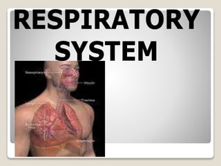
  • 2. RESPIRATORY SYSTEM Objectives: 1. To recognize, recall, and explain the functions of the respiratory system 2. To assess related pathology/ diseases of respiratory system 3. To learn accompanying diagnostic and therapeutic interventions
  • 3. RESPIRATORY SYSTEM (GAS EXCHANGE SYSTEM) WHY DO PEOPLE BREATH? HOW DOES BREATHING HAPPENS
  • 4. Neurochemical Control Respiratory center: lateral medulla oblongata Group of Respiratory Neurons a. Dorsal – autonomic rhythm b. Ventral – active when there is a demand for increased ventilatory effort c. Pneumotaxic and apneustic – modulate an established rhythm  Pneumotaxic – affects inspiratory effort by limiting the volume of air inspired  Apneustic – prevents excessive inflation of the lungs HERING-BREUER INFLATION REFLEX-inhibitory impulses sent to inspiratory neurons in the brainstem to prevent lung overdistention
  • 5. FUCTIONS OF RESPIRATION: DEFReSS 1. Deliver O2 to blood for transport to tissues/ cells 2. Excrete waste products of cellular metab_ 3. Filter, cleanse, warm, humidify air to the lungs 4. Regulate Blood Ph_ 5. Sound for speech, singing 6. Stimulus for Olfaction _
  • 6. EXTERNAL RESPIRATION- gas exchange between air in ALVEOLI and blood PULMONARY CAPILLARIES INTERNAL RESPIRATION- gas exchange between TISSUE CELLS and blood SYSTEMIC CAPILLARIES
  • 7. UPPER RESPIRATORY TRACT (N P L) LOWER RESPIRATORY TRACT (T B L)
  • 8. FUNCTIONS: Upper portion- filters, moistens, warms air  NOSE  PHANRYNX divisions: - NOL - laryngopharynx opens to esophagus and larynx - Phonation - TONSILS: Adenoids, Palatine, Lingual  LARYNX (Voice box) - composed of 9 cartilages in box-like formation - thyroid cartilage (Adam’s Apple) - epiglottis - cricoid - vocal cords: TRUE and FALSE
  • 9. FUNCTIONS: Lower Portion – gas exchange and regulation of pH, PO2, PaCO2  TRACHEA - smooth muscle, C-shaped cartilage - 10-11 cm (4 ½ inches)  BRONCHIAL TREE - bronchi: Left and Right - right: more vertical and larger than the left - bronchioles - terminal bronchioles
  • 10.  LUNGS - Lobes: Left and Right - mediastinum - carina - bronchi - bronchioles - alveoli - surfactant - Pleura: Visceral, Parietal - Pleurisy - Pleural Effusion
  • 11.
  • 12.
  • 13. Defense Mechanisms 1.Mucociliary system - Production of mucus and cilia action 2. Secretory immunity - Production of antibody in mucosal secretions that initiates immune response - Macrophage 3. Surfactant – keeps alveoli open 4. Irritant reflex - Reflex brochospasm, followed by coughing
  • 14. INSPIRATION 1.Respiratory Muscle Contract 2.Thorax increases in size 3.Lungs increases in size 4.Diaphragm flattens 5.Negative pressure in the pleural space_ 6.Gas rushes from higher atmospheric pressure to negative intrapulmonic pressure Inspiration is complete EXPIRATION relax decreases decreases Elevates Positive pressure in the pleural space high pressure in the lung to lower pressure in the atmosphere Expiration is complete PHYSIOLOGY OF RESPIRATION
  • 15. GAS EXCHANGE Ventilation - External respiration - Air flow Perfusion - Internal respiration - Blood flow
  • 16. TERMS Tidal Volume (V1) - Movement of air in and out of the lungs with each respiration - 500-700ml Hyperventilation - Excess 02 causing C02 to fall below normal - 38-42 mmHg Compliance- volume of air lung is able to accept vs pressure from properties that inhibit adequate lung expansion The greater the pressure needed to provide a given volume of air to overcome inhibiting properties, the lower is the lung compliance.
  • 17. Regulation of Breathing Factors: Lung volume and capacity Lung compliance (ability for expansion) Resistance to airflow
  • 18. Lung Compliance  Alteration may occur from the lung or chest wall from thoracic deformity, muscle spasm and abdominal distention_ Resistance  Changes may occur in lung tissues, chest wall or airways  Opposition to airflow
  • 19.  Refers to homeostasis of the hydrogen ion concentration in body fluids in the ECF  Acid – substance that donates hydrogen ions  Base – substance that accepts hydrogen ions
  • 20. Regulation of Acid-Base Balance 1. Buffer System- -control hydrogen ion concentration in the blood and kidney tubules Bicarbonate-carbonic acid system (Carbonate System) – body’s primary buffer system  Bicarbonate helps stabilize pH by combining reversibly with hydrogen ions ◦ Carbonic Acid (H2CO3)-dissolved C02 in water ◦ Sodium Bicarbonate (NaHCO3)- electrolyte, alkalinizing agent
  • 21. Production of Bicarbonate  Increase ◦H2CO3=CO2 + H2O (breakdown of carbonic acid)  Decrease ◦H2CO3= H+ HCO3 (formation of carbonic acid)
  • 22.  ARTERIAL BLOOD GAS – a diagnostic laboratory studies that measures the amount of dissolved O2 and CO2 in arterial blood and indicate the acid-base state. Blood pH– 7.35 to 7.45 PaCO2 – 35 to 45 mmHg PaO2 – 80 to 100 mmHg HCO3 – 22 to 26 meq/L O2 Sat – 96-100%
  • 23.
  • 24. Signs and Symptoms of Acid-Base Imbalance  Metabolic acidosis – kussmaul, headache, hyperkalemia)  Metabolic alkalosis – dizziness, confusion, tetany, convulsion  Respiratory alkalosis – lightheadedness, convulsions, tetany  Respiratory acidosis – hyperpnea, headache, drowsiness, coma, confusion
  • 25. Equipment:  Sterile tuberculin syringe  Heparin  Rubber stopper  Ice  Kidney basin
  • 26. Procedure:  Draw blood into heparinized syringe (90° angle)  Apply pressure to the puncture site for 5 to 10 mins  Sterile technique should be observed  SITE: right radial artery
  • 27. Normal Values: Blood pH – 7.35 to 7.45 PaCO2 – 35 to 45 mmHg PaO2 – 80 to 100 mmHg HCO3 – 22 to 26 meq/L O2 Sat – 96-100%
  • 28. HOW TO ANALYZE ABG 1. Check the pH  pH < 7.35 ( acidosis)  pH = 7.40 (normal)  pH > 7.45 (alkalosis)
  • 29. 2. Determine primary cause of disturbance Acidosis  If PCO2 > 40 – respiratory  If HCO3 < 24 – metabolic Alkalosis  If PCO2 < 40 – respiratory  If HCO3 > 24 – metabolic  ROME
  • 30. 3. Determine the degree of compensation Fully compensated -pH is normal Partially compensated -PCO2 & HCO3 are abnormal, pH abnormal Uncompensated -pH is abnormal, either of PCO2 or HCO3 is normal
  • 31.
  • 32.  Health History  Presenting problem ◦ Nose/nasal sinuses: symptoms may include colds, discharge, epistaxis, sinus problems (swelling, pain) ◦ Throat: symptoms may include sore throat, hoarseness, difficulty swallowing, strep throat ◦ Lungs: symptoms may include  Cough: note duration; frequency; type (dry, hacking, bubbly, barky, hoarse, congested);  sputum (productive vs nonproductive);  circumstances related to cough (time of day, positions, talking, anxiety)  treatment
  • 33.  Health History  Presenting problem  Dyspnea: note onset, severity, duration, efforts to treat,  if accompanied by cough or diaphoresis  time of day when it most likely occurs  interference with ADL  whether precipitated by any specific activities,  whether accompanied by cyanosis.  Wheezing  Chest pain  Hemoptysis  PAIN
  • 34.  Health History  Life-style:  smoking  occupation  geographical location  type and frequency of exercise/recreation ◦ Nutrition/diet: ◦ Past medical history:  immunizations (yearly immunizations for colds/flu  frequency and results of tuberculin skin testing)  allergies (foods, drugs, contact or inhalant allergens  precipitating factors, specific treatment, desensitization)
  • 35. Types of secretions  Sputum – an aggregation of secretions from the tracheobronchial tree, mouth, pharynx, nose and sinuses  Phlegm – refers to secretion of the tracheobronchial tree and lungs. Normal is 100 ml/24 hours
  • 36. Physical Assessment  Inspection (DOB, RR & Cyanosis)  Palpation- masses, respiratory excursion, fremitus  Percussion – resonance (normal lung)  Auscultation
  • 37.  Physical Examination  Inspect for configuration of the chest (kyphosis, scoliosis, barrel chest) and cyanosis.  Determine rate and pattern of breathing (normal rate 12- 18/minute); note tachypnea, hyperventilation, or labored breathing pattern.  Palpate skin, subcutaneous structures, and muscles for texture, temperature, and degree of development.
  • 38. FACTORS AFFECTING THE APPERANCE OF CYANOSIS 1. Pigmentation and thickness Cyanosis must be examined carefully Very thin skin, unpigmented and whose capillaries are superficial and numerous (e.g. tip of the tongue, buccal mucosa, cutaneous surfaces of lips, tips of fingers and toes, nail beds, earlobes and tips of the nose) must be observed Some areas are highly vascular (e.g. heels of the newborns) Mucous membranes (for dark skin patients)
  • 39.  Anemia – may not appear cyanotic  Polycythemia Vera – cyanotic with lesser degree of dissaturation than the normal individuals
  • 40. Breath Sounds & Patterns  Normal Breath Sounds ◦ Vesicular – heard over most of the lungs ◦ Bronchovesicular – main stem bronchi ◦ Bronchial / Tubular – trachea
  • 41.  Abnormal Breath Sounds  Rales – discrete, non-continuous sounds produce by moisture in the tracheobronchial tree. Heard best on inspiration Fine Crackles - COPD, CHF, Pneumonia Coarse Crackles – Pneumonia, pulmonary edema, bronchitis and atelectasis
  • 42. Rhonchi & Wheezes-passage of air in narrowed airways (inflammation, tumor, spastic). Prominent on expiration ◦Asthma, bronchitis, tumor, bronchiolar spasm, foreign body obstruction
  • 43. Friction rubs -cracking, grating sounds originating in an inflamed pleura -predominant in inspiration & expiration ◦Pleurisy, TB, pulmonary infarction, lung abscess
  • 44. Stridor – crowing. harsh, high pitched sounds usually associated with an obstruction in the upper trachea or vocal cords Predominantly in inspiration ◦Croup, foreign body obstruction, large airway obstruction
  • 45. Breathing Pattern Eupnea Tachypnea Bradypnea Apnea Cheyne-stokes – periodic breathing characteristic by periods of slowly waxing and wanning respirations separated by period of apnea Hyperpnea – increase rate and depth of breathing
  • 46. DIAGNOSTIC ASSESSMENTS  Radiographic Studies – used for screening and diagnostic purposes  Chest X-ray – identify pathologic changes in the lungs ◦ Also used to determine placement of catheters and tubes
  • 47. Possible Findings Infiltrates Solid masses (tumor) Areas of necrosis (TB) Excessive air trapping (Emphysema) Abnormal accumulation of air or fluid in the pleural space (pleural effusion or pneumothorax) Gross abnormalities
  • 48. ◦Tomography (Plamigraphy, Laminography or Sectional Radiograph) ◦ techniques used to demonstrate intrathoracic lesions (such as cavities, cysts or calcification) that are observed by overlying structures
  • 49. ◦Fluoroscopy – observation of the motion of the pulmonary and cardiac structures ◦Pulmonary Angiography – rapid injection of the radiopaque dye into the pulmonary circulation. Useful in the determination of the site of pulmonary embolism
  • 50. ◦Bronchography – refer to roent- geographic visualization of the size, shape, patency and number of bronchi
  • 51. ◦Lung scan (Pulmonary Scintiphotography) – use of scanning device that records and outline of the pulmonary radioactivity following injection or inhalation of radiopaque particles ◦Sinus X-ray – obtains information about the size and shape of the sinuses
  • 52.  Examination by Direct Visualization  Rhinoscopy – examination of the interior of the nasal cavities  Laryngoscopy b.1 Indirect – instructed to stick out the tongue and produce a sounds as in “ah” or “e-e-e” b.2 Direct – preparation
  • 53. ◦ Bronchoscopy – visualization of the larger bronchi of the tracheobronchial tree wherein a bronchoscope is inserted through the mouth Prep: NPO 6-8 hours, dentures removal, oral hygiene
  • 54. Post Procedure: ◦NPO until cough, swallowing and gag reflex returns. Talking should be discouraged (complication: laryngeal edema /hemorrhage)
  • 55. ◦ Bronchofiberoscopy (flexible fiber optic bronchoscopy –FFB) – direct visualization using fiber optic bronchoscope which are flexible tube that transmit light and a clear image around corners - Used to diagnose tumors, granulomatous lesions, to find hemorrhage sites, to evaluate trauma or nerve paralysis, obtain biopsy
  • 56. ◦Media Tinoscopy – a small incision is made in the supersternal notch where the media tinoscope is inserted usually performed under general anesthesia -Can identify carcinoma,sarcoidosis, histoplasmosis -Used for staging lung tumors
  • 57. ◦ Transillumination – method of examining the frontal and the maxillary sinuses by directing a beam ◦ Lung biopsy – aspiration of secretion of a needle
  • 58. 2 Approaches  Transtracheobronchial – performed during bronchoscopy with the aid of a fluoroscope  Transthoracic – includes percutaneous needle lung biopsy or aspiration and the open thorachotomy technique (open lung biopsy)
  • 59.  Pleural biopsy – specimen may be obtained from percutaneous needle lung biopsy
  • 60. Laboratory Studies ◦ Hematological ◦ Cytological Studies – sputum, tracheobronchial secretions and pleural fluid to detect the presence of CA cells (e.g. PAP Smear) ◦ Bacteriological – secretions from nasopharynx, chest and pleural cavities
  • 61. ◦ Sputum Studies – goblet cells normally produce 100 ml of mucus per day d.1 Culture of sputum d.2 Sensitivity studies
  • 62. Sputum Color Pathology •Mucoid •Tracheobronchitis, asthma •Yellow or green •Bacterial infection •Rust or blood tinge •Pneumonia, pulmonary infarction, TB
  • 63. Sputum Color Pathology •Black •Black lung disease •Pink •Pulmonary edema
  • 64. d.3 Cytologic Exam (CA)  5 to 10 ml of sputum is sufficient; c/s must be done prior to antimicrobial therapy. Occasionally all sputum collected over a period of 24-72 hours is needed and can be refrigerated in cases of delay
  • 65. e. Thoracentesis – aspiration of fluids or air from the pleural space. May be diagnostic or therapeutic purposes. SITE: 2nd or 3rd ICS MCL for air, 7th or 8th ICS PAL for fluid POSITION: Over bed table; seated in bed with the affected hand raised over the head
  • 66. NURSING RESPONSIBILITIES:  Inform patient to remain immobile  Pressure sensation during procedure  No anticipated discomforts after the procedure  Place patient on unaffected side approximately 1 hour to permit the pleural site to seal itself and prevent fluid seepage from cough or gravitational force
  • 67. f. Skin Test for TB – PPD (Purified Protein Derivatives) f. 1 Mantoux Test – read after 42- 72 hours Interpretation:  10 mm – positive (either infected or indicative of presence of antibodies)  5-9 mm – doubtful  <5mm – negative
  • 68. ASSESSMENT OF PULMONARY FUNCTION  Pulmonary Function Test ◦ to assess lung functions ◦ to rule-out non-organic types of dyspnea such as that cause by psychoneurotic disorders ◦ to differentiate diseases of the lungs (obstruction, restriction or both)
  • 69. Lung Capacities – sum of two or more primary non-overlapping lung volume a.1 Vital Capacity (VC) – quantity of air that a person can expel by a forcible expiration after the deepest inspiration possible; average: 4000-4800 ml for an adult man a.2. Normal Capacity (NC) average: 2900-3000 ml
  • 70. a.3 Total Lung Capacity (TLC) – total amount of air in the lungs after maximal expiration average: 5400 to 5800 ml a.4 Inspiratory Capacity (IC) – maximal amount of air that can be inspired after a normal expiration a.5 Functional Residual Capacity (FRC) amount of air left in the lungs after normal expiration
  • 71. Lung Volumes b.1 Tidal Volume (TV) – volume of air inspired and expired with a normal breath. Average is 500 ml. b.2 Inspiratory Reserve Volume (IRV) – maximal volume that can be inspired from the end of normal inspiration. 1800-2000 ml
  • 72. b.3 Expiratory Reserve Volumes (ERV) – maximal volume that can be exhaled by force expiration after a normal expiration. About 1400 ml
  • 73. b.4 Residual Volume (RV) – volume of air left in the lung after maximal expiration. Approx. 1200 ml or “Dead Space” (does not contribute to gas exchange) b.5 Minute Volume (MV) – volume inspired and expired in one minute of normal breathing. About 1000 ml
  • 74.
  • 75.  Spirometry – the means by which the lung capacity volumes and flow rate are measured.
  • 76. NURSING DIAGNOSIS  Ineffective Airway Clearance  Impaired Gas Exchange  Decrease Cardiac Output  Ineffective Tissue Perfusion OTHER NURSING DIAGNOSIS  Deficient knowledge  Activity intolerance  Disturbed Sleep Pattern  Imbalanced Nutrition  Acute pain  Anxiety
  • 77. Nursing Management:  Assess and monitor ◦ Fluid and electrolyte status ◦ Respiratory status  Nursing activities ◦ Administer oxygen as prescribed ◦ Maintain direct pressure over arterial puncture site for 5 to 10 minutes after drawing the sample ◦ Avoid O2 contamination of arterial specimen ◦ Report result as soon as possible
  • 78. PLANNING 1. Health Promotion  Adequate ventilation  Prevent inhalation of dust and fumes discourage over use of inhalers, sprays and nose drops
  • 79. 2. Restoration and Maintenance ◦ Coughing techniques – 5x q1-2 hours ◦ Suctioning ◦ Reducing metabolic demands  Rest  Decreasing effort of breathing  Maintaining nutrition and hydration  Maintaining elimination
  • 80. 3. Preventing and controlling infection  Medical asepsis  Prophylaxis (e.g. vaccines)  Medications
  • 81.  Oxygen therapy Purposes:  Improved tissue oxygenation  Decrease work of breathing in dyspneic clients  Decrease work of the heart in clients with cardiac disease
  • 82.  Hypoxemia  Hypoxia Types  Hypoxic hypoxia – results from decrease in the diffusion of O2 from the lungs into the arterial blood (e.g. decrease O2 in inspired air (high altitude): lung problems – Atelectasis, pneumonia and pulmonary edema
  • 83.  Anemic hypoxia – occurs in conditions where there is insufficient hgb as in anemic problems and CO poisoning  Ischemic hypoxia – results from decrease tissue perfusion (e.g. MI, CHF, hypovolemic shock, vascular diseases and thrombosis
  • 84.  Histotoxic Hypoxia / Cellular Hypoxia – conditions in which cells have an increase demand for O2 (e.g. hypermetabolism)
  • 85. Assessment for the Need for Oxygen  Increase HR  Dyspnea, rapid, shallow breathing  cardiac dysrhythmias  Drowsiness  Headache  disorientation, excitement, apprehension
  • 86.  flaring nostrils  yawning  restlessness  cyanosis  ICS and sternal retractions
  • 87. GOALS OF O2 THERAPY  Psychological ad physical comfort ◦ Information regarding purposes ◦ Comfort measures such as hygiene (skin. Oral, nasal at least q 2 hours ◦ Proper positioning and repositioning
  • 88.  Promoting safety  Knowledge dissemination regarding O2 properties such as odorless, colorless, tasteless and heavier than air  O2 supports combustion (no smoking rule, electrical equipment should be far from O2 proximity  Drying effect of O2
  • 89.  Maintaining adequate oxygen Mode of Supply  Low-flow System – system that delivers O2 at a rate less than the inspiratory rate requirement
  • 90. Types  Nasal Cannula (Nasal Prong) – O2 flow 1- 6 lpm which delivers 21-24%. Mouth breathing is discouraged  Face mask – O2 flow rates from 35-60 % between 5-10 lpm. Inspired air should be equal or higher than minute ventilation of the client. The following may be used:
  • 91. ◦ Partial re breathing mask (50 to 70%) ◦ Face tents or hoods ◦ Incubators (22 – 40%) ◦ Humidifying tent – provide mists to transport O2 to terminal alveoli ◦ O2 tent – suitable for administration of moderately high concentration of O2. (croup disease – 21 to 30%)
  • 93. SIMPLE O2 MASK RESERVOIR MASK
  • 94.  High-flow – provides the total volume of inspired gas for the client; that is the client only breathes the gas that is being supplied
  • 95. TYPES:  Non-rebreathing mask – concentration is high as 95%. Useful therapy for MI
  • 96.  Venturi Mask – deliver a precise, fixed concentration of O2 ranging from 24-50% ◦ Humidifier is not required to 30% below ◦ Used in COPD because increase concentration of O2 might depress ventilation. It prevents abrupt changes on PaO2 and PCO2 and is thereby an effective method to control the amount of inspired air.
  • 97. Other ways  Tracheostomy  Portable O2  Hyperbaric oxygenation – delivering a 100% O2 in an environment of increase atmospheric pressure. Used to treat carbon monoxide poisoning, air embolism, acute cyanide poisoning
  • 98.  Incentive Spirometry - The client will blow air out of the lungs and then to inhale deeply through a mouth piece attached to a device that measures the client’s maximum inspiration.
  • 99.
  • 101. Aerosol Therapy  To add moisture to oxygen delivery systems  To hydrate thick sputum and prevent mucus plugging  To administer various drugs in the airway ◦ NSS ◦ Detergents – e.g. propyl glycol or glycerine to decrease viscosity of secretions by reducing surface tension
  • 103.  Mucolytics – e.g. acetylcysteine change the physical characteristics of bronchial secretions and increase mobilization  Others – antibiotics, steroids, vasoconstricrtors, bronchodilators
  • 106.  IPPB – refers to pressure greater than the atmospheric pressure at the airway opening during the inspiration. The client’s inspiratory effort triggers the ventilator which pushes air to the lungs  Pressure assisted  Client controlled  Prescribed QID adults – 10 to 15 minutes children – 10 to 15 minutes
  • 107.  Observe for signs of  Hyperventilation: ◦ Headache, chest pain, tingling of the fingers and toes, numbness, vertigo, syncope  Gastric distention  Dangers of worsening pneumothorax  Possible air trapping in clients with obstructive diseases
  • 108.  Artificial airway Functions:  Ensure open airway  Facilitate administration of high concentration of oxygen and humidification  Facilitate mechanical ventilation
  • 109. TYPES:  Oropharyngeal airway – prevents the tongue from falling back and blocking the airway  Endotracheal intubation – inserted through the mouth (orotracheal) or nostril (nasotracheal)
  • 111. ORAL AIRWAYS NASAL TRUMPHETS
  • 112.  Tracheostomy – inflated with at least 1-3 cc of air to seal off. A slight air leak should always be left to decrease pressure in the trachea and prevent ischemic necrosis thus preventing the development of tracheo- esophageal fistulas.
  • 113.
  • 114. Signs to Determine Adequate Amount of Air  Aphonia because air flow in the vocal cords  Absence of an audible escape of air from the nose, mouth and tracheostomy tube when occluded
  • 115. TRACHEOSTOMY CARE 3 MAIN PRINCIPLES 1. maintain patent airway – suction 10-15 minutes during the first 24 hours Sign of mental occlusion  changes in vital signs  changes in mental attitude
  • 116. 2. Prevent infection  Remove inner cannula and cleanse it as often as necessary  Dressing should be change and sites must be inspected for inflammation 3. Prevent drying and crusting of the mucosa  Provide adequate hydration  Installation of at least 2-3 cc of NSS
  • 117.
  • 118.
  • 119.  Ventilation Therapy (Mechanical Ventilation) – use of mechanical device Purposes:  To maintain adequate alveolar ventilation  To provide pulmonary system the mechanical power to maintain physiologic ventilation
  • 120.  To manipulate ventilatory patterns and airway pressures to improve efficiency of ventilation  To decrease myocardial workload by diminishing the work of breathing
  • 121. Indications: 1. Impaired ventilation  Chronic airway obstruction  Restrictive defects  Neuromuscular defects  Respiratory center damage or depression
  • 122. 2. Impaired gas exchange and diffusion  ARDS  Atelectasis  Pneumonia  Tumor
  • 123. 3. Respiratory Failure as evidenced by ABG  Continuous decrease in oxygenation  An increase in arterial carbon dioxide  Persistence of acidosis
  • 124. Types: POSITIVE-PRESSURE VENTILATORS  Pressure cycled – permits air to flow into the client’s lung until a predetermined pressure is reached (e.g. Bird and Bennet, PR-1, PR-2) - The major limitation is that the volume of air can vary as the patient’s airway resistance or compliance change
  • 125.  Volume cycled ◦ delivers a predetermined volume of gas into the patient’s lung with each breath and exhalation occurs passively ◦ Intended for long term use for patient with primary pulmonary disease ◦ Volume may range up to 2000 cc or more (e.g. Bennet MA-1, Engstron, Ohio and Emerson Critical Care Ventilators)
  • 126.  Time cycled ventilators ◦Terminates or control inspiration after preset time ◦Volume of air the patient receives is regulated by the length of inspiration and flow rate of the air ◦Used in newborns and infants
  • 127. Accessory Attachments:  Intermittent Mandatory Ventilation (IMV) – a method for providing both spontaneous breathing and breathing by machine. This is also used for weaning.
  • 128.  Continuous Positive Airway Pressure (CPAP) - a non-mechanical means of ventilation. ◦ It provides continuous positive airway pressure by maintaining pressure above zero at the end of expiratory phase thereby preventing alveolar collapse ◦ An endotracheal tube or face mask may be used to delivered measured concentration of O2.
  • 129.  Positive End-Expiratory Pressure (PEEP) – a method of maintaining a pressure higher than the atmospheric pressure in the lungs at the end of expiration ◦ - CPAP differs from PEEP in that in the client in CPAP is breathing spontaneously and may not be on ventilator whereby in PEEP, it is controlled by a ventilator.
  • 131. Ventilator Setting  FiO2 – fraction of inspired oxygen; the concentration of O2 delivered (21 – 100%)  Tidal Volume – the amount of air the machine has been set to deliver with each ventilator breath ◦ Pt’s wt. in kilograms multiplied by 10-15 cc  Respiratory rate – no. of positive pressure per minute  Sigh – additional volumes of air delivered several times each hour. Average is 10 to 15 per hour  Respirator mode
  • 132. Potential Complications:  ET displacement or trache extubation  Infections  Barotrauma – leads to rupture of over distended alveolus  Central nervous system disturbances due to decrease cerebral venous return  Psychological trauma – fear, anxiety, inability to communicate  Oxygen toxicity – high alveolar oxygen tensions can impair respiratory function
  • 133.  Hemodynamic alterations caused by ◦ Restriction of left ventricular filling ◦ Limitation of expansion and filling of the ventricles during systole ◦ Limitation of the intrathoracic pump  Musculoskeletal complications ◦ Tissue necrosis on mouth (ET), trache stoma, infection and pressure sores
  • 134. CARE OF MECHANICALLY VENTILATED PATIENTS  Basic airway care  Prevention of respiratory complications ◦ Humidification, USN, suctioning, turning, deep breathing, CPT  Prevention of infection ◦ Aseptic technique ◦ Clean possible sources of contamination
  • 135. ◦ Use sterile supplies ◦ Daily dressing ◦ Nutrition and hygiene  Prevention of Oxygen toxicity ◦ Limit the use of 100% O2 to brief periods ◦ Decrease FiO2 to lowest possible level to maintain PaO2 at 60 to 90 mmHg ◦ Up to 70% O2 may be used safely for 24 hours
  • 136. ◦ After 2 days, an FiO2 about $0% is potentially toxic ◦ Prolonged use of FiO2 below 40% rarely causes O2 toxicity ◦ Up to 50% O2 may be used safely for 2 days
  • 137.  Prevention of other complications ◦ Bedsore formation Areas of Concerns:  Nutrition  Fluid and electrolyte balance  Muscle tone and skin integrity  Communication, emotional and psychological support  Environmental factors
  • 138. TROUBLE SHOOTING VENTILATOR PROBLEMS CAUSES INTERVENTIONS Increased secretions Suction the patient Kinked ventilator tubing or ET tube Unkink the tubing Assess & check the cm markings on the ET at the lips or nares Check for air entry bilaterally ALARM TYPE: HIGH PRESSURE
  • 139. CAUSES INTERVENTIONS Patient biting the ET Place an oral airway Water in the ventilator tubing Disconnect the tubing at a connection point near the patient and drain water using special receptacle for contaminated drainage
  • 140. CAUSES INTERVENTIONS ET advance into the right main stem bronchus Notify doctor CXR or pulling back Barotrauma (rupture of over distended alveolus) Assess for palpable SQ emphysema, tracheal deviation, unequal breath sounds, shock and cardiac or respiratory arrest from tension pneumothorax and inform MD
  • 141. CAUSES INTERVENTIONS Brochospasm from constricted bronchioles Ensure proper ET position by auscultating the lungs and abdomen. Check marking cm Rule out other causes of high alarms and notify doctors for sedation and bronchodilators
  • 142. CAUSES INTERVENTIONS Patient fighting (“bucking”) the ventilator Manual ventilation. Determine the cause of discomforts. Provide health teachings If with hypoxemia, hyper oxygenate the patient and monitor pulse and ABG.
  • 143. CAUSES INTERVENTIONS Decrease pulmonary compliance Rule possible problems. If persistent, suspect ARDS, pneumonia, or pleural effusion. Refer to MD.
  • 144. ALARM TYPE: LOW PRESSURE CAUSES INTERVENTIONS Disconnected tubing Secure all disconnections Accidental extubation Reposition the tube if slightly disconnected. If completely blocking the airway, remove tube and manually ventilate the patient. Re- intubation.
  • 145. CAUSES INTERVENTIONS Cuff leak Deflate and re-inflate the cuff. If persistent, change the tube Accidental removal of trache tube Insert a new tube with the obturator and then remove the obturator. Always have an extra trache tube at the bedside.
  • 146. CAUSES INTERVENTIONS The patient being weaned fails to breath during preset periods Assess respirations, ABG’s as indicated during weaning. Consult respiratory therapist and MD to determine if weaning will be discontinued. If prolonged, check for respiratory arrest. Stimulate and ventilate the patient ALARM TYPE: APNEA
  • 147. ALARM TYPE: LOW EXHALED TIDAL VOLUME CAUSES INTERVENTIONS The ventilator detects a single shallow low exhaled volume during weaning Assess respirations and ABG’s as indicated during weaning. consult
  • 148. CAUSES INTERVENTIONS Exhaled volume per minute is low during weaning Assess respirations and ABG’s ALARM TYPE: LOW EXHALED MINUTE VOLUME
  • 149. CAUSES INTERVENTIONS The O2 supply is insufficient or isn’t properly connected Correct malfunction and manually ventilate the patient. Monitor O2 saturation. ALARM TYPE: OXYGEN
  • 150. CAUSES INTERVENTIONS Ventilator inoperable Plug ventilator to an outlet connected to back up electrical source. Check the patient. ALARM TYPE: POWER FAILURE
  • 151. Criteria for termination of weaning:  Decease in LOC  DBP 100 mmHg  Fall in SBP  Increase HR  pH 7.35  PaCO2 increased by 8  PVC’s
  • 152.  Chest Surgery Pre-op care:  Diagnostic exams  Nutritional status – CHON and Vitamin C  CPT
  • 153. Post-op:  Maintain patent airway and promoting comfort  Maintain hydration and nutrition  Maintain activity