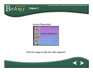
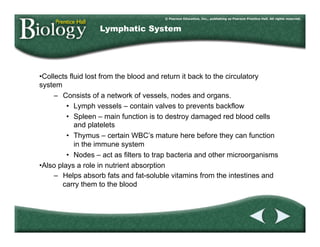
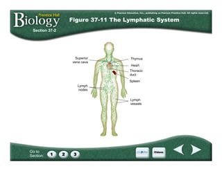
The document provides information about the human respiratory system, including:
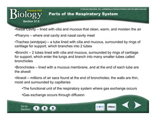
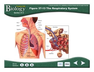
- The parts of the respiratory system including the nose, pharynx, larynx, trachea, bronchi, bronchioles, and alveoli.
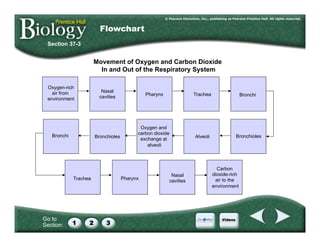
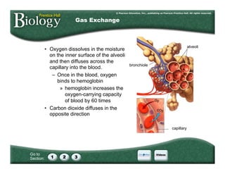
- How oxygen and carbon dioxide are exchanged through diffusion between the alveoli and capillaries in the lungs.
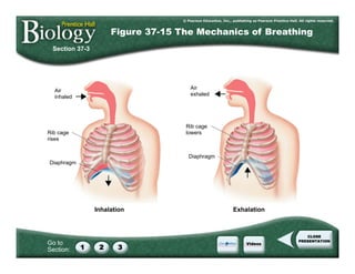
- The mechanics of breathing which is controlled by the diaphragm and rib cage and regulated by carbon dioxide levels in the blood.
- Common respiratory diseases such as bronchitis, asthma, and emphysema.


![Polymer [ बहुलक ] Chemistry Notes PDF - Irfanullah Mehar - JJ Sir Chemistry.pdf](https://cdn.slidesharecdn.com/ss_thumbnails/polymerchemistrynotespdf-irfanullahmehar-jjsirchemistry-260210172118-3f9b37f7-thumbnail.jpg?width=640&height=640&fit=bounds)


