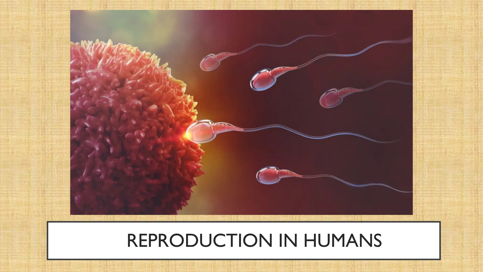
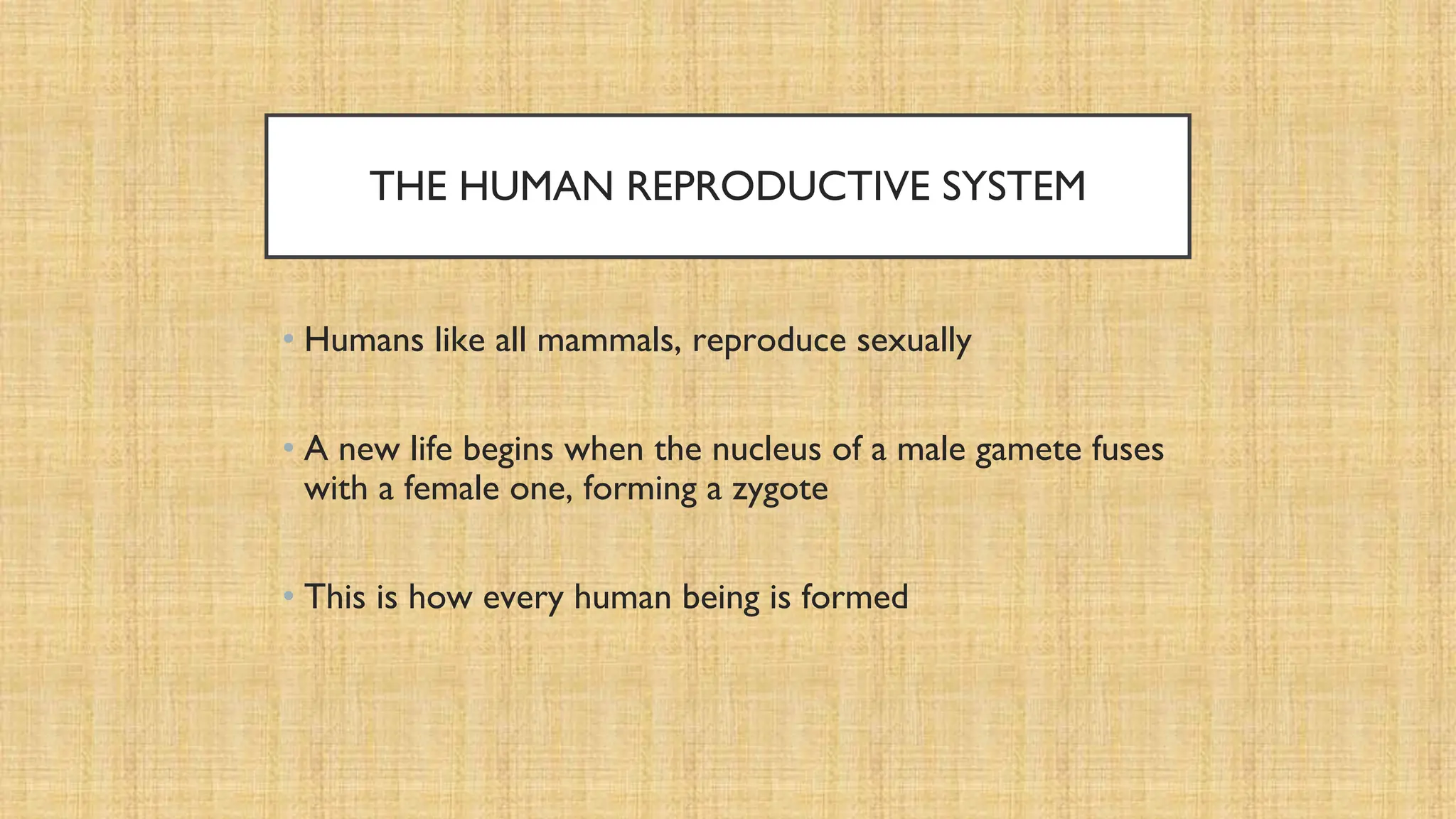
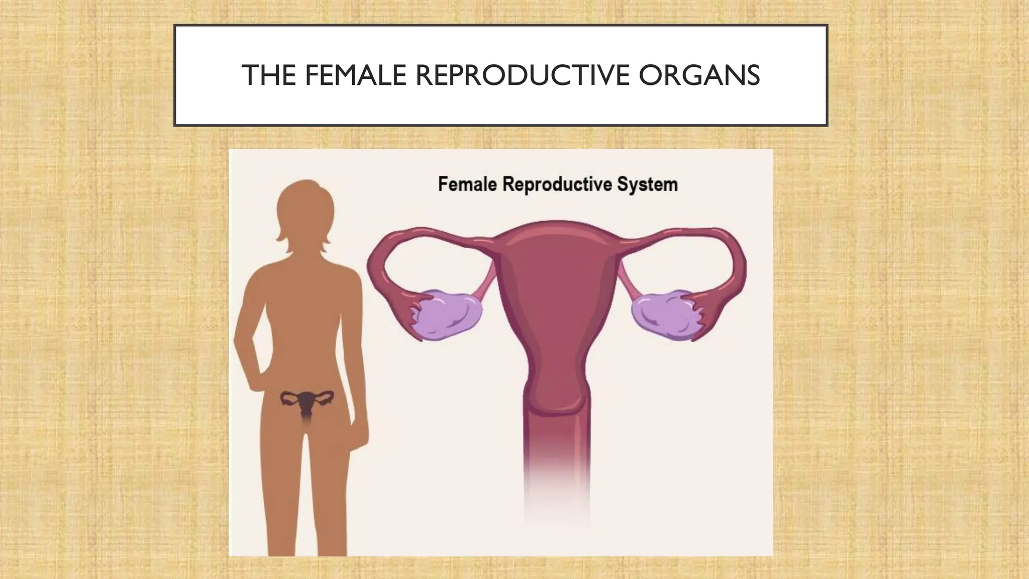
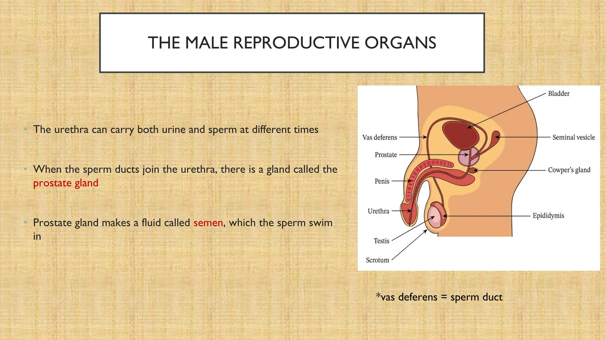
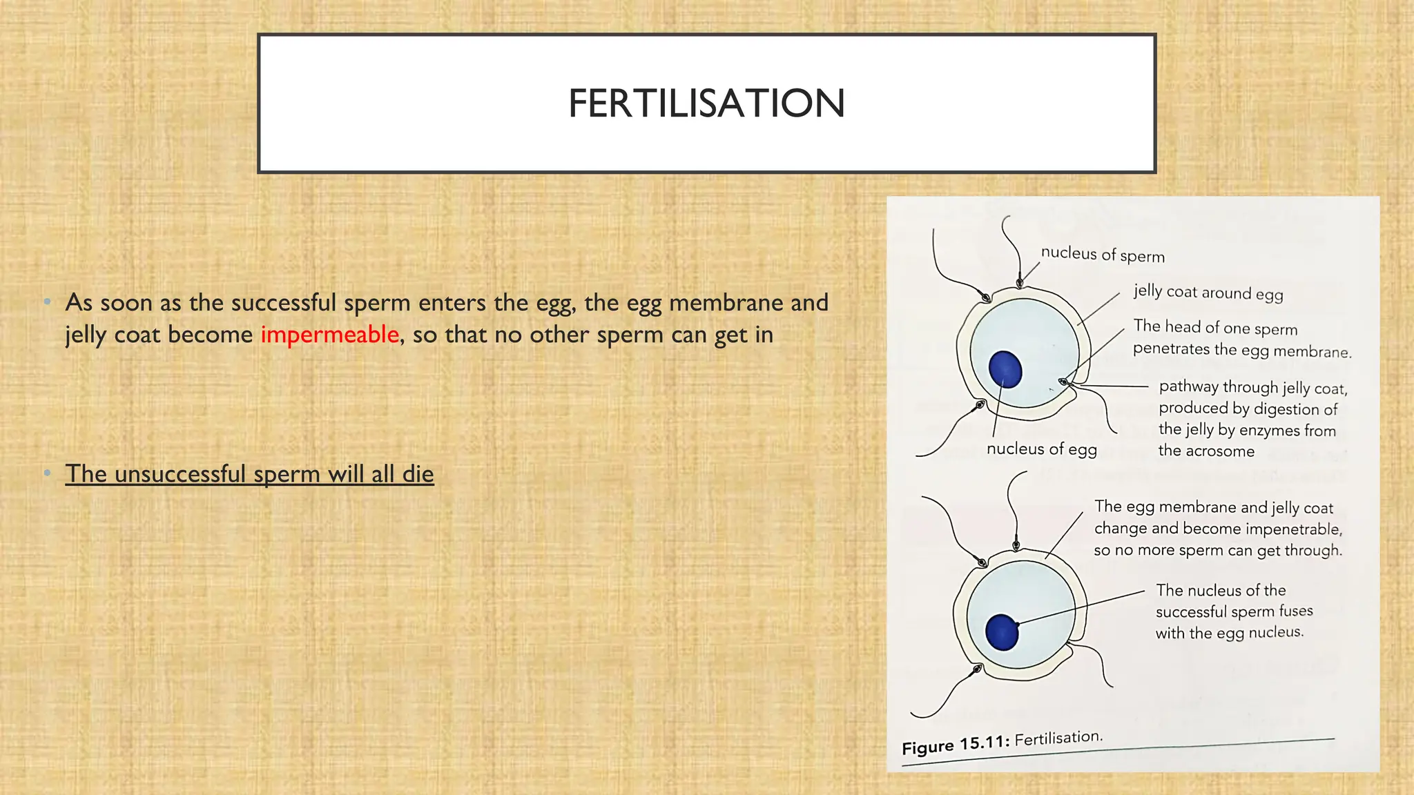
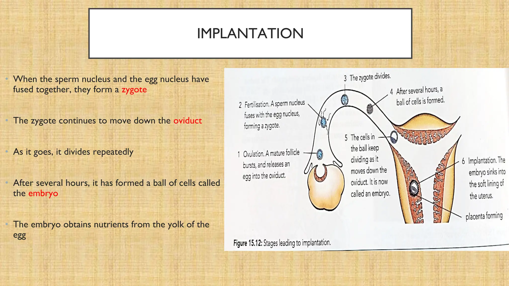
REPRODUCTION IN HUMANS
VIDEO LINKS
• https://www.youtube.com/watch?v=-ekRRuSa_UQ (introduction)
• https://www.youtube.com/watch?v=5EvnCzOpy2o (fertilization till 1:34)
THE HUMAN REPRODUCTIVE SYSTEM
• Humans like all mammals, reproduce sexually
• A new life begins when the nucleus of a male gamete fuses
with a female one, forming a zygote
• This is how every human being is formed
THE FEMALE REPRODUCTIVE ORGANS
• The female gametes called eggs or egg cells, are made in the
two ovaries
• Leading away from the ovaries are the oviducts (also called
Fallopian tubes)
• The tubes do not connect directly to the ovaries but have a
funnel shaped opening just a short distance away
• The two oviducts lead to the womb or the uterus
• Uterus has very thick walls, made of muscle
THE FEMALE REPRODUCTIVE ORGANS
• Uterus is very small – only about the size of a clenched fist, but
it can stretch a great deal when a woman is pregnant
• At the base of the uterus is a narrow opening, guarded by
muscles
• This is the neck of the uterus, or the cervix
• The narrow opening connects to the vagina, which then leads
to the outside of the body
THE FEMALE REPRODUCTIVE ORGANS
• The opening from the bladder, called the urethra, is a tube
that is in front of the vagina
• While the rectum is just behind the vagina
• These three tubes open quite separately to the outside
THE MALE REPRODUCTIVE ORGANS
• The male gametes called spermatozoa or sperm are made in two
testes
• Testes are outside the body in two sacs of skin called the scrotum
• The sperm are carried away from each testes in a tube called the
sperm duct (vas deferens)
• The sperm ducts from the testes join up with the urethra just
below the bladder
• The urethra continues downwards and opens at the tip of the
penis
*vas deferens = sperm duct
THE MALE REPRODUCTIVE ORGANS
• The urethra can carry both urine and sperm at different times
• When the sperm ducts join the urethra, there is a gland called the
prostate gland
• Prostate gland makes a fluid called semen, which the sperm swim
in
*vas deferens = sperm duct
THE MALE REPRODUCTIVE ORGANS
HUMAN GAMETES
• Eggs begin to be formed inside a girl’s ovaries before she is born
• At birth, she will already have thousands of partly developed eggs inside her ovaries
• When she reaches puberty, some of these eggs begin to mature
• Usually, only one develops at a time
HUMAN GAMETES
• An egg is a single cell
• When it is fully developed, it bursts out of the ovary and into the funnel
at the end of the oviduct
• This is called ovulation
• In humans, it happens once a month throughout a woman’s reproductive
cycle
HUMAN GAMETES
• Figure shows a section through a testis
• It contains thousands of very narrow, coiled tubes or tubules
• These are where the sperm are made
• Sperm are then stored in the epididymis
• Sperm develop from cells in the walls of the tubules
• Sperm are continually made from puberty onwards
HUMAN GAMETES
• Sperm production is very sensitive to heat
• If they get too hot, the cells in the tubules will not develop into sperm
• This is why the testes are positioned outside the body, where they are
cooler then they would be inside
Figure shows the structure of a sperm and how it is adapted for its function of fertilizing an egg
HUMAN GAMETES
• Eggs and sperm are haploid cells
• Each human egg and each human sperm has a single set of 23 chromosomes in its nucleus
• Eggs and sperm are made when cells in the ovaries and testes divide by meiosis
COMPARING MALE AND FEMALE GAMETES
• An egg cell usually millions of times larger than a sperm cell
• Egg cell and sperm cells differ greatly in their structures (as already seen)
• Usually one egg cell is released during one reproductive cycle of a woman, while millions of sperms are
released at once in a man
• Egg cells are non-motile while sperm cells are motile
FERTILISATION
• After ovulation, the egg is caught in the funnel of the oviduct
• Very slowly, the egg travels towards the uterus
• Cilia lining the oviduct help it to sweep along
• Muscles in the walls of the oviduct also help to move it, producing gentle
rippling movements
Movement of egg from the oviduct into the uterus
FERTILISATION
• If the egg is not fertilized by a sperm within 8 to 24 hours after ovulation, it
dies
• By this time it has only travelled a short way along the oviduct
• This means that a sperm must reach an egg while it is quite near the top of
the oviduct if fertilization is to be successful
FERTILISATION
• To bring the sperm as close as possible to the egg, the man’s penis is
placed inside the vagina of the woman
• Sperm travel out of the penis into the vagina
• This happens when muscles in the walls of the tubes containing the sperm
contract rhythmically
• The wave of contraction begins in the testes, travels along the sperm ducts
and into the penis
• The sperm are squeezed along the tubes, out of the man’s urethra and into
the woman’s vagina
FERTILISATION
• The fluid containing the sperm is called semen
• The liquid part of the semen is produced by the prostate gland, and it
contains sugars and other nutrients to provide the sperm with energy
• The semen is deposited at the top of the vagina, near the cervix
• The sperm are still quite a long way from the egg
• They swim (using their flagella and energy released in their mitochondria)
up through the cervix, through the uterus and into the oviduct
FERTILISATION
• Sperm can only swim at a rate of about 4 mm per minute, so it takes
quite a while for them to get as far as the oviducts
• Many will never get there at all
• But about a million sperm are deposited in the vagina at once,
so there is a good chance that some of them will reach the egg
FERTILISATION
• When a sperm cell contacts the jelly coat surrounding an egg cell, the
acrosome is activated
• It releases its digestive enzymes, and these enzymes digest the jelly coat
• This allows the head of the sperm to push through and get into the cytoplasm
of the egg
• Only the head of the sperm goes in; the flagellum is left outside
• The nucleus of the sperm fuses with the nucleus of the egg
• This is fertilisation
FERTILISATION
• As soon as the successful sperm enters the egg, the egg membrane and
jelly coat become impermeable, so that no other sperm can get in
• The unsuccessful sperm will all die
A sperm as it makes its way into and egg cell
IMPLANTATION
• When the sperm nucleus and the egg nucleus have
fused together, they form a zygote
• The zygote continues to move down the oviduct
• As it goes, it divides repeatedly
• After several hours, it has formed a ball of cells called
the embryo
• The embryo obtains nutrients from the yolk of the
egg
IMPLANTATION
• It takes several hours for the embryo to reach the uterus, and by this time it is a ball of 16 or 32 cells
• The uterus has a thick, spongy lining, and the embryo sinks into it
• This is called implantation
THE PLACENTA
• The cells in the embryo, now buried in the soft wall of the uterus, continue
to divide
• As the embryo grows, a placenta also grows, which connects it to the wall of
the uterus
• The placenta is soft, dark red, and has finger-like projections called
villi
• The villi fit closely into the uterus wall
• The placenta is where substances are exchanged between the mother’s
blood and the embryo’s blood
• It is the embryo’s life support system
THE PLACENTA
• After 11 weeks, the embryo has developed most of its organs, and is
now called a fetus
• The placenta is joined to the fetus by the umbilical cord
• Inside the cord are two arteries and a vein
• The arteries take blood from the fetus into the placenta, and
the vein returns the blood to the fetus
THE PLACENTA
• The placenta contains capillaries filled with the fetus’s blood
• The lining of the uterus contains large spaces filled with the
mother’s blood
• The fetus’s blood and the mother’s blood do not mix
• They are separated by the wall of the placenta
• But they are brought very close together, because the wall of the
placenta is very thin
THE PLACENTA
• Oxygen and dissolved nutrients in the mother’s blood diffuse
across the placenta into the fetus’s blood and are then carried
along the umbilical cord to the fetus
• Carbon dioxide and other excretory products diffuse in the
other direction and are carried away in the mother’s blood
• Some unwanted substances can also cross the placenta, these
include toxins (alcohol, carbon monoxide)
• Some pathogens such as the virus that causes rubella, can also
pass across the placenta
THE PLACENTA
• The arteries in the uterus lining, dissolve and release their blood, forming a
pool of blood
• Hence, the mother’s blood found in the uterus lining is no longer enclosed in
blood vessels
• This reduces one membrane that the materials from the mother’s blood have
to pass through, to the reach the fetus’s blood
THE PLACENTA
• As the fetus grows, the placenta grows too
• By the time the baby is born, the placenta is a
flat disc
• It is, at that time, about 12 cm in diameter and 3
cm thick
THE AMNIOTIC SAC
• The fetus is surrounded by a strong membrane, called the
amniotic sac
• This makes a liquid called the amniotic fluid
• Amniotic fluid helps to support the embryo, and to
protect it from mechanical injury
• The embryo floats in the fluid and is able to freely move its
arms and legs, which helps the muscle and skeleton to
develop correctly
THE AMNIOTIC SAC
• After nine months, when the fetus is ready to be born,
contractions of muscles in the uterus wall often cause the
amniotic sac to break
• This releases the amniotic fluid which flows into the vagina
• This is often the first sign that a woman is about to give
birth
• Mother’s blood to the embryo’s blood: oxygen, glucose, amino acids, fatty
acids, glycerol, minerals and water
• Embryo’s blood to the mother’s blood: carbon dioxide and urea
• https://www.youtube.com/watch?v=pX55X9au6zQ (video shows a very rare condition in which a baby is shown
born through a C-section with the amniotic sac still intact)
• https://www.youtube.com/watch?v=izOa3-AX8zQ (how a wonder is born) A very detailed video with amazing
animations! You must watch this till the end and focus on the details that you have covered in class.

REPRODUCTION IN HUMANS.pdfhjhjwiowwowjwww

  • 1.
  • 2.
    VIDEO LINKS • https://www.youtube.com/watch?v=-ekRRuSa_UQ(introduction) • https://www.youtube.com/watch?v=5EvnCzOpy2o (fertilization till 1:34)
  • 3.
    THE HUMAN REPRODUCTIVESYSTEM • Humans like all mammals, reproduce sexually • A new life begins when the nucleus of a male gamete fuses with a female one, forming a zygote • This is how every human being is formed
  • 4.
  • 5.
    THE FEMALE REPRODUCTIVEORGANS • The female gametes called eggs or egg cells, are made in the two ovaries • Leading away from the ovaries are the oviducts (also called Fallopian tubes) • The tubes do not connect directly to the ovaries but have a funnel shaped opening just a short distance away • The two oviducts lead to the womb or the uterus • Uterus has very thick walls, made of muscle
  • 6.
    THE FEMALE REPRODUCTIVEORGANS • Uterus is very small – only about the size of a clenched fist, but it can stretch a great deal when a woman is pregnant • At the base of the uterus is a narrow opening, guarded by muscles • This is the neck of the uterus, or the cervix • The narrow opening connects to the vagina, which then leads to the outside of the body
  • 7.
    THE FEMALE REPRODUCTIVEORGANS • The opening from the bladder, called the urethra, is a tube that is in front of the vagina • While the rectum is just behind the vagina • These three tubes open quite separately to the outside
  • 8.
    THE MALE REPRODUCTIVEORGANS • The male gametes called spermatozoa or sperm are made in two testes • Testes are outside the body in two sacs of skin called the scrotum • The sperm are carried away from each testes in a tube called the sperm duct (vas deferens) • The sperm ducts from the testes join up with the urethra just below the bladder • The urethra continues downwards and opens at the tip of the penis *vas deferens = sperm duct
  • 9.
    THE MALE REPRODUCTIVEORGANS • The urethra can carry both urine and sperm at different times • When the sperm ducts join the urethra, there is a gland called the prostate gland • Prostate gland makes a fluid called semen, which the sperm swim in *vas deferens = sperm duct
  • 10.
  • 11.
    HUMAN GAMETES • Eggsbegin to be formed inside a girl’s ovaries before she is born • At birth, she will already have thousands of partly developed eggs inside her ovaries • When she reaches puberty, some of these eggs begin to mature • Usually, only one develops at a time
  • 12.
    HUMAN GAMETES • Anegg is a single cell • When it is fully developed, it bursts out of the ovary and into the funnel at the end of the oviduct • This is called ovulation • In humans, it happens once a month throughout a woman’s reproductive cycle
  • 13.
    HUMAN GAMETES • Figureshows a section through a testis • It contains thousands of very narrow, coiled tubes or tubules • These are where the sperm are made • Sperm are then stored in the epididymis • Sperm develop from cells in the walls of the tubules • Sperm are continually made from puberty onwards
  • 14.
    HUMAN GAMETES • Spermproduction is very sensitive to heat • If they get too hot, the cells in the tubules will not develop into sperm • This is why the testes are positioned outside the body, where they are cooler then they would be inside
  • 15.
    Figure shows thestructure of a sperm and how it is adapted for its function of fertilizing an egg
  • 16.
    HUMAN GAMETES • Eggsand sperm are haploid cells • Each human egg and each human sperm has a single set of 23 chromosomes in its nucleus • Eggs and sperm are made when cells in the ovaries and testes divide by meiosis
  • 17.
    COMPARING MALE ANDFEMALE GAMETES • An egg cell usually millions of times larger than a sperm cell • Egg cell and sperm cells differ greatly in their structures (as already seen) • Usually one egg cell is released during one reproductive cycle of a woman, while millions of sperms are released at once in a man • Egg cells are non-motile while sperm cells are motile
  • 18.
    FERTILISATION • After ovulation,the egg is caught in the funnel of the oviduct • Very slowly, the egg travels towards the uterus • Cilia lining the oviduct help it to sweep along • Muscles in the walls of the oviduct also help to move it, producing gentle rippling movements
  • 19.
    Movement of eggfrom the oviduct into the uterus
  • 20.
    FERTILISATION • If theegg is not fertilized by a sperm within 8 to 24 hours after ovulation, it dies • By this time it has only travelled a short way along the oviduct • This means that a sperm must reach an egg while it is quite near the top of the oviduct if fertilization is to be successful
  • 21.
    FERTILISATION • To bringthe sperm as close as possible to the egg, the man’s penis is placed inside the vagina of the woman • Sperm travel out of the penis into the vagina • This happens when muscles in the walls of the tubes containing the sperm contract rhythmically • The wave of contraction begins in the testes, travels along the sperm ducts and into the penis • The sperm are squeezed along the tubes, out of the man’s urethra and into the woman’s vagina
  • 22.
    FERTILISATION • The fluidcontaining the sperm is called semen • The liquid part of the semen is produced by the prostate gland, and it contains sugars and other nutrients to provide the sperm with energy • The semen is deposited at the top of the vagina, near the cervix • The sperm are still quite a long way from the egg • They swim (using their flagella and energy released in their mitochondria) up through the cervix, through the uterus and into the oviduct
  • 24.
    FERTILISATION • Sperm canonly swim at a rate of about 4 mm per minute, so it takes quite a while for them to get as far as the oviducts • Many will never get there at all • But about a million sperm are deposited in the vagina at once, so there is a good chance that some of them will reach the egg
  • 25.
    FERTILISATION • When asperm cell contacts the jelly coat surrounding an egg cell, the acrosome is activated • It releases its digestive enzymes, and these enzymes digest the jelly coat • This allows the head of the sperm to push through and get into the cytoplasm of the egg • Only the head of the sperm goes in; the flagellum is left outside • The nucleus of the sperm fuses with the nucleus of the egg • This is fertilisation
  • 26.
    FERTILISATION • As soonas the successful sperm enters the egg, the egg membrane and jelly coat become impermeable, so that no other sperm can get in • The unsuccessful sperm will all die
  • 27.
    A sperm asit makes its way into and egg cell
  • 29.
    IMPLANTATION • When thesperm nucleus and the egg nucleus have fused together, they form a zygote • The zygote continues to move down the oviduct • As it goes, it divides repeatedly • After several hours, it has formed a ball of cells called the embryo • The embryo obtains nutrients from the yolk of the egg
  • 30.
    IMPLANTATION • It takesseveral hours for the embryo to reach the uterus, and by this time it is a ball of 16 or 32 cells • The uterus has a thick, spongy lining, and the embryo sinks into it • This is called implantation
  • 31.
    THE PLACENTA • Thecells in the embryo, now buried in the soft wall of the uterus, continue to divide • As the embryo grows, a placenta also grows, which connects it to the wall of the uterus • The placenta is soft, dark red, and has finger-like projections called villi • The villi fit closely into the uterus wall • The placenta is where substances are exchanged between the mother’s blood and the embryo’s blood • It is the embryo’s life support system
  • 32.
    THE PLACENTA • After11 weeks, the embryo has developed most of its organs, and is now called a fetus • The placenta is joined to the fetus by the umbilical cord • Inside the cord are two arteries and a vein • The arteries take blood from the fetus into the placenta, and the vein returns the blood to the fetus
  • 33.
    THE PLACENTA • Theplacenta contains capillaries filled with the fetus’s blood • The lining of the uterus contains large spaces filled with the mother’s blood • The fetus’s blood and the mother’s blood do not mix • They are separated by the wall of the placenta • But they are brought very close together, because the wall of the placenta is very thin
  • 34.
    THE PLACENTA • Oxygenand dissolved nutrients in the mother’s blood diffuse across the placenta into the fetus’s blood and are then carried along the umbilical cord to the fetus • Carbon dioxide and other excretory products diffuse in the other direction and are carried away in the mother’s blood • Some unwanted substances can also cross the placenta, these include toxins (alcohol, carbon monoxide) • Some pathogens such as the virus that causes rubella, can also pass across the placenta
  • 35.
    THE PLACENTA • Thearteries in the uterus lining, dissolve and release their blood, forming a pool of blood • Hence, the mother’s blood found in the uterus lining is no longer enclosed in blood vessels • This reduces one membrane that the materials from the mother’s blood have to pass through, to the reach the fetus’s blood
  • 36.
    THE PLACENTA • Asthe fetus grows, the placenta grows too • By the time the baby is born, the placenta is a flat disc • It is, at that time, about 12 cm in diameter and 3 cm thick
  • 37.
    THE AMNIOTIC SAC •The fetus is surrounded by a strong membrane, called the amniotic sac • This makes a liquid called the amniotic fluid • Amniotic fluid helps to support the embryo, and to protect it from mechanical injury • The embryo floats in the fluid and is able to freely move its arms and legs, which helps the muscle and skeleton to develop correctly
  • 38.
    THE AMNIOTIC SAC •After nine months, when the fetus is ready to be born, contractions of muscles in the uterus wall often cause the amniotic sac to break • This releases the amniotic fluid which flows into the vagina • This is often the first sign that a woman is about to give birth
  • 40.
    • Mother’s bloodto the embryo’s blood: oxygen, glucose, amino acids, fatty acids, glycerol, minerals and water • Embryo’s blood to the mother’s blood: carbon dioxide and urea
  • 41.
    • https://www.youtube.com/watch?v=pX55X9au6zQ (videoshows a very rare condition in which a baby is shown born through a C-section with the amniotic sac still intact) • https://www.youtube.com/watch?v=izOa3-AX8zQ (how a wonder is born) A very detailed video with amazing animations! You must watch this till the end and focus on the details that you have covered in class.