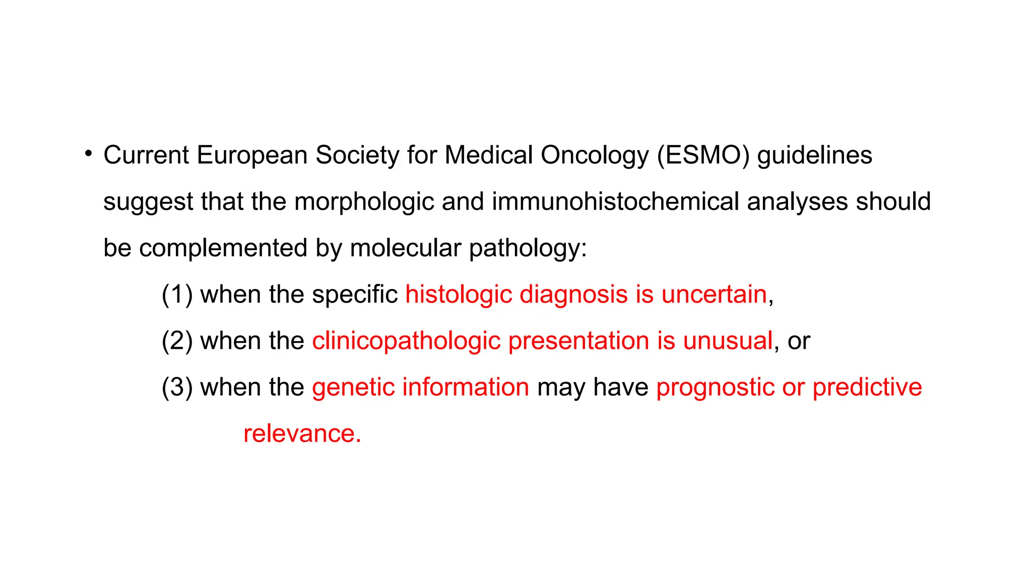
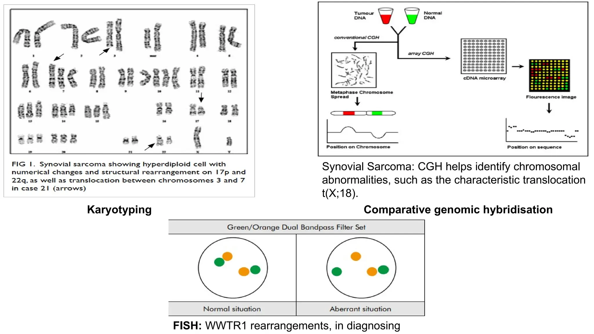
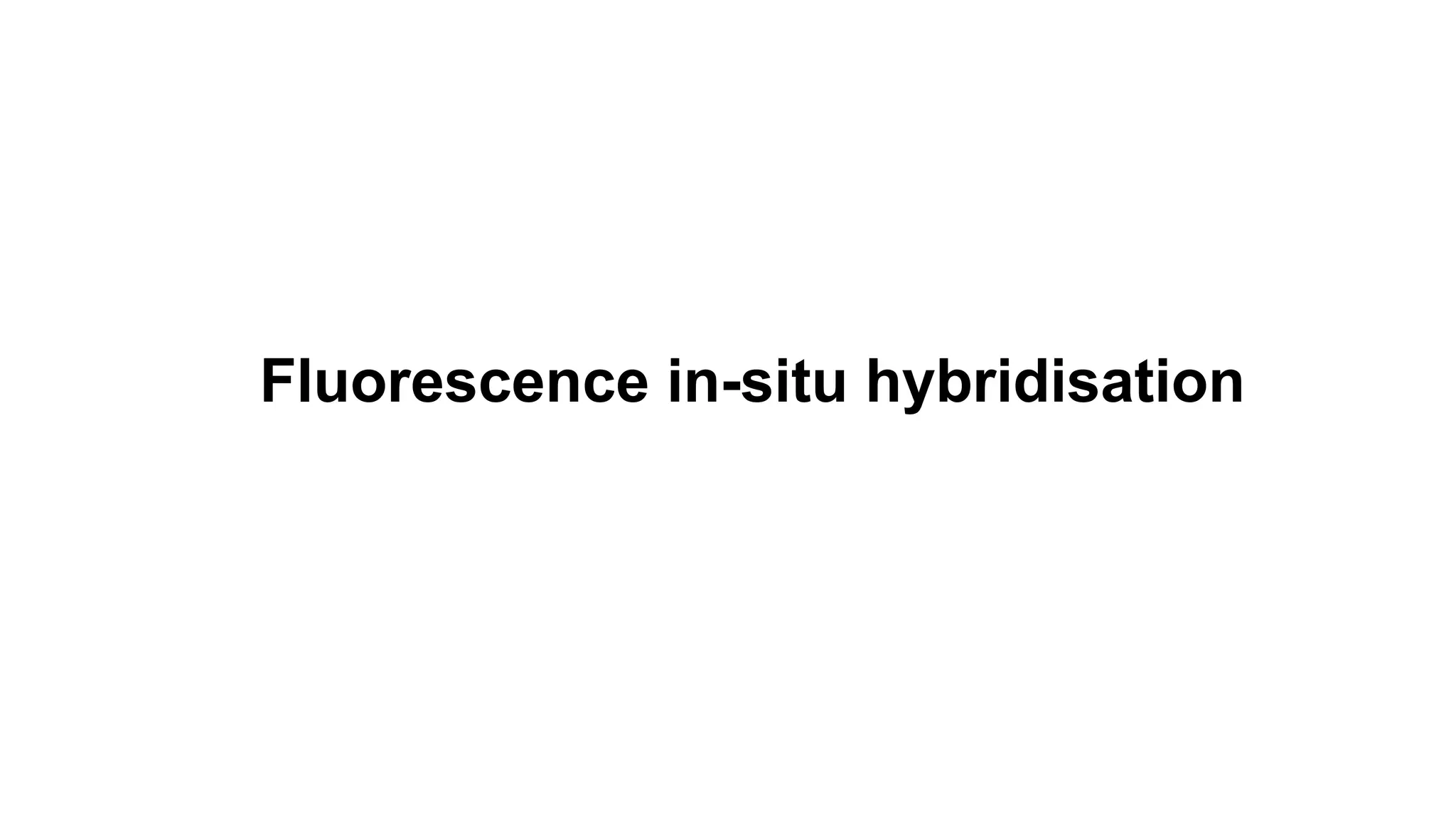
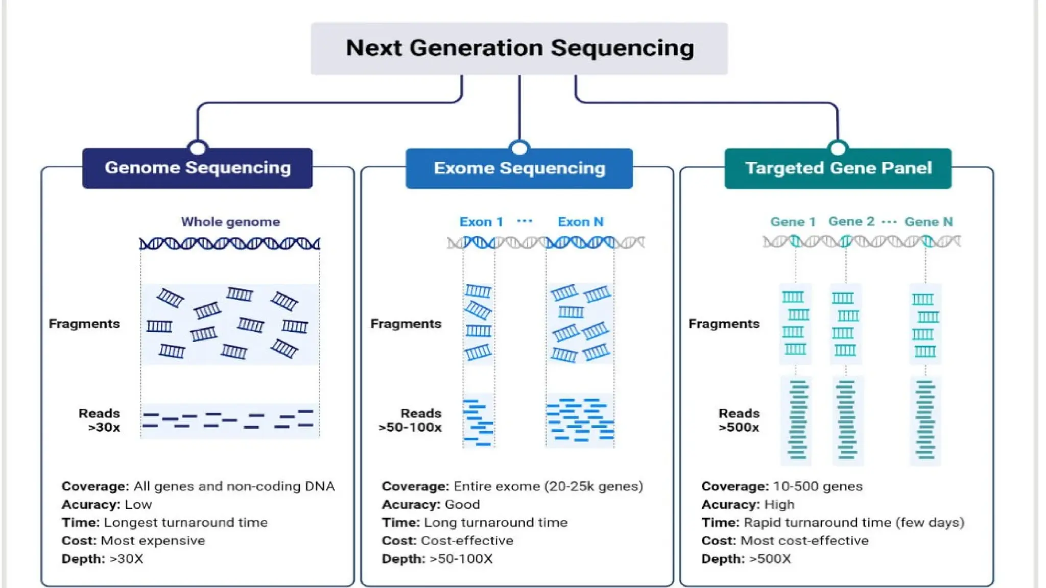
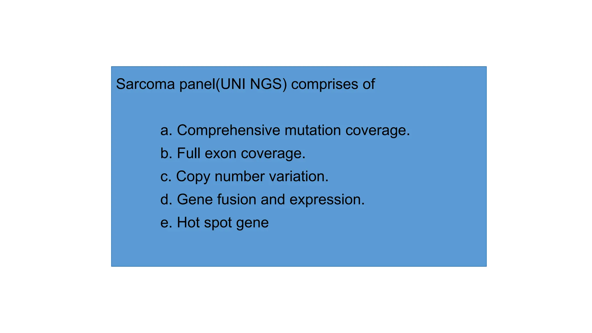
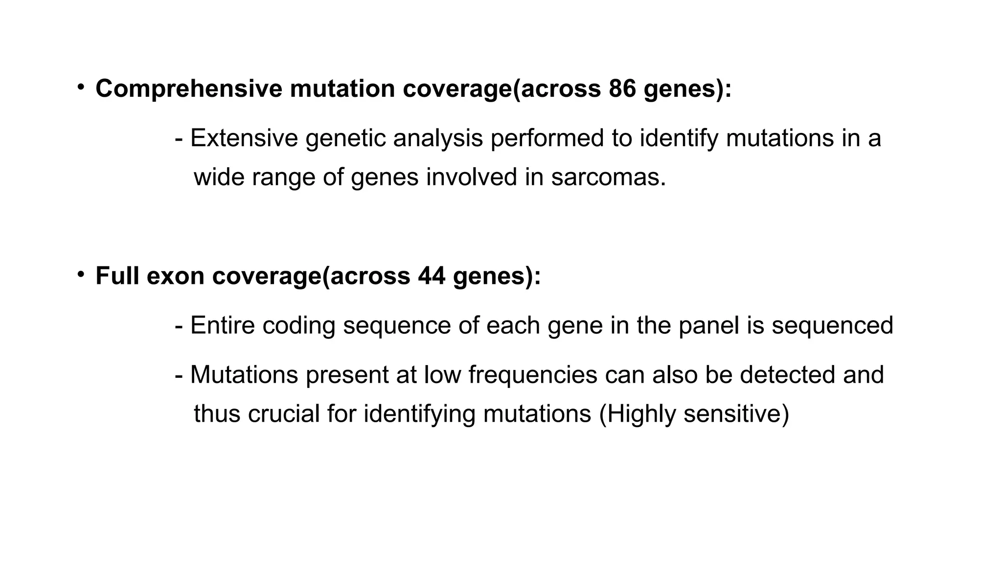
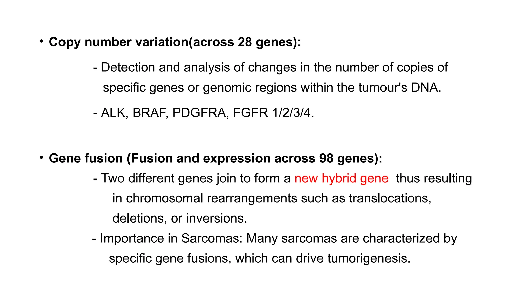
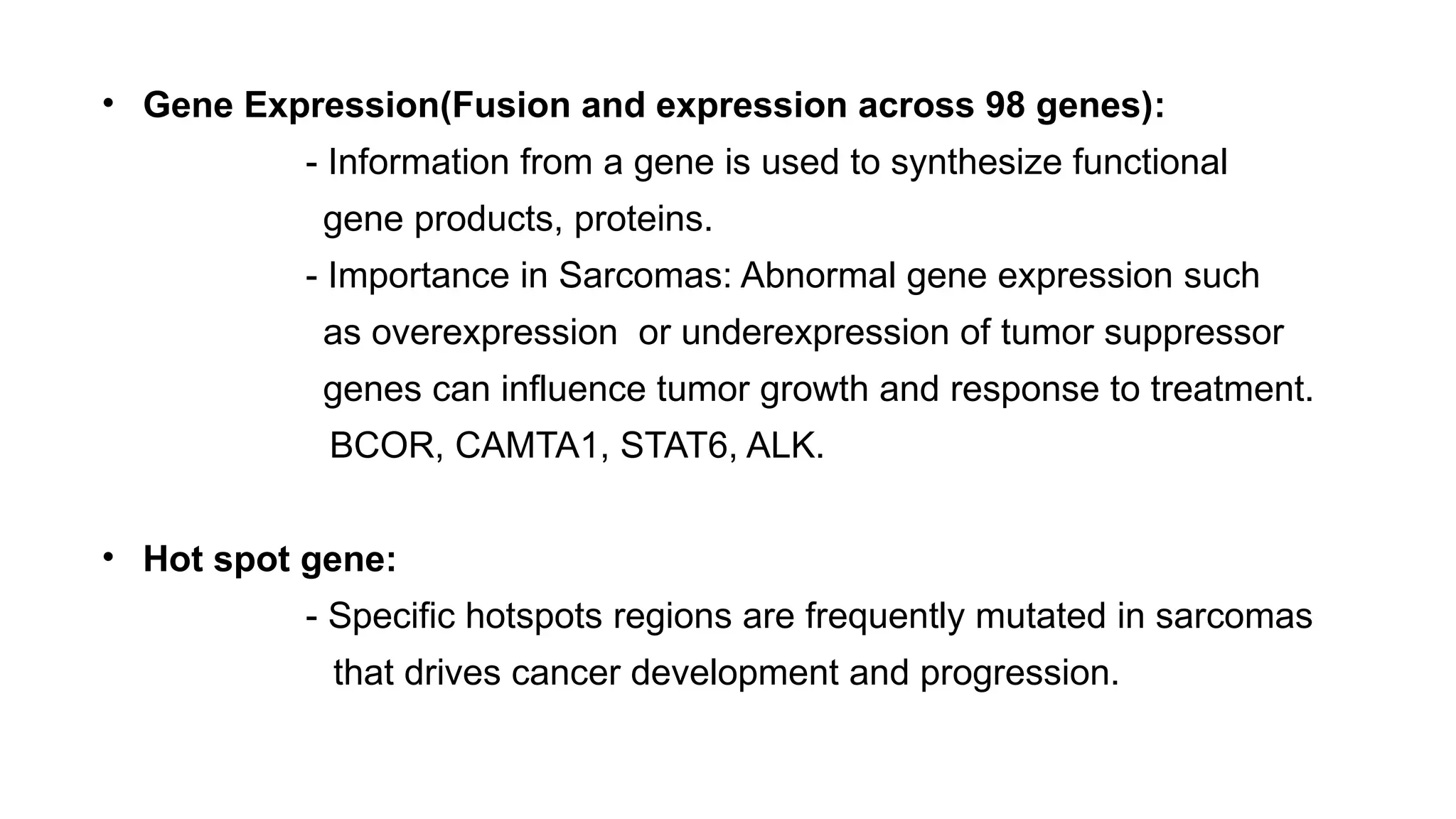
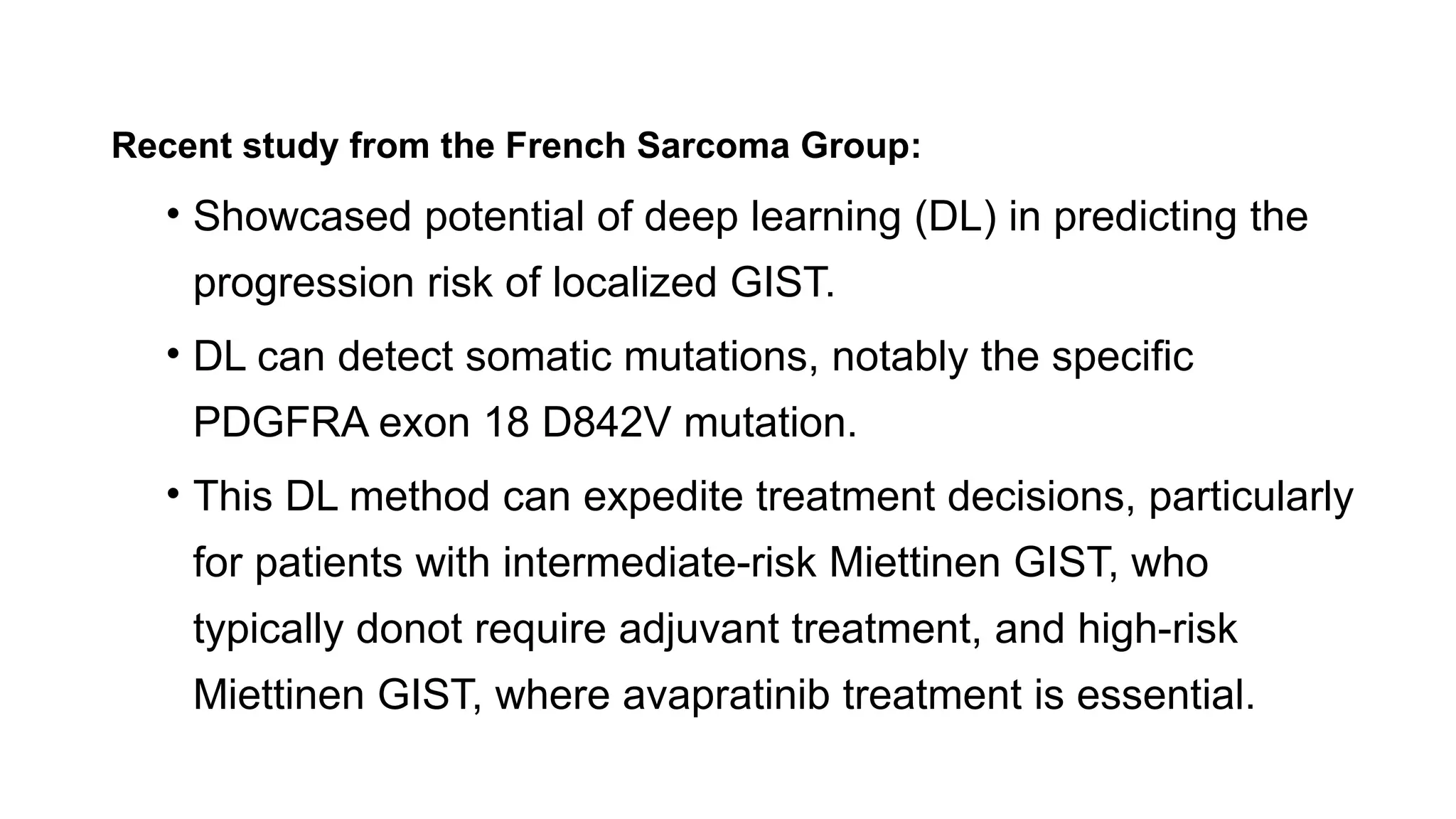
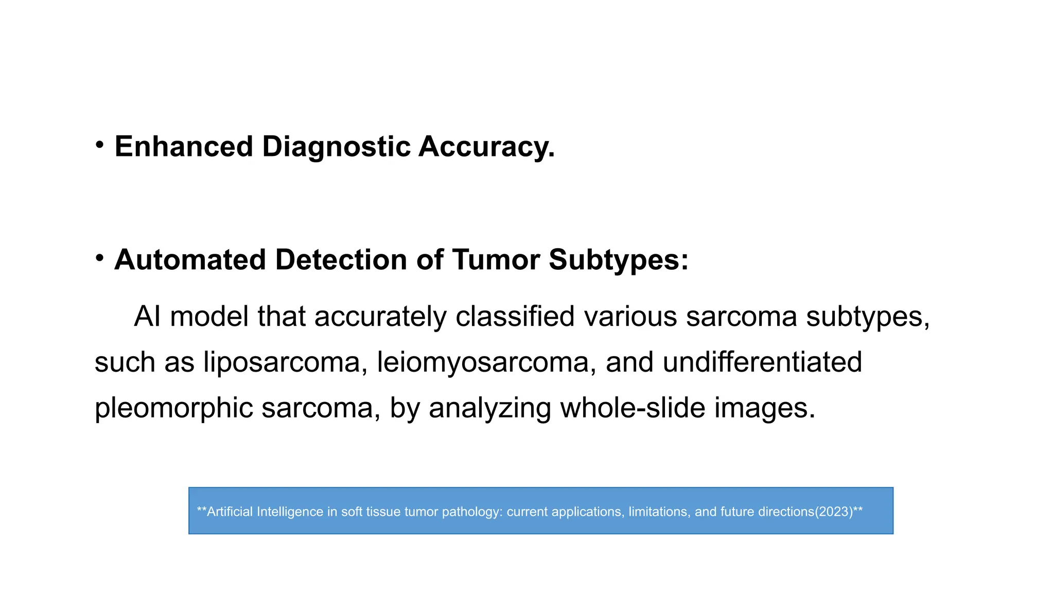
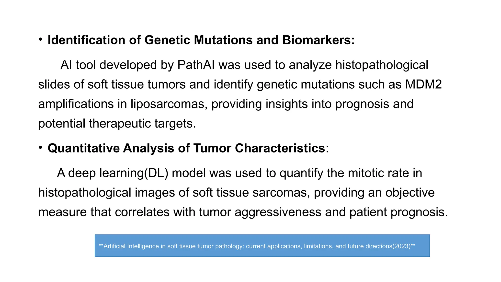
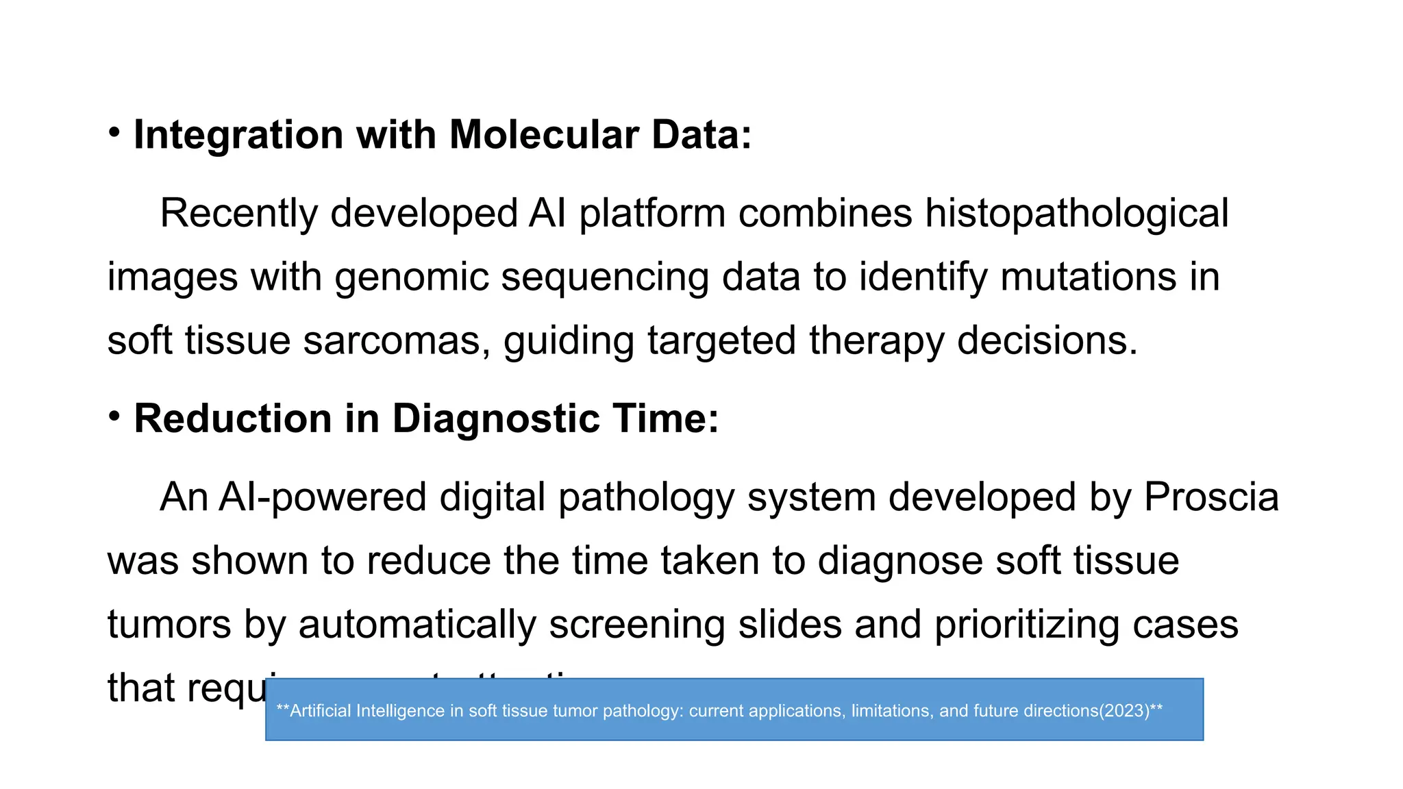
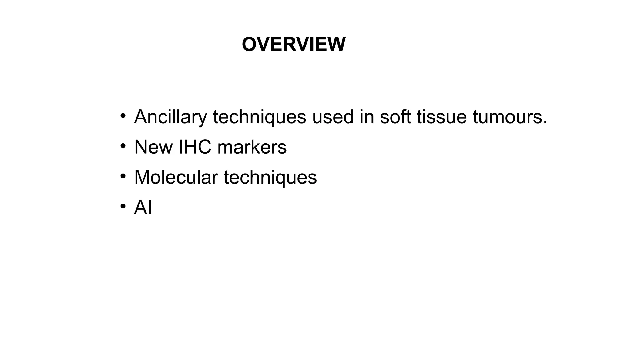
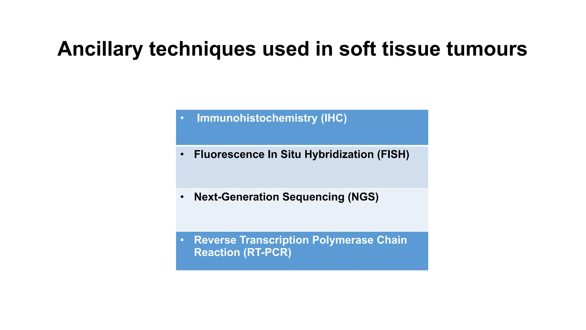
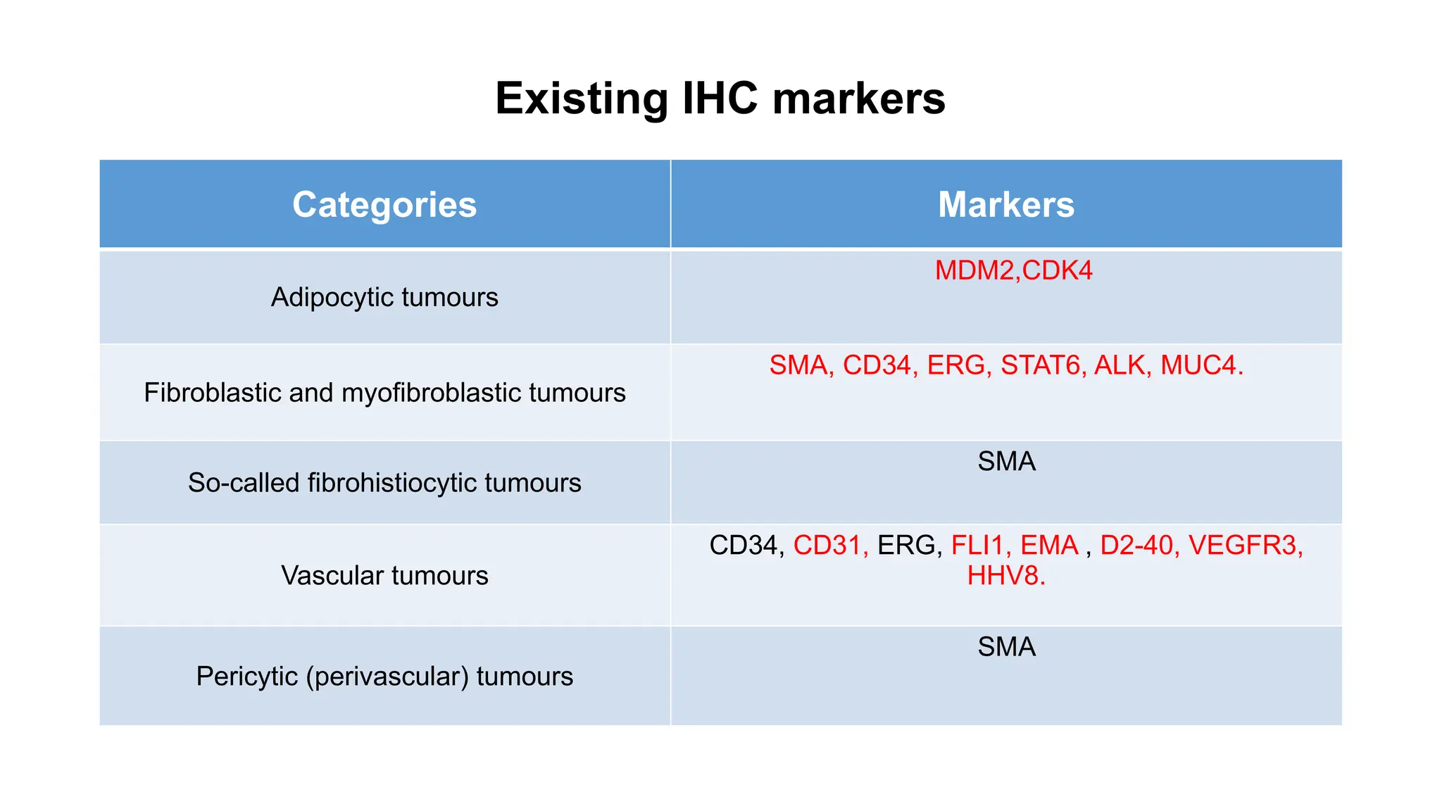
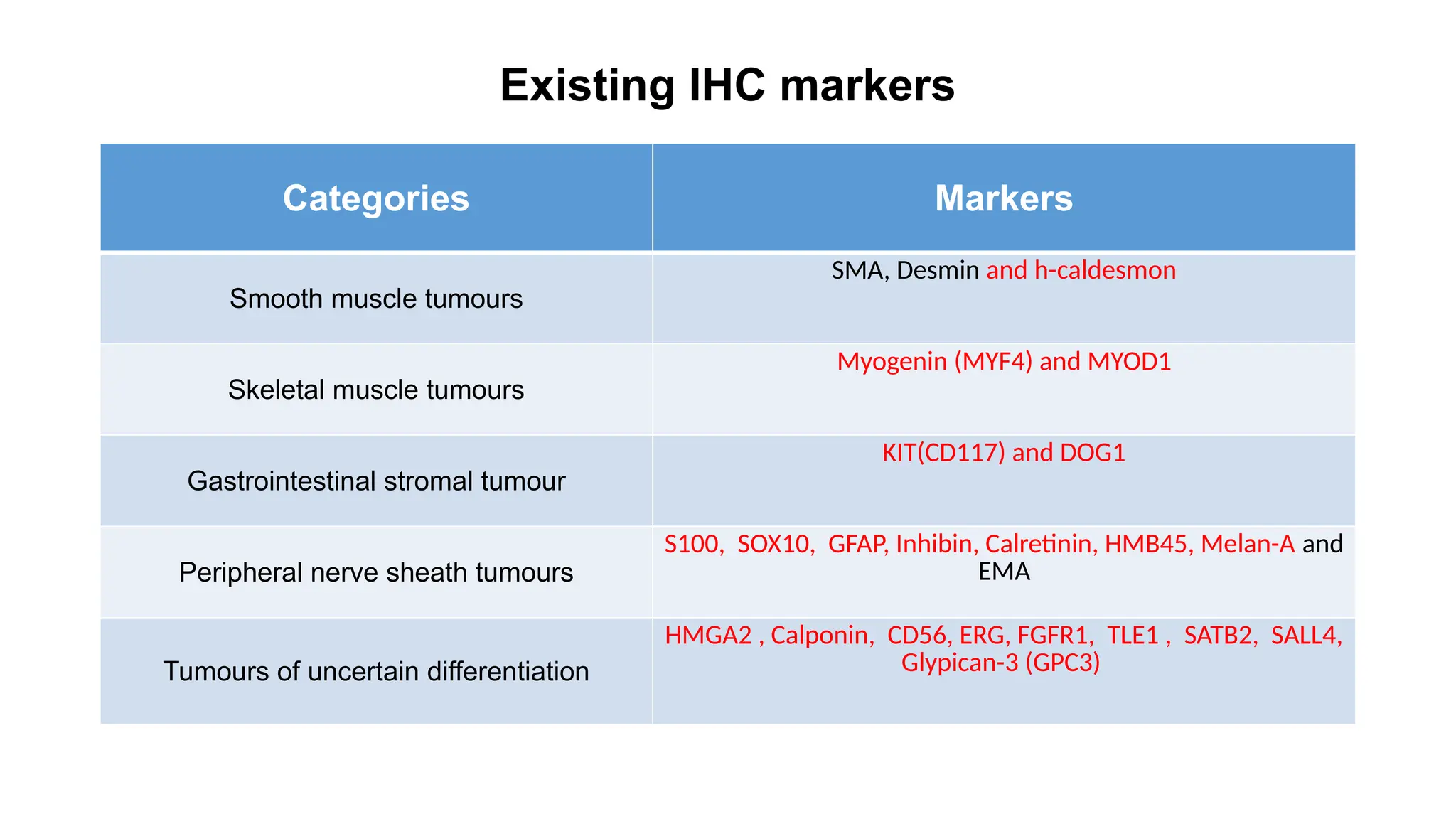
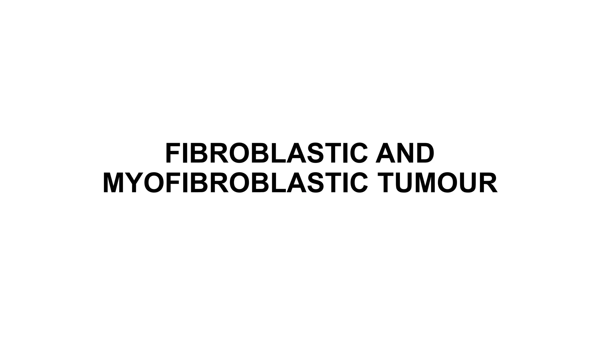
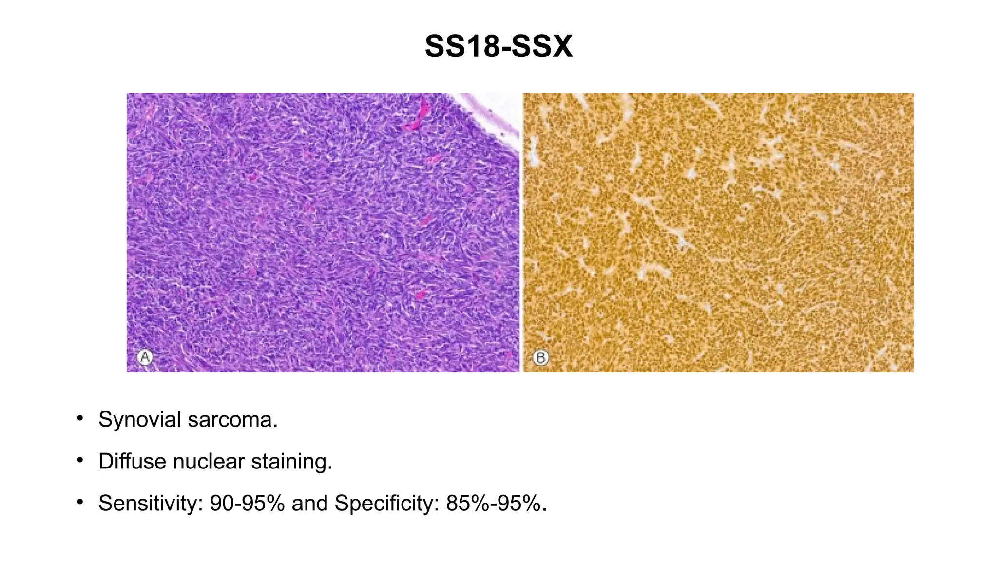
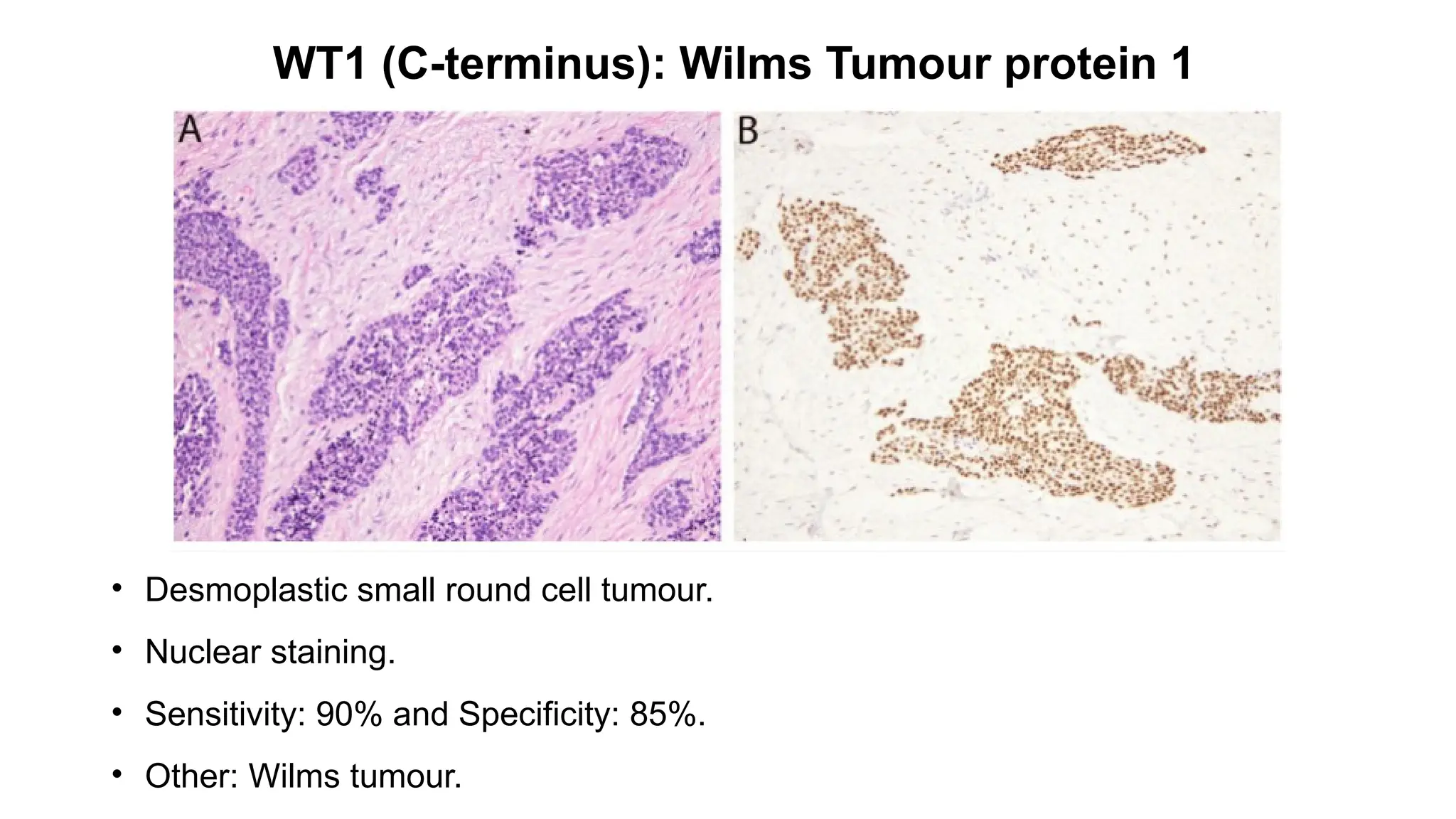
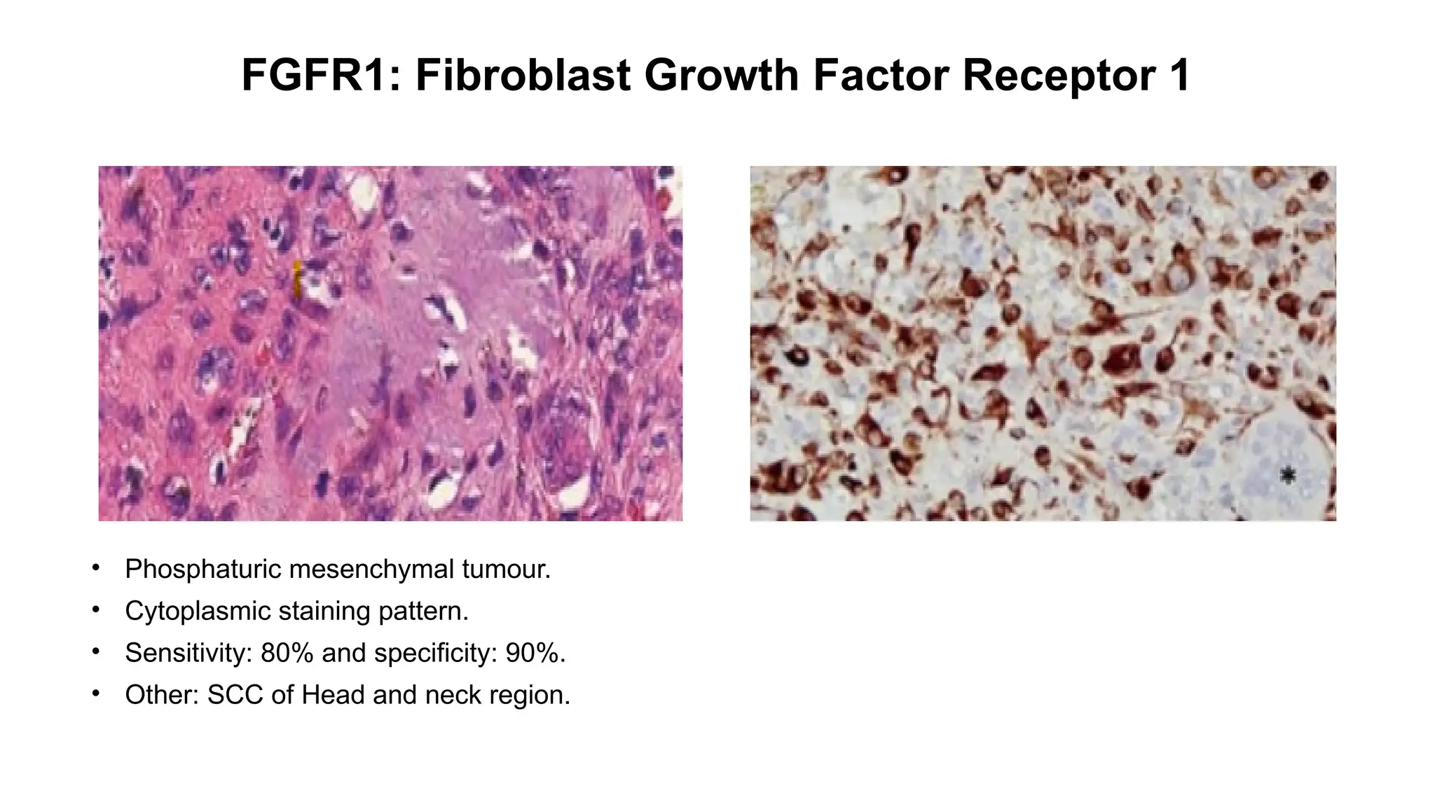
RECENT ADVANCES IN
ANCILLARY TECHNIQUES IN
SOFT TISSUE
SARCOMA
- D.VINOTH KUMAR (Ist year PG)
OVERVIEW
• Ancillary techniques used in soft tissue tumours.
• New IHC markers
• Molecular techniques
• AI
Ancillary techniques used in soft tissue tumours
• Reverse Transcription Polymerase Chain
Reaction (RT-PCR)
• Immunohistochemistry (IHC)
• Fluorescence In Situ Hybridization (FISH)
• Next-Generation Sequencing (NGS)
Existing IHC markers
Categories Markers
Adipocytic tumours
MDM2,CDK4
Fibroblastic and myofibroblastic tumours
SMA, CD34, ERG, STAT6, ALK, MUC4.
So-called fibrohistiocytic tumours
SMA
Vascular tumours
CD34, CD31, ERG, FLI1, EMA , D2-40, VEGFR3,
HHV8.
Pericytic (perivascular) tumours
SMA
Existing IHC markers
Categories Markers
Smooth muscle tumours
SMA, Desmin and h-caldesmon
Skeletal muscle tumours
Myogenin (MYF4) and MYOD1
Gastrointestinal stromal tumour
KIT(CD117) and DOG1
Peripheral nerve sheath tumours
S100, SOX10, GFAP, Inhibin, Calretinin, HMB45, Melan-A and
EMA
Tumours of uncertain differentiation
HMGA2 , Calponin, CD56, ERG, FGFR1, TLE1 , SATB2, SALL4,
Glypican-3 (GPC3)
Newer IHC markers
ADIPOCYTIC TUMOUR
DDIT3: DNA Damage Inducible Transcript 3
• Myxoid liposarcoma.
• Diffuse nuclear positivity.
• Sensitivity: 85% and Specificity: 89%.
• Other: Ewings sarcoma.
FIBROBLASTIC AND
MYOFIBROBLASTIC TUMOUR
pan-TRK: pan - Tyrosine Receptor Kinase
• Infantile fibrosarcoma.
• Nuclear expression in a tumour with ETV6-NTRK3 fusion.
• Sensitivity: 92% and Specificity: 95%.
• Other: Paediatric spindle cell tumour.
Vascular tumors
FOSB
• Pseudomyogenic hemangioendothelioma(PHE).
• Diffuse nuclear positivity.
• Sensitivity: 96% and Specficity: 100% .
• Other: EHE(Epithelioid haemangioendothelioma).
Skeletal muscle tumors
CAMTA1: Calmodulin-binding transcription activator 1
• Epithelioid hemangioendothelioma [WWTR1::CAMTA1 (85–90%) ]
• Strong nuclear.
• Sensitivity: 91.2% and Specificity: 100%.
• Other: Gliomas
GIST
SDHB: Succinate Dehydrogenase B
• SDHB-mutant GIST(d).
• Loss of cytoplasmic expression of SDHB (e).
• Sensitivity: 100% and Specificity: 100%.
• Other: Paragangliomas.
Peripheral nerve sheath tumours
PRAME: PReferentially expressed Antigen in MElanoma
.
• Primary cutaneous melanoma.
• Diffuse nuclear staining .
• Sensitivity: 67-91.7% and Specificity: 93.5-100%.
• Other: Leukaemias and Lymphomas.
PRKAR1A: Protein Kinase cAMP dependent type I
Regulatory subunit Alpha
• Melanotic malignant nerve sheath tumour.
• Complete loss of cytoplasmic expression.
• Sensitivity: 60-80% and Specificity: 70-90%.
• Other: Carcinoid tumours.
H3K27me3: Trimethylation of histone H3 at lysine 27.
• Malignant peripheral nerve sheath tumour.
• Loss of nuclear staining pattern.
• Sensitivity: 44% and Specificity: 92%.
• Other: DIPG (Diffuse Intrinsic Pontine Gliomas).
Tumours of uncertain differentiation
SS18-SSX
• Synovial sarcoma.
• Diffuse nuclear staining.
• Sensitivity: 90-95% and Specificity: 85%-95%.
WT1 (C-terminus): Wilms Tumour protein 1
• Desmoplastic small round cell tumour.
• Nuclear staining.
• Sensitivity: 90% and Specificity: 85%.
• Other: Wilms tumour.
FGFR1: Fibroblast Growth Factor Receptor 1
• Phosphaturic mesenchymal tumour.
• Cytoplasmic staining pattern.
• Sensitivity: 80% and specificity: 90%.
• Other: SCC of Head and neck region.
Undifferentiated Small Round Cell
Sarcomas
DUX4
• CIC-rearranged sarcoma[CIC::DUX4 (95%)].
• Nuclear staining.
• Sensitivity: 90% and Specificity: 85% .
• Other: Ewing sarcoma, Desmoplastic small round cell tumour.
ETV4
• CIC-rearranged sarcoma.
• Diffuse Nuclear staining.
• Sensitivity: 70-80% and Specificity: 80-90%.
• Other: Prostate cancer.
BCOR: BCL 6 interacting Corepressor
• BCOR CCNB3 –rearranged sarcoma.
• Nuclear staining.
• Sensitivity: 80-90% and Specificity: 85-95%.
• Other: Ovarian tumours.
Molecular techniques
• Current European Society for Medical Oncology (ESMO) guidelines
suggest that the morphologic and immunohistochemical analyses should
be complemented by molecular pathology:
(1) when the specific histologic diagnosis is uncertain,
(2) when the clinicopathologic presentation is unusual, or
(3) when the genetic information may have prognostic or predictive
relevance.
Karyotyping Comparative genomic hybridisation
FISH: WWTR1 rearrangements, in diagnosing
Synovial Sarcoma: CGH helps identify chromosomal
abnormalities, such as the characteristic translocation
t(X;18).
Fluorescence in-situ hybridisation
Multiplex FISH:
• Multiple fluorescent probes to detect several genetic alterations
simultaneously within the same tissue sample.
• Improves diagnostic accuracy
• Detecting complex genetic alterations within a single assay.
Automated FISH Analysis:
• Incorporates automated image analysis software to assist in the
interpretation.
• Reduces human error, increases consistency, accuracy and reliability of
genetic diagnostics.
**Three-Dimensional Fluorescence In Situ Hybridization for the Diagnosis of Soft Tissue Tumors" published in Human Pathology (2022)**
**Automated Fluorescence In Situ Hybridization Analysis in Soft Tissue Sarcomas: Enhancements in Diagnostic Accuracy" published in The
American Journal of Surgical Pathology (2021)**
3D FISH:
• Provides 3D imaging of chromosomal abnormalities.
• Improves understanding of the structural organization of chromosomal
aberrations.
• Used in research and diagnostic purposes, for understanding tumor
heterogeneity and complex chromosomal arrangements.
Integration with NGS:
• Provide comprehensive genetic profiling.
• Simultaneous detection of gene fusions, mutations, and other genomic
alterations
• Powerful diagnostic tool that enhances the precision of genetic classification
and guides personalized treatment strategies .
**Three-Dimensional Fluorescence In Situ Hybridization (3D FISH) for the Study of Chromosomal Abnormalities in Soft Tissue Sarcomas" published in Modern Pathology
(2023)**
**Integration of NGS and FISH for Comprehensive Analysis of Genetic Alterations in Soft Tissue Sarcomas" published in Journal of Molecular Diagnostics (2023)**
Reverse Transcription Polymerase Chain Reaction
(RT-PCR)
• High-Throughput RT-PCR:
Simultaneous analysis of multiple gene expressions in a single sample,
improving the efficiency and scope of gene expression profiling in soft tissue
tumors.
• Digital PCR:
Higher precision and sensitivity. It partitions the sample into many small
reactions and counts the number of positive reactions. Thus useful for detecting
low-abundance transcripts or rare mutations.
Reverse Transcription Polymerase Chain Reaction
(RT-PCR)
• Integration with NGS:
It enhances the ability to identify novel biomarkers and genetic alterations.
• Improved Targeting of Fusion Genes.
• Quantitative RT-PCR (qRT-PCR):
Improved accuracy and reproducibility, allowing more precise measurement
of gene expression levels, which aids in prognosis and treatment planning.
NEXT GENERATION SEQUENCING
Sarcoma panel(UNI NGS) comprises of
a. Comprehensive mutation coverage.
b. Full exon coverage.
c. Copy number variation.
d. Gene fusion and expression.
e. Hot spot gene
• Comprehensive mutation coverage(across 86 genes):
- Extensive genetic analysis performed to identify mutations in a
wide range of genes involved in sarcomas.
• Full exon coverage(across 44 genes):
- Entire coding sequence of each gene in the panel is sequenced
- Mutations present at low frequencies can also be detected and
thus crucial for identifying mutations (Highly sensitive)
• Copy number variation(across 28 genes):
- Detection and analysis of changes in the number of copies of
specific genes or genomic regions within the tumour's DNA.
- ALK, BRAF, PDGFRA, FGFR 1/2/3/4.
• Gene fusion (Fusion and expression across 98 genes):
- Two different genes join to form a new hybrid gene thus resulting
in chromosomal rearrangements such as translocations,
deletions, or inversions.
- Importance in Sarcomas: Many sarcomas are characterized by
specific gene fusions, which can drive tumorigenesis.
• Gene Expression(Fusion and expression across 98 genes):
- Information from a gene is used to synthesize functional
gene products, proteins.
- Importance in Sarcomas: Abnormal gene expression such
as overexpression or underexpression of tumor suppressor
genes can influence tumor growth and response to treatment.
BCOR, CAMTA1, STAT6, ALK.
• Hot spot gene:
- Specific hotspots regions are frequently mutated in sarcomas
that drives cancer development and progression.
• NGS, is utilised in the development of targeted therapies.
eg., Detecting NTRK fusions enables the use of TRK inhibitors, which have
shown high efficacy in treating tumors with these genetic alterations.
• Tumor Mutation Burden (TMB):
It is a measure of the total no. of mutations / megabase of tumor DNA.
It is used as a biomarker to predict response to immunotherapies.
High TMB is associated with better responses to immune checkpoint
inhibitors.
The application of NGS in measuring TMB helps in selecting suitable
candidates for these therapies.
Non-targeted massive sequencing
• This techniques can decode any genetic rearrangement without
prior knowledge.
• It encompasses the holistic RNA-Seq, WES, and WGS
techniques.
Nanopore
• The “nanopore - long read sequencing” method uses to drive DNA
strands or single nucleotides through a very small hole embedded in a
membrane.
• An enzyme motor controls the rate at which a DNA molecule passes
through the nanopore.
• The sequence is determined in real-time based on the extent to which the
nucleotides disrupt the current flowing through a nanopore sensor.
• Advantages: No RNA splicing / amplication.
Nanostring technology
• It is a single molecule counting system.
• It works by attaching molecular barcodes to target molecules of
interest by nucleic acid base pairing.
• This method quantifies RNA directly, bypassing the need of prior
amplification.
• Adapts to analyse even subpar RNA samples.
Methylome studies
• These studies deals with analysing the methylation patterns that offers a
more resilient and granular analysis.
• Their binary approach, focusing on methylation or its absence, furnishes a
unique diagnostic approach to a particular tumour.
• The emerging field of liquid biopsies includes-
• a. Cell free DNA(cfDNA)
• b. Circulating tumour DNA(ctDNA)
• c. Circulating tumour cells (CTCs)
• These non invasive tests are primarily derived from blood samples,
without need for tissue biopsies.
• These liquid biopsies aid in early diagnosis, monitoring treatment
responses and detecting recurrences.
• Sensitivity: affected by deep seated nature of many sarcomas because the
amount of ctDNA shed into the blood stream may be low.
Circulating biomarkers in sarcomas
**Circulating Biomarkers in Soft Tissue Sarcomas: Current Applications and Future Directions" published in Cancer Research (2023)**
AI in soft tissue pathology
Recent study from the French Sarcoma Group:
• Showcased potential of deep learning (DL) in predicting the
progression risk of localized GIST.
• DL can detect somatic mutations, notably the specific
PDGFRA exon 18 D842V mutation.
• This DL method can expedite treatment decisions, particularly
for patients with intermediate-risk Miettinen GIST, who
typically donot require adjuvant treatment, and high-risk
Miettinen GIST, where avapratinib treatment is essential.
• Enhanced Diagnostic Accuracy.
• Automated Detection of Tumor Subtypes:
AI model that accurately classified various sarcoma subtypes,
such as liposarcoma, leiomyosarcoma, and undifferentiated
pleomorphic sarcoma, by analyzing whole-slide images.
**Artificial Intelligence in soft tissue tumor pathology: current applications, limitations, and future directions(2023)**
• Identification of Genetic Mutations and Biomarkers:
AI tool developed by PathAI was used to analyze histopathological
slides of soft tissue tumors and identify genetic mutations such as MDM2
amplifications in liposarcomas, providing insights into prognosis and
potential therapeutic targets.
• Quantitative Analysis of Tumor Characteristics:
A deep learning(DL) model was used to quantify the mitotic rate in
histopathological images of soft tissue sarcomas, providing an objective
measure that correlates with tumor aggressiveness and patient prognosis.
**Artificial Intelligence in soft tissue tumor pathology: current applications, limitations, and future directions(2023)**
• Integration with Molecular Data:
Recently developed AI platform combines histopathological
images with genomic sequencing data to identify mutations in
soft tissue sarcomas, guiding targeted therapy decisions.
• Reduction in Diagnostic Time:
An AI-powered digital pathology system developed by Proscia
was shown to reduce the time taken to diagnose soft tissue
tumors by automatically screening slides and prioritizing cases
that require urgent attention.
**Artificial Intelligence in soft tissue tumor pathology: current applications, limitations, and future directions(2023)**
References:
1. The Recent advances in molecular diagnosis of soft tissue tumors.
2. Current challenges and practical aspects of molecular pathology.
3. Immunohistochemistry Update in Dermatopathology and Bone and Soft Tissue Pathology.
4. Diagnostic Immunohistochemistry of Soft Tissue and Bone Tumors: An Update on Biomarkers That Correlate
with Molecular Alterations.
THANK YOU

Recent advances in soft tissue sarcoma and its applications

  • 1.
    RECENT ADVANCES IN ANCILLARYTECHNIQUES IN SOFT TISSUE SARCOMA - D.VINOTH KUMAR (Ist year PG)
  • 2.
    OVERVIEW • Ancillary techniquesused in soft tissue tumours. • New IHC markers • Molecular techniques • AI
  • 3.
    Ancillary techniques usedin soft tissue tumours • Reverse Transcription Polymerase Chain Reaction (RT-PCR) • Immunohistochemistry (IHC) • Fluorescence In Situ Hybridization (FISH) • Next-Generation Sequencing (NGS)
  • 4.
    Existing IHC markers CategoriesMarkers Adipocytic tumours MDM2,CDK4 Fibroblastic and myofibroblastic tumours SMA, CD34, ERG, STAT6, ALK, MUC4. So-called fibrohistiocytic tumours SMA Vascular tumours CD34, CD31, ERG, FLI1, EMA , D2-40, VEGFR3, HHV8. Pericytic (perivascular) tumours SMA
  • 5.
    Existing IHC markers CategoriesMarkers Smooth muscle tumours SMA, Desmin and h-caldesmon Skeletal muscle tumours Myogenin (MYF4) and MYOD1 Gastrointestinal stromal tumour KIT(CD117) and DOG1 Peripheral nerve sheath tumours S100, SOX10, GFAP, Inhibin, Calretinin, HMB45, Melan-A and EMA Tumours of uncertain differentiation HMGA2 , Calponin, CD56, ERG, FGFR1, TLE1 , SATB2, SALL4, Glypican-3 (GPC3)
  • 6.
  • 7.
  • 8.
    DDIT3: DNA DamageInducible Transcript 3 • Myxoid liposarcoma. • Diffuse nuclear positivity. • Sensitivity: 85% and Specificity: 89%. • Other: Ewings sarcoma.
  • 9.
  • 10.
    pan-TRK: pan -Tyrosine Receptor Kinase • Infantile fibrosarcoma. • Nuclear expression in a tumour with ETV6-NTRK3 fusion. • Sensitivity: 92% and Specificity: 95%. • Other: Paediatric spindle cell tumour.
  • 11.
  • 12.
    FOSB • Pseudomyogenic hemangioendothelioma(PHE). •Diffuse nuclear positivity. • Sensitivity: 96% and Specficity: 100% . • Other: EHE(Epithelioid haemangioendothelioma).
  • 13.
  • 14.
    CAMTA1: Calmodulin-binding transcriptionactivator 1 • Epithelioid hemangioendothelioma [WWTR1::CAMTA1 (85–90%) ] • Strong nuclear. • Sensitivity: 91.2% and Specificity: 100%. • Other: Gliomas
  • 15.
  • 16.
    SDHB: Succinate DehydrogenaseB • SDHB-mutant GIST(d). • Loss of cytoplasmic expression of SDHB (e). • Sensitivity: 100% and Specificity: 100%. • Other: Paragangliomas.
  • 17.
  • 18.
    PRAME: PReferentially expressedAntigen in MElanoma . • Primary cutaneous melanoma. • Diffuse nuclear staining . • Sensitivity: 67-91.7% and Specificity: 93.5-100%. • Other: Leukaemias and Lymphomas.
  • 19.
    PRKAR1A: Protein KinasecAMP dependent type I Regulatory subunit Alpha • Melanotic malignant nerve sheath tumour. • Complete loss of cytoplasmic expression. • Sensitivity: 60-80% and Specificity: 70-90%. • Other: Carcinoid tumours.
  • 20.
    H3K27me3: Trimethylation ofhistone H3 at lysine 27. • Malignant peripheral nerve sheath tumour. • Loss of nuclear staining pattern. • Sensitivity: 44% and Specificity: 92%. • Other: DIPG (Diffuse Intrinsic Pontine Gliomas).
  • 21.
    Tumours of uncertaindifferentiation
  • 22.
    SS18-SSX • Synovial sarcoma. •Diffuse nuclear staining. • Sensitivity: 90-95% and Specificity: 85%-95%.
  • 23.
    WT1 (C-terminus): WilmsTumour protein 1 • Desmoplastic small round cell tumour. • Nuclear staining. • Sensitivity: 90% and Specificity: 85%. • Other: Wilms tumour.
  • 24.
    FGFR1: Fibroblast GrowthFactor Receptor 1 • Phosphaturic mesenchymal tumour. • Cytoplasmic staining pattern. • Sensitivity: 80% and specificity: 90%. • Other: SCC of Head and neck region.
  • 25.
  • 26.
    DUX4 • CIC-rearranged sarcoma[CIC::DUX4(95%)]. • Nuclear staining. • Sensitivity: 90% and Specificity: 85% . • Other: Ewing sarcoma, Desmoplastic small round cell tumour.
  • 27.
    ETV4 • CIC-rearranged sarcoma. •Diffuse Nuclear staining. • Sensitivity: 70-80% and Specificity: 80-90%. • Other: Prostate cancer.
  • 28.
    BCOR: BCL 6interacting Corepressor • BCOR CCNB3 –rearranged sarcoma. • Nuclear staining. • Sensitivity: 80-90% and Specificity: 85-95%. • Other: Ovarian tumours.
  • 29.
  • 30.
    • Current EuropeanSociety for Medical Oncology (ESMO) guidelines suggest that the morphologic and immunohistochemical analyses should be complemented by molecular pathology: (1) when the specific histologic diagnosis is uncertain, (2) when the clinicopathologic presentation is unusual, or (3) when the genetic information may have prognostic or predictive relevance.
  • 31.
    Karyotyping Comparative genomichybridisation FISH: WWTR1 rearrangements, in diagnosing Synovial Sarcoma: CGH helps identify chromosomal abnormalities, such as the characteristic translocation t(X;18).
  • 32.
  • 33.
    Multiplex FISH: • Multiplefluorescent probes to detect several genetic alterations simultaneously within the same tissue sample. • Improves diagnostic accuracy • Detecting complex genetic alterations within a single assay. Automated FISH Analysis: • Incorporates automated image analysis software to assist in the interpretation. • Reduces human error, increases consistency, accuracy and reliability of genetic diagnostics. **Three-Dimensional Fluorescence In Situ Hybridization for the Diagnosis of Soft Tissue Tumors" published in Human Pathology (2022)** **Automated Fluorescence In Situ Hybridization Analysis in Soft Tissue Sarcomas: Enhancements in Diagnostic Accuracy" published in The American Journal of Surgical Pathology (2021)**
  • 34.
    3D FISH: • Provides3D imaging of chromosomal abnormalities. • Improves understanding of the structural organization of chromosomal aberrations. • Used in research and diagnostic purposes, for understanding tumor heterogeneity and complex chromosomal arrangements. Integration with NGS: • Provide comprehensive genetic profiling. • Simultaneous detection of gene fusions, mutations, and other genomic alterations • Powerful diagnostic tool that enhances the precision of genetic classification and guides personalized treatment strategies . **Three-Dimensional Fluorescence In Situ Hybridization (3D FISH) for the Study of Chromosomal Abnormalities in Soft Tissue Sarcomas" published in Modern Pathology (2023)** **Integration of NGS and FISH for Comprehensive Analysis of Genetic Alterations in Soft Tissue Sarcomas" published in Journal of Molecular Diagnostics (2023)**
  • 35.
    Reverse Transcription PolymeraseChain Reaction (RT-PCR) • High-Throughput RT-PCR: Simultaneous analysis of multiple gene expressions in a single sample, improving the efficiency and scope of gene expression profiling in soft tissue tumors. • Digital PCR: Higher precision and sensitivity. It partitions the sample into many small reactions and counts the number of positive reactions. Thus useful for detecting low-abundance transcripts or rare mutations.
  • 36.
    Reverse Transcription PolymeraseChain Reaction (RT-PCR) • Integration with NGS: It enhances the ability to identify novel biomarkers and genetic alterations. • Improved Targeting of Fusion Genes. • Quantitative RT-PCR (qRT-PCR): Improved accuracy and reproducibility, allowing more precise measurement of gene expression levels, which aids in prognosis and treatment planning.
  • 37.
  • 39.
    Sarcoma panel(UNI NGS)comprises of a. Comprehensive mutation coverage. b. Full exon coverage. c. Copy number variation. d. Gene fusion and expression. e. Hot spot gene
  • 40.
    • Comprehensive mutationcoverage(across 86 genes): - Extensive genetic analysis performed to identify mutations in a wide range of genes involved in sarcomas. • Full exon coverage(across 44 genes): - Entire coding sequence of each gene in the panel is sequenced - Mutations present at low frequencies can also be detected and thus crucial for identifying mutations (Highly sensitive)
  • 41.
    • Copy numbervariation(across 28 genes): - Detection and analysis of changes in the number of copies of specific genes or genomic regions within the tumour's DNA. - ALK, BRAF, PDGFRA, FGFR 1/2/3/4. • Gene fusion (Fusion and expression across 98 genes): - Two different genes join to form a new hybrid gene thus resulting in chromosomal rearrangements such as translocations, deletions, or inversions. - Importance in Sarcomas: Many sarcomas are characterized by specific gene fusions, which can drive tumorigenesis.
  • 42.
    • Gene Expression(Fusionand expression across 98 genes): - Information from a gene is used to synthesize functional gene products, proteins. - Importance in Sarcomas: Abnormal gene expression such as overexpression or underexpression of tumor suppressor genes can influence tumor growth and response to treatment. BCOR, CAMTA1, STAT6, ALK. • Hot spot gene: - Specific hotspots regions are frequently mutated in sarcomas that drives cancer development and progression.
  • 43.
    • NGS, isutilised in the development of targeted therapies. eg., Detecting NTRK fusions enables the use of TRK inhibitors, which have shown high efficacy in treating tumors with these genetic alterations. • Tumor Mutation Burden (TMB): It is a measure of the total no. of mutations / megabase of tumor DNA. It is used as a biomarker to predict response to immunotherapies. High TMB is associated with better responses to immune checkpoint inhibitors. The application of NGS in measuring TMB helps in selecting suitable candidates for these therapies.
  • 44.
    Non-targeted massive sequencing •This techniques can decode any genetic rearrangement without prior knowledge. • It encompasses the holistic RNA-Seq, WES, and WGS techniques.
  • 45.
    Nanopore • The “nanopore- long read sequencing” method uses to drive DNA strands or single nucleotides through a very small hole embedded in a membrane. • An enzyme motor controls the rate at which a DNA molecule passes through the nanopore. • The sequence is determined in real-time based on the extent to which the nucleotides disrupt the current flowing through a nanopore sensor. • Advantages: No RNA splicing / amplication.
  • 47.
    Nanostring technology • Itis a single molecule counting system. • It works by attaching molecular barcodes to target molecules of interest by nucleic acid base pairing. • This method quantifies RNA directly, bypassing the need of prior amplification. • Adapts to analyse even subpar RNA samples.
  • 48.
    Methylome studies • Thesestudies deals with analysing the methylation patterns that offers a more resilient and granular analysis. • Their binary approach, focusing on methylation or its absence, furnishes a unique diagnostic approach to a particular tumour.
  • 49.
    • The emergingfield of liquid biopsies includes- • a. Cell free DNA(cfDNA) • b. Circulating tumour DNA(ctDNA) • c. Circulating tumour cells (CTCs) • These non invasive tests are primarily derived from blood samples, without need for tissue biopsies. • These liquid biopsies aid in early diagnosis, monitoring treatment responses and detecting recurrences. • Sensitivity: affected by deep seated nature of many sarcomas because the amount of ctDNA shed into the blood stream may be low. Circulating biomarkers in sarcomas **Circulating Biomarkers in Soft Tissue Sarcomas: Current Applications and Future Directions" published in Cancer Research (2023)**
  • 50.
    AI in softtissue pathology
  • 51.
    Recent study fromthe French Sarcoma Group: • Showcased potential of deep learning (DL) in predicting the progression risk of localized GIST. • DL can detect somatic mutations, notably the specific PDGFRA exon 18 D842V mutation. • This DL method can expedite treatment decisions, particularly for patients with intermediate-risk Miettinen GIST, who typically donot require adjuvant treatment, and high-risk Miettinen GIST, where avapratinib treatment is essential.
  • 52.
    • Enhanced DiagnosticAccuracy. • Automated Detection of Tumor Subtypes: AI model that accurately classified various sarcoma subtypes, such as liposarcoma, leiomyosarcoma, and undifferentiated pleomorphic sarcoma, by analyzing whole-slide images. **Artificial Intelligence in soft tissue tumor pathology: current applications, limitations, and future directions(2023)**
  • 53.
    • Identification ofGenetic Mutations and Biomarkers: AI tool developed by PathAI was used to analyze histopathological slides of soft tissue tumors and identify genetic mutations such as MDM2 amplifications in liposarcomas, providing insights into prognosis and potential therapeutic targets. • Quantitative Analysis of Tumor Characteristics: A deep learning(DL) model was used to quantify the mitotic rate in histopathological images of soft tissue sarcomas, providing an objective measure that correlates with tumor aggressiveness and patient prognosis. **Artificial Intelligence in soft tissue tumor pathology: current applications, limitations, and future directions(2023)**
  • 54.
    • Integration withMolecular Data: Recently developed AI platform combines histopathological images with genomic sequencing data to identify mutations in soft tissue sarcomas, guiding targeted therapy decisions. • Reduction in Diagnostic Time: An AI-powered digital pathology system developed by Proscia was shown to reduce the time taken to diagnose soft tissue tumors by automatically screening slides and prioritizing cases that require urgent attention. **Artificial Intelligence in soft tissue tumor pathology: current applications, limitations, and future directions(2023)**
  • 55.
    References: 1. The Recentadvances in molecular diagnosis of soft tissue tumors. 2. Current challenges and practical aspects of molecular pathology. 3. Immunohistochemistry Update in Dermatopathology and Bone and Soft Tissue Pathology. 4. Diagnostic Immunohistochemistry of Soft Tissue and Bone Tumors: An Update on Biomarkers That Correlate with Molecular Alterations.
  • 56.