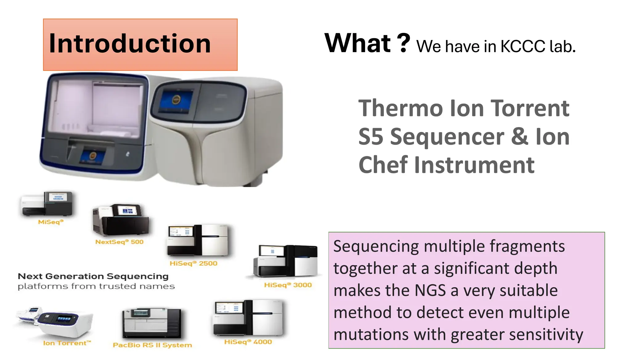
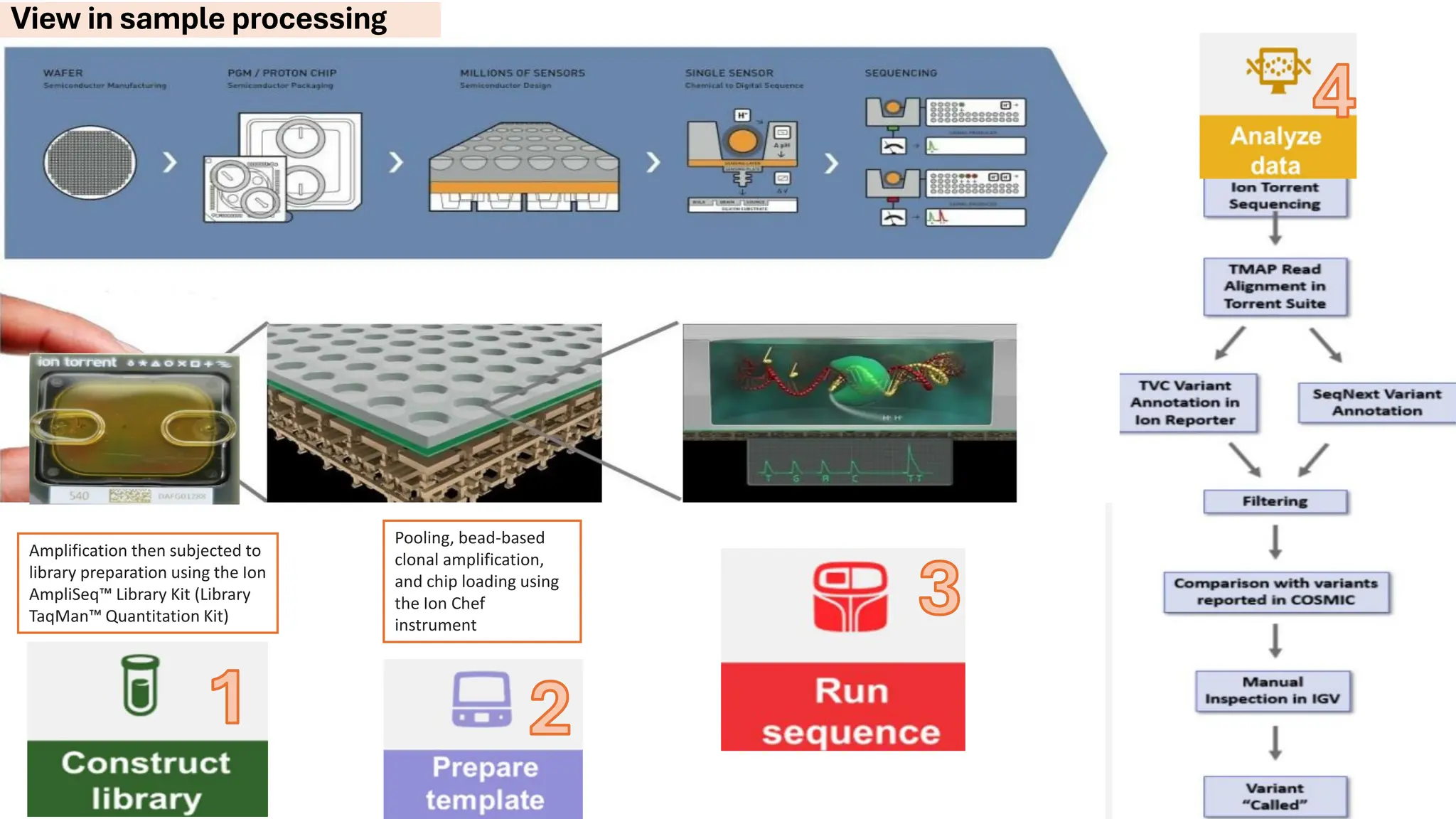
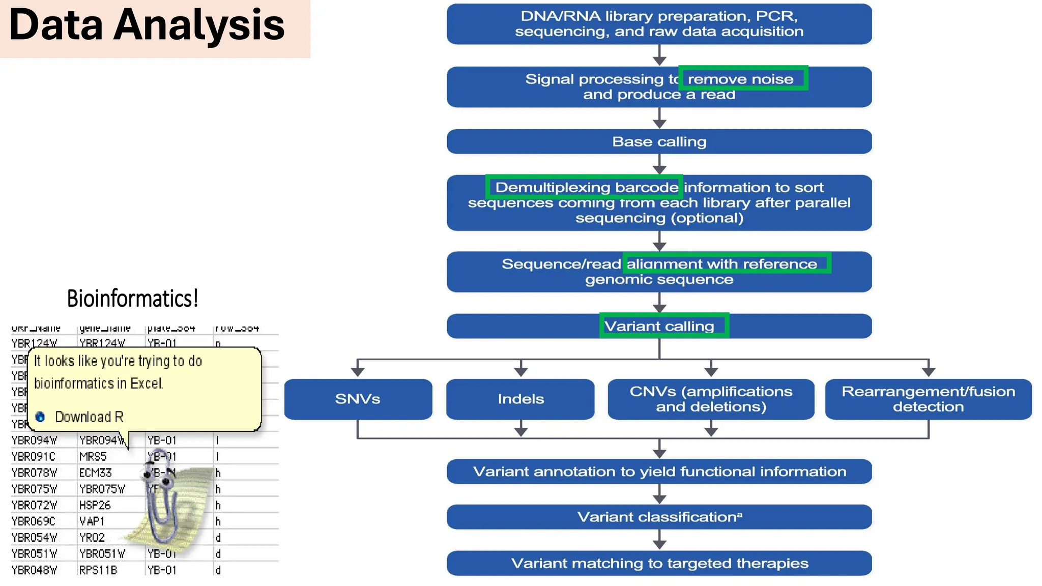
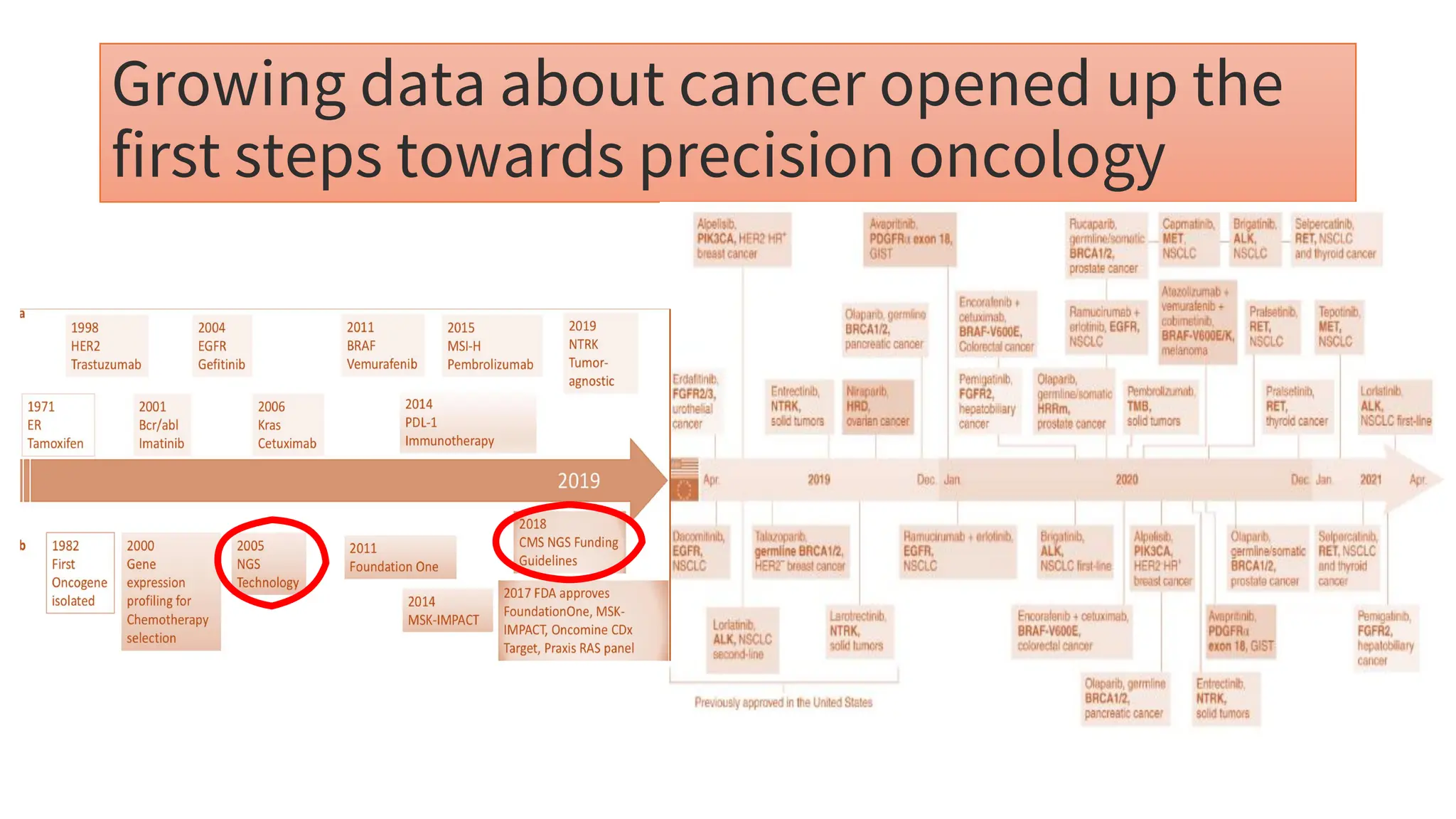
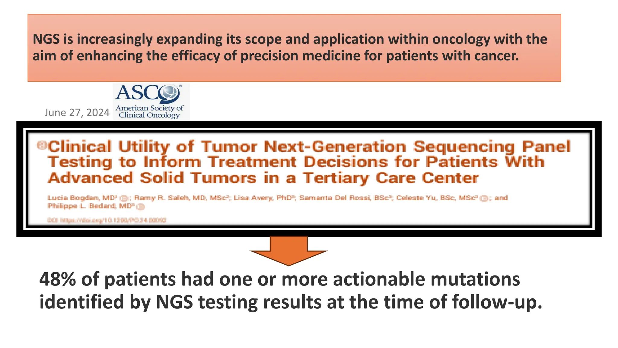
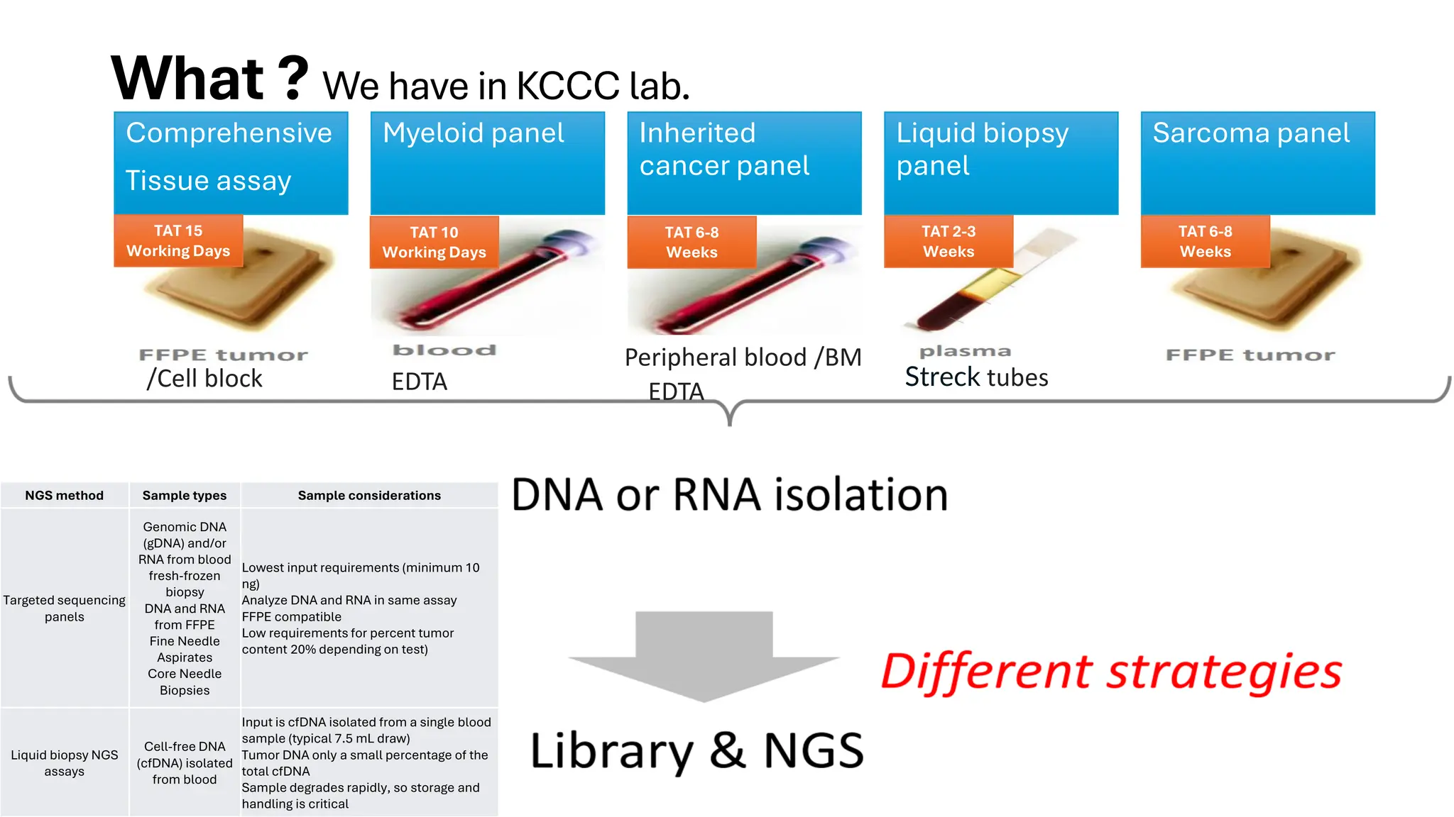
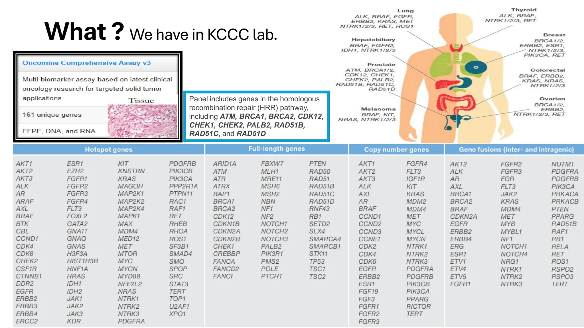
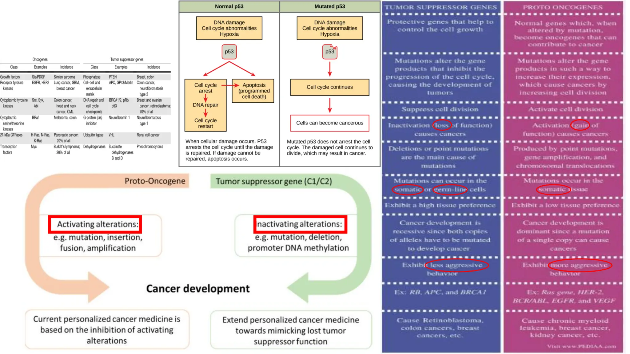
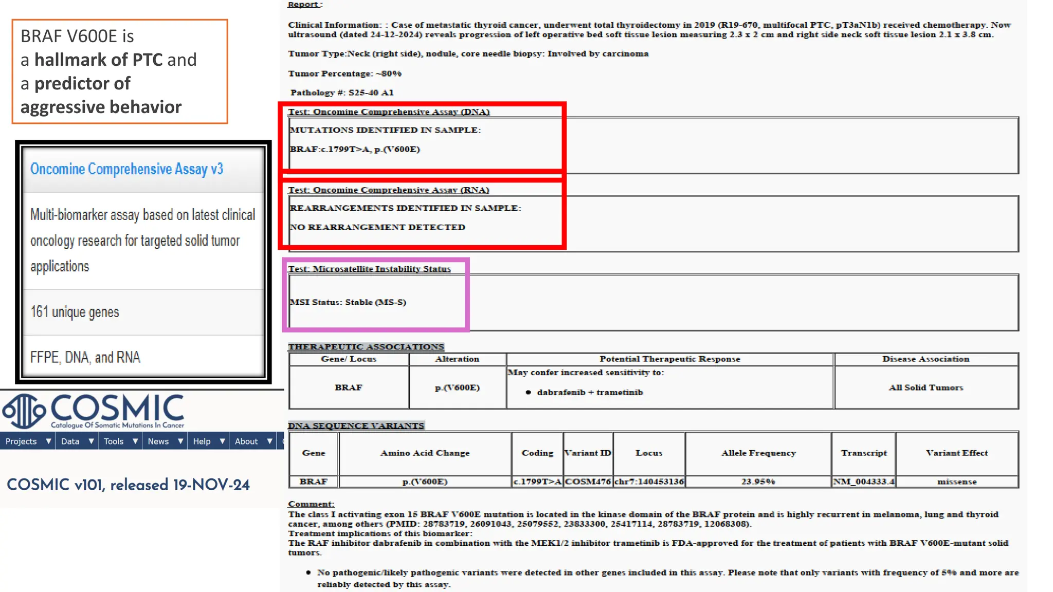
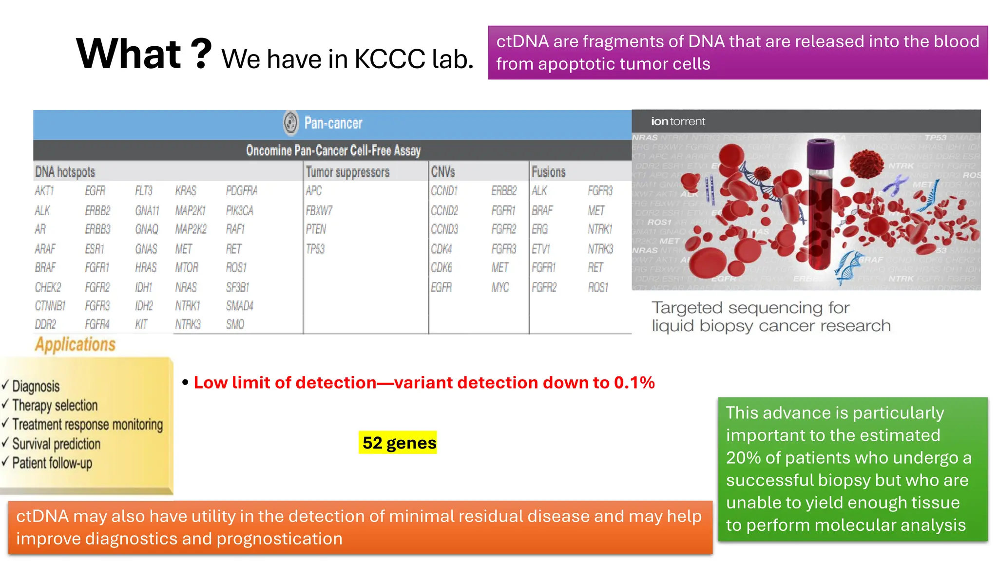
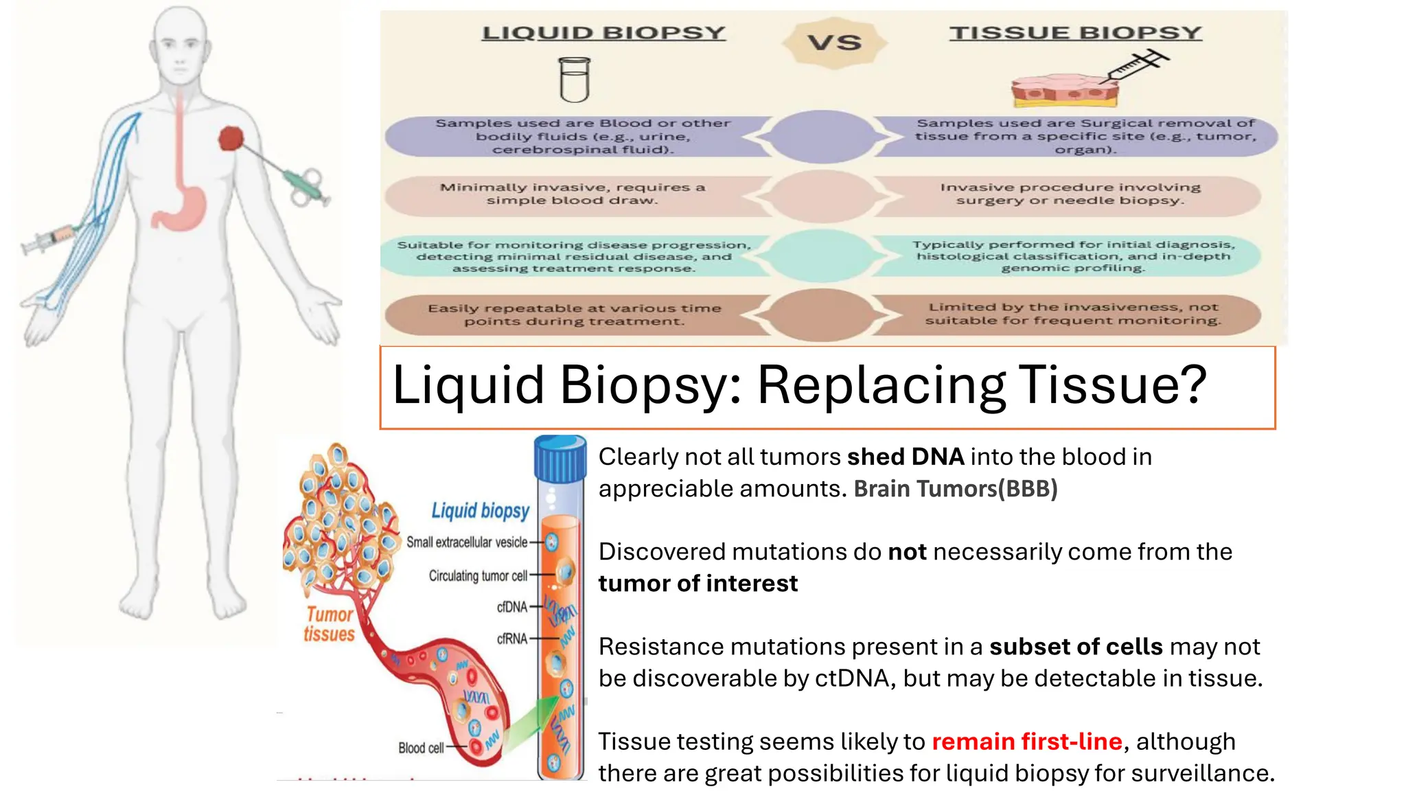
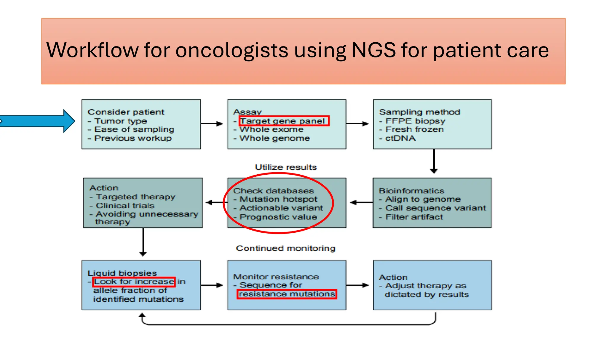
NGS: Expanding Scope and
Applications in Oncology at KCCC
Dr.Amir A Ahmed , MD
Biochemical and molecular pathology
Section Time
1. Intro 3 min
2. Why NGS 4 min
3. Workflow 5 min
4. Clinical Apps 8 min
5. Case Studies 5 min
6. Liquid Biopsy 3 min
7. Future + Q&A 2 min + 5 min
Time Allocation Guide
1. Introduction to Personalized Medicine & NGS
2. Why NGS? Key Advantages & Challenges
3. NGS Workflow at KCCC
4. Clinical Applications in Oncology
5. Case Studies: NGS in Action
6. Liquid Biopsy: Opportunities & Limits
7. Future Directions & Conclusion
Table of Contents
Personalized Medical Care
• The genetic information of any given patient is used as part of their
clinical care to help predict how they will respond to a given
treatment regimen
• Personalized medicine has the potential to offer new possibilities:
from prediction of a patient’s cancer risk to earlier diagnoses and
development of novel targeted therapies
Introduction
Translate a patient’s genomic information in a clinically meaningful way
DNA
sequencing
timeline
Introduction
What is
NGS?
Introduction
(a) Immunohistochemistry (IHC)
(b) Fluorescence in situ hybridization (FISH)
(c) polymerase chain reaction (PCR)
(d) Sanger sequencing.
Fluorescence in situ hybridization (FISH) , and polymerase chain reaction (PCR) can analyze small numbers of tumor
markers by searching for known“hotspots”
Sanger sequencing, the historic gold standard, can detect single nucleotide variations (SNVs) and small insertions and
deletions, but cannot sequence multiple types of genetic alterations or simultaneously screen for multiple genes in a
single assay
None of these
traditional methods
are scalable or
capable of high
throughput
NGS is the performance of high-throughput sequencing—the ability to sequence millions of small DNA fragments in parallel.
NGS can analyze more detailed information about the molecular makers of a tumor than any previous technology
NGS - Effective at All Size Scales
Introduction
NEXT GENERATION SEQUENCING
Also Known As "High Throughput Sequencing for
millions of reads ".
➢ "Faster Sequencing Rate" And "Cost
Effective" Procedure.
➢ Improvised Sequencing Technology Of
"Sanger Sequencing".
➢ Capability to sequence multiple individuals
at the same time.
➢ Providing high depth to deliver accurate data
and an insight into unexpected DNA variation
➢ Captures a broader spectrum of mutations
than Sanger sequencing
Sanger Sequencing is the current gold standard
method. SS is a faster and more cost-effective
method, but due to its low sensitivity with a
mutation detection limit of 10 to 20%
Potential for discovery of novel actionable targets.
Why ?NGS…..
Improved turn-around time by avoiding sequential testing
Tissue preservation - many genes simultaneously assessed rom single extraction
Introduction
• High-throughput sequencing enables simultaneous
analysis of multiple genes.
• Detects a wide range of mutations, including SNVs,
indels, fusions, and CNVs.
• Lower DNA input required compared to traditional
sequencing.
• Faster turnaround time by eliminating sequential
testing.
• Personalized therapy selection based on actionable
mutations.
• False positives/negatives – sequencing errors
can affect variant calling.
• Distinguishing germline vs. somatic mutations
requires additional filtering.
• Tumor heterogeneity – sampling bias can miss
subclonal mutations.
• Data interpretation complexity – bioinformatics
pipelines are evolving.
• Cost and accessibility – despite cost reductions,
affordability varies globally.
Advantages
Challenges
Advantages and challenges of clinical NGS
Introduction
What ? We have in KCCC lab.
Thermo Ion Torrent
S5 Sequencer & Ion
Chef Instrument
Sequencing multiple fragments
together at a significant depth
makes the NGS a very suitable
method to detect even multiple
mutations with greater sensitivity
Introduction
Introduction
The exome contains the portions of genes that
encode proteins.
It represents only 15% to 20% percent of the genome.
Reasonable approach, because over 85 % of known
disease-causing mutations are found in exons.
This approach substantially reduces cost and data
storage requirements compared with whole genome
sequencing.
Exome sequencing also simplifies clinical reporting,
because the significance of variants in exons is easier
to interpret in most cases.
Introduction
Comprehensive
Tissue assay
What ? We have in KCCC lab.
Myeloid panel Inherited
cancer panel
Liquid biopsy
panel
Sarcoma panel
NGS method Sample types Sample considerations
Targeted sequencing
panels
Genomic DNA
(gDNA) and/or
RNA from blood
fresh-frozen
biopsy
DNA and RNA
from FFPE
Fine Needle
Aspirates
Core Needle
Biopsies
Lowest input requirements (minimum 10
ng)
Analyze DNA and RNA in same assay
FFPE compatible
Low requirements for percent tumor
content 20% depending on test)
Liquid biopsy NGS
assays
Cell-free DNA
(cfDNA) isolated
from blood
Input is cfDNA isolated from a single blood
sample (typical 7.5 mL draw)
Tumor DNA only a small percentage of the
total cfDNA
Sample degrades rapidly, so storage and
handling is critical
Streck tubes
EDTA
/Cell block EDTA
TAT 15
Working Days
TAT 10
Working Days
TAT 6-8
Weeks
TAT 2-3
Weeks
TAT 6-8
Weeks
View in sample processing
using the QIAamp
DNA/RNA extraction Kit
NGS Reverse
Transcription Kit
Peripheral blood /BM
1 2
3
4
View in sample processing
Amplification then subjected to
library preparation using the Ion
AmpliSeq Library Kit (Library
TaqMan Quantitation Kit)
Pooling, bead-based
clonal amplification,
and chip loading using
the Ion Chef
instrument
Data Analysis
Data Analysis
All the reported variants were verified
by integrated genome viewer (IGV)
analysis. (using BAM files)
The sequencing coverage of genes
was confirmed by torrent suit
software, and it was observed
sequencing gene reading were
captured without any gaps.
Data Analysis
Challenge: Finding Low%
Mutations in NGS Data
Data Analysis
Data Analysis
Increased Depth Improves Mutation Detection
Data Analysis
Data Analysis
Analysis was performed manually to
check the hotspot mutations analyzed by
the IonReporter (IR) Software using bed
files targeting the desired regions of
genes as a reference and non
target/novel checked manual
SEE VIDEO
The annotation for variants was derived using various disease databases like
ClinVar. The population frequency information from 1000 genomes (ExAC,
GnomAD, and ESP) was used for the elimination of common variants and
polymorphisms.
the prediction of the possible impact of coding non-synonymous SNVs on the
structure and function of a protein, PolyPhen-2 and SIFT scores were used.
Data Analysis
Variant Classification
Data Analysis
• Variant
Classification
(ClinVar, COSMIC,
PolyPhen, SIFT).
• ESCAT & NCCN
Guidelines used to
assess clinical
actionability.
ESMO Scale for Clinical Actionability of
molecular Targets (ESCAT)
A collaborative project initiated by the ESMO Translational Research and Precision
Medicine Working Group provides a systematic framework to rank molecular targets
based on evidence available supporting their value as clinical targets.
Tiers:
Tier I: Targets with validated clinical utility (e.g., BRCA1/2 mutations
→ PARP inhibitors).
Tier II: Targets with investigational/emerging evidence (e.g., HER2
amplifications in non-breast cancers).
Tier III: Preclinical evidence or analogy to validated targets.
Tier IV/V: Insufficient/no evidence or evidence against
actionability.
•Key Features:
• Evidence-based prioritization for treatment selection.
• Facilitates interpretation of NGS reports in clinical practice.
• Aligns with precision oncology goals (right drug, right patient).
Genomic alterations according to ESCAT in advanced breast cancer
Genomic alterations according to ESCAT in advanced colorectal cancer (CRC)
HER2
HER2
Genomic alterations according to ESCAT in advanced breast cancer
BRCA
ESCAT in advanced prostate cancer
ESCAT in pancreatic ductal adenocarcinoma
BRCA
BRCA
Further Oncomine Reporter software was used for annotating variants with
a curated list of relevant labels, guidelines, and global clinical trials.
Data Analysis
NGS is changing cancer classification
• Traditionally, tumors have been classified
through histology. However, morphology alone
cannot detect the mutational signatures that
have been shown to be crucial in the
development of these tumors
new biomarkers are predict a given patient’s
treatment response and outcome.
targeted therapy survival rate.
non-small cell lung cancer (NSCLC). Molecular testing
for mutations in the epidermal growth factor receptor
(EGFR) has become the standard of care prior to
initiation of tyrosine kinase inhibitors (TKIs, such as
erlotinib) that can typically lead to a higher response
rate and longer progression-free survival . Also their
potential for developing drug resistance
Example
Growing data about cancer opened up the
first steps towards precision oncology
Sequencing is essential for development of
personalized immunotherapies
• When tumor-specfic DNA mutations alter the function
of proteins, cancer cells acquire antigens on their
surface that are absent from the normal genome
(neoantigens)
• It is believed that tumors known to be highly mutated are
more likely to be populated with neoantigens, which
may make them targetable by active immune cells
• Small sets of selected neoantigens can then be used for
vaccine development or cell transfer
In a recent study, a higher TMB was shown to be predictive
of more durable clinical benets, such as in patients with
NSCLC who have been treated with programmed death
receptor 1 (PD-1) inhibitors.
Example
48% of patients had one or more actionable mutations
identified by NGS testing results at the time of follow-up.
June 27, 2024
NGS is increasingly expanding its scope and application within oncology with the
aim of enhancing the efficacy of precision medicine for patients with cancer.
Comprehensive
Tissue assay
What ? We have in KCCC lab.
Myeloid panel Inherited
cancer panel
Liquid biopsy
panel
Sarcoma panel
NGS method Sample types Sample considerations
Targeted sequencing
panels
Genomic DNA
(gDNA) and/or
RNA from blood
fresh-frozen
biopsy
DNA and RNA
from FFPE
Fine Needle
Aspirates
Core Needle
Biopsies
Lowest input requirements (minimum 10
ng)
Analyze DNA and RNA in same assay
FFPE compatible
Low requirements for percent tumor
content 20% depending on test)
Liquid biopsy NGS
assays
Cell-free DNA
(cfDNA) isolated
from blood
Input is cfDNA isolated from a single blood
sample (typical 7.5 mL draw)
Tumor DNA only a small percentage of the
total cfDNA
Sample degrades rapidly, so storage and
handling is critical
Streck tubes
EDTA
/Cell block EDTA
Peripheral blood /BM
TAT 15
Working Days
TAT 10
Working Days
TAT 6-8
Weeks
TAT 2-3
Weeks
TAT 6-8
Weeks
What ? We have in KCCC lab.
Panel includes genes in the homologous
recombination repair (HRR) pathway,
including ATM, BRCA1, BRCA2, CDK12,
CHEK1, CHEK2, PALB2, RAD51B,
RAD51C, and RAD51D
Cancer – Low % Mutations
Cancer – Difficult Specimens
Challenges and practical aspect
Samples may be: ◦ Small ◦ Old ◦ Low number of tumoral
cells ◦ Limited material to obtain DNA & RNA
Samples may be:
◦ Small
◦ Old
◦ Low number of tumoral cells
◦ Limited material to obtain DNA & RNA
BRAF V600E is
a hallmark of PTC and
a predictor of
aggressive behavior
What ? We have in KCCC lab.
• Low limit of detection—variant detection down to 0.1%
52 genes
ctDNA are fragments of DNA that are released into the blood
from apoptotic tumor cells
This advance is particularly
important to the estimated
20% of patients who undergo a
successful biopsy but who are
unable to yield enough tissue
to perform molecular analysis
ctDNA may also have utility in the detection of minimal residual disease and may help
improve diagnostics and prognostication
Liquid Biopsy: Replacing Tissue?
Clearly not all tumors shed DNA into the blood in
appreciable amounts. Brain Tumors(BBB)
Discovered mutations do not necessarily come from the
tumor of interest
Resistance mutations present in a subset of cells may not
be discoverable by ctDNA, but may be detectable in tissue.
Tissue testing seems likely to remain first-line, although
there are great possibilities for liquid biopsy for surveillance.
KIT mutations confirm GIST
diagnosis
KIT :Exon 11 Mutation
• Imatinib (1st-line TKI)
achieves high response
rates
• After imatinib failure:
Regorafenib (3rd-line)
or ripretinib (4th-line)
Sunitinib (2nd-line) has less
activity against primary exon
11 mutations
ESR1 encodes the estrogen
receptor alpha (ERα)
Predicts Endocrine Therapy
Resistance
Switch to SERDs (e.g.,
fulvestrant) or SERD-CDK4/6
inhibitor combinations (e.g.,
elacestrant)
correlates with aggressive
disease
What ? We have in KCCC lab.
• more accurate and more cost-effective
fusion detection
• can identify known and novel fusions
• open-ended targeted amplification to
identify gene fusions whether or not the
fusion partner is known.
Genes target: 63
Targeted RNA panel
Archer FUSIONPlex Sarcoma v2 panel
Archer
FUSIONPlex
Sarcoma v2 panel
WWTR1-CAMTA1 Fusion
•Translocation: t(1;3)(p36;q25)
fuses WWTR1 (1p36)
with CAMTA1 (3q25).
•Prevalence: Present in ~90% of
EHE cases.
novel
Archer
FUSIONPlex
Sarcoma v2 panel
Distinguishes Lipoblastoma from Mimics:
•Liposarcoma: Lacks PLAG1 rearrangement
•PLAG1 rearrangements are diagnostic of lipoblastoma
•a benign diagnosis, critical for avoiding overtreatment
Inherited Cancer panel
AIP, AKT1, ALK, ANTXR1, APC, ASCC1, ATM, ATR, AXIN2, BAP1, BARD1, BLM,
BMPR1A, BRCA1, BRCA2, BRIP1, BUB1, BUB1B, CACNA1D, CBL, CDC73, CDH1,
CDK4, CDKN1B, CDKN2A, CHEK2, CTHRC1, CYLD, DDB2, DICER1, EGFR, ELAC2,
EPCAM, ERCC2, ERCC3, ERCC4, ERCC5, EXT1, EXT2, FANCA, FANCB, FANCC,
FANCD2, FANCE, FANCF, FANCG, FANCI, FANCL, FANCM, FH, FLCN, GPC3, HNF1A,
HNF1B HOXB13, KDR, KIT, KLLN, LIG4, MAX, MEN1, MET, MITF, MLH1, MLH3,
MRE11A, MSH2, MSH6, MSR1, MTAP, MUTYH, MYH8, NBN, NCOA4, NF1, NF2,
NTRK1, PALB2, PALLD, PDGFRB, PHOX2B, PIK3CA, PMS2, POLD1, POLE, POLH,
PPM1D, PRF1, PRKAR1A, PRSS1, PTCH1, PTCH2, PTEN, PTPN11, RAD50, RAD51C,
RAD51D, RB1, RECQL4, RET, RHBDF2, RNASEL, RNF168, RSPO1, RUNX1, SETBP1,
SBDS, SDHA, SDHAF2, SDHB, SDHC, SDHD, SH2D1A, SLX4, SMAD4, SMARCB1,
SMARCE1, SPINK1, SPRED1, STK11, SUFU, TERT, TGFBR1, TMEM127, TP53, TSC1,
TSC2, VHL, WAS, WRN, WT1, XPA, XPC, and XRCC2
134 germline genes
Patient consent needed
Inherited
Cancer
panel
confer a 45–80% lifetime
risk of breast cancer and
a 40–60% risk of ovarian
cancer
BRCA1 loss
disrupts homologous
recombination repair
(HRR), leading to defective
DNA repair and
chromosomal instability.
•PARP Inhibitors (e.g.,
Olaparib, Talazoparib)
ClinVar database
What ? We have in KCCC lab.
40 key DNA genes and 29
RNA
fusion transcript driver
genes
Gene content on the panel has been
curated to cover relevant targets for
all the major myeloid disorders—
AML, MDS, MPN, CML, CMML, and
JMML.
Role of NGS in Hematologic Cancers
• Acute Myeloid Leukemia (AML) – Identifies FLT3, IDH1/2, NPM1, TP53
mutations for risk stratification.
• Myelodysplastic Syndromes (MDS) – Detects SF3B1, ASXL1, and TET2
mutations for prognostic evaluation.
• Chronic Myeloid Leukemia (CML) – Detects BCR-ABL resistance
mutations guiding TKI therapy.
• Myeloproliferative Neoplasms (MPN) – Identifies JAK2, CALR, and MPL
mutations for disease classification.
Minimal Residual Disease (MRD) Detection with NGS Higher sensitivity than flow cytometry for
MRD tracking. Detects low-frequency mutations down to 0.1% variant allele frequency (VAF).
Not
available
Workflow for oncologists using NGS for patient care
Case study(1): NGS application
50 y.o. female, Diagnosis of primary lung cancer
with brain metastasis ((Median survival for this
diagnosis historically has been 5-6 months))
Genomic sequencing was done by NGS
Sequencing result: EGFR c.2573T>G: p.L858R
Patient was put on a therapy targeting EGFR L858R.
•Erlotinib is a tyrosine kinase inhibitor (TKI).
•Tumors exhibited rapid reduction in size.
•Patient still alive ~2 years later
At ~2 year mark, new scans revealed that patient tumors
now progressing again.
•Sequencing of new biopsy sample reveals the presence of
EGFR T790M mutation.
•T790M is a common acquired resistance mechanism for
TKI therapies.
Case study(2): NGS application
RNA
RNA
Emerging Trends & Future Directions in NGS
• Whole-Genome Sequencing (WGS) → Beyond exome sequencing, identifies non-coding regulatory
mutations.
• Single-Cell Sequencing → Captures intratumoral heterogeneity, detecting rare cancer clones.
• Spatial Transcriptomics → Maps gene expression in tissue sections for tumor microenvironment
analysis.
• AI-Driven Bioinformatics → Speeds up variant interpretation & clinical decision-making.
• Personalized vaccine development using NGS-identified neoantigens
• CRISPR-based functional genomics for identifying new drug target
• Tumor-informed liquid biopsy panels for screening programs/ real-time monitoring
Cancer pharmacogenomics
Inherited (or, germline)
mutations can affect the
pharmacokinetics and
pharmacodynamics of a
selected treatment,
which may in turn impact
a patient’s response to
that treatment.
Therefore, the DNA
sequence of certain
genes can help
determine the amount of
drug to be prescribed or
what adverse events
might be anticipated in
certain phenotypes
Factors contributing to drug response.
Summary
NGS continues to revolutionize personalized diagnostics
in oncology.
NGS allows for the rapid sequencing of millions of DNA
fragments simultaneously, providing comprehensive insights
into genome structure
The discovery of these unique molecular ‘signatures’
has added a new level of complexity to developing
potential cancer treatments and is driving the growth of
precision medicine.
Thanks

NGS application in Kuwait cancer control center

  • 1.
    NGS: Expanding Scopeand Applications in Oncology at KCCC Dr.Amir A Ahmed , MD Biochemical and molecular pathology
  • 2.
    Section Time 1. Intro3 min 2. Why NGS 4 min 3. Workflow 5 min 4. Clinical Apps 8 min 5. Case Studies 5 min 6. Liquid Biopsy 3 min 7. Future + Q&A 2 min + 5 min Time Allocation Guide 1. Introduction to Personalized Medicine & NGS 2. Why NGS? Key Advantages & Challenges 3. NGS Workflow at KCCC 4. Clinical Applications in Oncology 5. Case Studies: NGS in Action 6. Liquid Biopsy: Opportunities & Limits 7. Future Directions & Conclusion Table of Contents
  • 3.
    Personalized Medical Care •The genetic information of any given patient is used as part of their clinical care to help predict how they will respond to a given treatment regimen • Personalized medicine has the potential to offer new possibilities: from prediction of a patient’s cancer risk to earlier diagnoses and development of novel targeted therapies Introduction Translate a patient’s genomic information in a clinically meaningful way
  • 4.
  • 5.
    What is NGS? Introduction (a) Immunohistochemistry(IHC) (b) Fluorescence in situ hybridization (FISH) (c) polymerase chain reaction (PCR) (d) Sanger sequencing. Fluorescence in situ hybridization (FISH) , and polymerase chain reaction (PCR) can analyze small numbers of tumor markers by searching for known“hotspots” Sanger sequencing, the historic gold standard, can detect single nucleotide variations (SNVs) and small insertions and deletions, but cannot sequence multiple types of genetic alterations or simultaneously screen for multiple genes in a single assay None of these traditional methods are scalable or capable of high throughput NGS is the performance of high-throughput sequencing—the ability to sequence millions of small DNA fragments in parallel. NGS can analyze more detailed information about the molecular makers of a tumor than any previous technology
  • 6.
    NGS - Effectiveat All Size Scales Introduction
  • 7.
    NEXT GENERATION SEQUENCING AlsoKnown As "High Throughput Sequencing for millions of reads ". ➢ "Faster Sequencing Rate" And "Cost Effective" Procedure. ➢ Improvised Sequencing Technology Of "Sanger Sequencing". ➢ Capability to sequence multiple individuals at the same time. ➢ Providing high depth to deliver accurate data and an insight into unexpected DNA variation ➢ Captures a broader spectrum of mutations than Sanger sequencing Sanger Sequencing is the current gold standard method. SS is a faster and more cost-effective method, but due to its low sensitivity with a mutation detection limit of 10 to 20% Potential for discovery of novel actionable targets. Why ?NGS….. Improved turn-around time by avoiding sequential testing Tissue preservation - many genes simultaneously assessed rom single extraction Introduction
  • 8.
    • High-throughput sequencingenables simultaneous analysis of multiple genes. • Detects a wide range of mutations, including SNVs, indels, fusions, and CNVs. • Lower DNA input required compared to traditional sequencing. • Faster turnaround time by eliminating sequential testing. • Personalized therapy selection based on actionable mutations. • False positives/negatives – sequencing errors can affect variant calling. • Distinguishing germline vs. somatic mutations requires additional filtering. • Tumor heterogeneity – sampling bias can miss subclonal mutations. • Data interpretation complexity – bioinformatics pipelines are evolving. • Cost and accessibility – despite cost reductions, affordability varies globally. Advantages Challenges Advantages and challenges of clinical NGS Introduction
  • 9.
    What ? Wehave in KCCC lab. Thermo Ion Torrent S5 Sequencer & Ion Chef Instrument Sequencing multiple fragments together at a significant depth makes the NGS a very suitable method to detect even multiple mutations with greater sensitivity Introduction
  • 10.
    Introduction The exome containsthe portions of genes that encode proteins. It represents only 15% to 20% percent of the genome. Reasonable approach, because over 85 % of known disease-causing mutations are found in exons. This approach substantially reduces cost and data storage requirements compared with whole genome sequencing. Exome sequencing also simplifies clinical reporting, because the significance of variants in exons is easier to interpret in most cases.
  • 11.
  • 12.
    Comprehensive Tissue assay What ?We have in KCCC lab. Myeloid panel Inherited cancer panel Liquid biopsy panel Sarcoma panel NGS method Sample types Sample considerations Targeted sequencing panels Genomic DNA (gDNA) and/or RNA from blood fresh-frozen biopsy DNA and RNA from FFPE Fine Needle Aspirates Core Needle Biopsies Lowest input requirements (minimum 10 ng) Analyze DNA and RNA in same assay FFPE compatible Low requirements for percent tumor content 20% depending on test) Liquid biopsy NGS assays Cell-free DNA (cfDNA) isolated from blood Input is cfDNA isolated from a single blood sample (typical 7.5 mL draw) Tumor DNA only a small percentage of the total cfDNA Sample degrades rapidly, so storage and handling is critical Streck tubes EDTA /Cell block EDTA TAT 15 Working Days TAT 10 Working Days TAT 6-8 Weeks TAT 2-3 Weeks TAT 6-8 Weeks View in sample processing using the QIAamp DNA/RNA extraction Kit NGS Reverse Transcription Kit Peripheral blood /BM
  • 13.
    1 2 3 4 View insample processing Amplification then subjected to library preparation using the Ion AmpliSeq Library Kit (Library TaqMan Quantitation Kit) Pooling, bead-based clonal amplification, and chip loading using the Ion Chef instrument
  • 14.
  • 15.
  • 16.
    All the reportedvariants were verified by integrated genome viewer (IGV) analysis. (using BAM files) The sequencing coverage of genes was confirmed by torrent suit software, and it was observed sequencing gene reading were captured without any gaps. Data Analysis
  • 17.
    Challenge: Finding Low% Mutationsin NGS Data Data Analysis
  • 18.
  • 19.
    Increased Depth ImprovesMutation Detection Data Analysis
  • 20.
    Data Analysis Analysis wasperformed manually to check the hotspot mutations analyzed by the IonReporter (IR) Software using bed files targeting the desired regions of genes as a reference and non target/novel checked manual SEE VIDEO
  • 21.
    The annotation forvariants was derived using various disease databases like ClinVar. The population frequency information from 1000 genomes (ExAC, GnomAD, and ESP) was used for the elimination of common variants and polymorphisms. the prediction of the possible impact of coding non-synonymous SNVs on the structure and function of a protein, PolyPhen-2 and SIFT scores were used. Data Analysis
  • 22.
    Variant Classification Data Analysis •Variant Classification (ClinVar, COSMIC, PolyPhen, SIFT). • ESCAT & NCCN Guidelines used to assess clinical actionability.
  • 23.
    ESMO Scale forClinical Actionability of molecular Targets (ESCAT) A collaborative project initiated by the ESMO Translational Research and Precision Medicine Working Group provides a systematic framework to rank molecular targets based on evidence available supporting their value as clinical targets. Tiers: Tier I: Targets with validated clinical utility (e.g., BRCA1/2 mutations → PARP inhibitors). Tier II: Targets with investigational/emerging evidence (e.g., HER2 amplifications in non-breast cancers). Tier III: Preclinical evidence or analogy to validated targets. Tier IV/V: Insufficient/no evidence or evidence against actionability. •Key Features: • Evidence-based prioritization for treatment selection. • Facilitates interpretation of NGS reports in clinical practice. • Aligns with precision oncology goals (right drug, right patient).
  • 24.
    Genomic alterations accordingto ESCAT in advanced breast cancer Genomic alterations according to ESCAT in advanced colorectal cancer (CRC) HER2 HER2
  • 25.
    Genomic alterations accordingto ESCAT in advanced breast cancer BRCA ESCAT in advanced prostate cancer ESCAT in pancreatic ductal adenocarcinoma BRCA BRCA
  • 26.
    Further Oncomine Reportersoftware was used for annotating variants with a curated list of relevant labels, guidelines, and global clinical trials. Data Analysis
  • 28.
    NGS is changingcancer classification • Traditionally, tumors have been classified through histology. However, morphology alone cannot detect the mutational signatures that have been shown to be crucial in the development of these tumors new biomarkers are predict a given patient’s treatment response and outcome. targeted therapy survival rate. non-small cell lung cancer (NSCLC). Molecular testing for mutations in the epidermal growth factor receptor (EGFR) has become the standard of care prior to initiation of tyrosine kinase inhibitors (TKIs, such as erlotinib) that can typically lead to a higher response rate and longer progression-free survival . Also their potential for developing drug resistance Example
  • 29.
    Growing data aboutcancer opened up the first steps towards precision oncology
  • 30.
    Sequencing is essentialfor development of personalized immunotherapies • When tumor-specfic DNA mutations alter the function of proteins, cancer cells acquire antigens on their surface that are absent from the normal genome (neoantigens) • It is believed that tumors known to be highly mutated are more likely to be populated with neoantigens, which may make them targetable by active immune cells • Small sets of selected neoantigens can then be used for vaccine development or cell transfer In a recent study, a higher TMB was shown to be predictive of more durable clinical benets, such as in patients with NSCLC who have been treated with programmed death receptor 1 (PD-1) inhibitors. Example
  • 31.
    48% of patientshad one or more actionable mutations identified by NGS testing results at the time of follow-up. June 27, 2024 NGS is increasingly expanding its scope and application within oncology with the aim of enhancing the efficacy of precision medicine for patients with cancer.
  • 32.
    Comprehensive Tissue assay What ?We have in KCCC lab. Myeloid panel Inherited cancer panel Liquid biopsy panel Sarcoma panel NGS method Sample types Sample considerations Targeted sequencing panels Genomic DNA (gDNA) and/or RNA from blood fresh-frozen biopsy DNA and RNA from FFPE Fine Needle Aspirates Core Needle Biopsies Lowest input requirements (minimum 10 ng) Analyze DNA and RNA in same assay FFPE compatible Low requirements for percent tumor content 20% depending on test) Liquid biopsy NGS assays Cell-free DNA (cfDNA) isolated from blood Input is cfDNA isolated from a single blood sample (typical 7.5 mL draw) Tumor DNA only a small percentage of the total cfDNA Sample degrades rapidly, so storage and handling is critical Streck tubes EDTA /Cell block EDTA Peripheral blood /BM TAT 15 Working Days TAT 10 Working Days TAT 6-8 Weeks TAT 2-3 Weeks TAT 6-8 Weeks
  • 33.
    What ? Wehave in KCCC lab. Panel includes genes in the homologous recombination repair (HRR) pathway, including ATM, BRCA1, BRCA2, CDK12, CHEK1, CHEK2, PALB2, RAD51B, RAD51C, and RAD51D
  • 35.
    Cancer – Low% Mutations Cancer – Difficult Specimens Challenges and practical aspect Samples may be: ◦ Small ◦ Old ◦ Low number of tumoral cells ◦ Limited material to obtain DNA & RNA Samples may be: ◦ Small ◦ Old ◦ Low number of tumoral cells ◦ Limited material to obtain DNA & RNA
  • 36.
    BRAF V600E is ahallmark of PTC and a predictor of aggressive behavior
  • 37.
    What ? Wehave in KCCC lab. • Low limit of detection—variant detection down to 0.1% 52 genes ctDNA are fragments of DNA that are released into the blood from apoptotic tumor cells This advance is particularly important to the estimated 20% of patients who undergo a successful biopsy but who are unable to yield enough tissue to perform molecular analysis ctDNA may also have utility in the detection of minimal residual disease and may help improve diagnostics and prognostication
  • 38.
    Liquid Biopsy: ReplacingTissue? Clearly not all tumors shed DNA into the blood in appreciable amounts. Brain Tumors(BBB) Discovered mutations do not necessarily come from the tumor of interest Resistance mutations present in a subset of cells may not be discoverable by ctDNA, but may be detectable in tissue. Tissue testing seems likely to remain first-line, although there are great possibilities for liquid biopsy for surveillance.
  • 39.
    KIT mutations confirmGIST diagnosis KIT :Exon 11 Mutation • Imatinib (1st-line TKI) achieves high response rates • After imatinib failure: Regorafenib (3rd-line) or ripretinib (4th-line) Sunitinib (2nd-line) has less activity against primary exon 11 mutations
  • 40.
    ESR1 encodes theestrogen receptor alpha (ERα) Predicts Endocrine Therapy Resistance Switch to SERDs (e.g., fulvestrant) or SERD-CDK4/6 inhibitor combinations (e.g., elacestrant) correlates with aggressive disease
  • 41.
    What ? Wehave in KCCC lab. • more accurate and more cost-effective fusion detection • can identify known and novel fusions • open-ended targeted amplification to identify gene fusions whether or not the fusion partner is known. Genes target: 63 Targeted RNA panel Archer FUSIONPlex Sarcoma v2 panel
  • 42.
    Archer FUSIONPlex Sarcoma v2 panel WWTR1-CAMTA1Fusion •Translocation: t(1;3)(p36;q25) fuses WWTR1 (1p36) with CAMTA1 (3q25). •Prevalence: Present in ~90% of EHE cases.
  • 43.
    novel Archer FUSIONPlex Sarcoma v2 panel DistinguishesLipoblastoma from Mimics: •Liposarcoma: Lacks PLAG1 rearrangement •PLAG1 rearrangements are diagnostic of lipoblastoma •a benign diagnosis, critical for avoiding overtreatment
  • 44.
    Inherited Cancer panel AIP,AKT1, ALK, ANTXR1, APC, ASCC1, ATM, ATR, AXIN2, BAP1, BARD1, BLM, BMPR1A, BRCA1, BRCA2, BRIP1, BUB1, BUB1B, CACNA1D, CBL, CDC73, CDH1, CDK4, CDKN1B, CDKN2A, CHEK2, CTHRC1, CYLD, DDB2, DICER1, EGFR, ELAC2, EPCAM, ERCC2, ERCC3, ERCC4, ERCC5, EXT1, EXT2, FANCA, FANCB, FANCC, FANCD2, FANCE, FANCF, FANCG, FANCI, FANCL, FANCM, FH, FLCN, GPC3, HNF1A, HNF1B HOXB13, KDR, KIT, KLLN, LIG4, MAX, MEN1, MET, MITF, MLH1, MLH3, MRE11A, MSH2, MSH6, MSR1, MTAP, MUTYH, MYH8, NBN, NCOA4, NF1, NF2, NTRK1, PALB2, PALLD, PDGFRB, PHOX2B, PIK3CA, PMS2, POLD1, POLE, POLH, PPM1D, PRF1, PRKAR1A, PRSS1, PTCH1, PTCH2, PTEN, PTPN11, RAD50, RAD51C, RAD51D, RB1, RECQL4, RET, RHBDF2, RNASEL, RNF168, RSPO1, RUNX1, SETBP1, SBDS, SDHA, SDHAF2, SDHB, SDHC, SDHD, SH2D1A, SLX4, SMAD4, SMARCB1, SMARCE1, SPINK1, SPRED1, STK11, SUFU, TERT, TGFBR1, TMEM127, TP53, TSC1, TSC2, VHL, WAS, WRN, WT1, XPA, XPC, and XRCC2 134 germline genes Patient consent needed
  • 46.
    Inherited Cancer panel confer a 45–80%lifetime risk of breast cancer and a 40–60% risk of ovarian cancer BRCA1 loss disrupts homologous recombination repair (HRR), leading to defective DNA repair and chromosomal instability. •PARP Inhibitors (e.g., Olaparib, Talazoparib)
  • 47.
  • 49.
    What ? Wehave in KCCC lab. 40 key DNA genes and 29 RNA fusion transcript driver genes Gene content on the panel has been curated to cover relevant targets for all the major myeloid disorders— AML, MDS, MPN, CML, CMML, and JMML.
  • 50.
    Role of NGSin Hematologic Cancers • Acute Myeloid Leukemia (AML) – Identifies FLT3, IDH1/2, NPM1, TP53 mutations for risk stratification. • Myelodysplastic Syndromes (MDS) – Detects SF3B1, ASXL1, and TET2 mutations for prognostic evaluation. • Chronic Myeloid Leukemia (CML) – Detects BCR-ABL resistance mutations guiding TKI therapy. • Myeloproliferative Neoplasms (MPN) – Identifies JAK2, CALR, and MPL mutations for disease classification. Minimal Residual Disease (MRD) Detection with NGS Higher sensitivity than flow cytometry for MRD tracking. Detects low-frequency mutations down to 0.1% variant allele frequency (VAF). Not available
  • 53.
    Workflow for oncologistsusing NGS for patient care
  • 54.
    Case study(1): NGSapplication 50 y.o. female, Diagnosis of primary lung cancer with brain metastasis ((Median survival for this diagnosis historically has been 5-6 months)) Genomic sequencing was done by NGS Sequencing result: EGFR c.2573T>G: p.L858R Patient was put on a therapy targeting EGFR L858R. •Erlotinib is a tyrosine kinase inhibitor (TKI). •Tumors exhibited rapid reduction in size. •Patient still alive ~2 years later At ~2 year mark, new scans revealed that patient tumors now progressing again. •Sequencing of new biopsy sample reveals the presence of EGFR T790M mutation. •T790M is a common acquired resistance mechanism for TKI therapies.
  • 56.
    Case study(2): NGSapplication
  • 57.
  • 58.
  • 59.
    Emerging Trends &Future Directions in NGS • Whole-Genome Sequencing (WGS) → Beyond exome sequencing, identifies non-coding regulatory mutations. • Single-Cell Sequencing → Captures intratumoral heterogeneity, detecting rare cancer clones. • Spatial Transcriptomics → Maps gene expression in tissue sections for tumor microenvironment analysis. • AI-Driven Bioinformatics → Speeds up variant interpretation & clinical decision-making. • Personalized vaccine development using NGS-identified neoantigens • CRISPR-based functional genomics for identifying new drug target • Tumor-informed liquid biopsy panels for screening programs/ real-time monitoring
  • 60.
    Cancer pharmacogenomics Inherited (or,germline) mutations can affect the pharmacokinetics and pharmacodynamics of a selected treatment, which may in turn impact a patient’s response to that treatment. Therefore, the DNA sequence of certain genes can help determine the amount of drug to be prescribed or what adverse events might be anticipated in certain phenotypes Factors contributing to drug response.
  • 61.
    Summary NGS continues torevolutionize personalized diagnostics in oncology. NGS allows for the rapid sequencing of millions of DNA fragments simultaneously, providing comprehensive insights into genome structure The discovery of these unique molecular ‘signatures’ has added a new level of complexity to developing potential cancer treatments and is driving the growth of precision medicine.
  • 62.