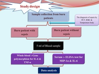
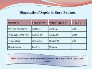
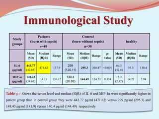
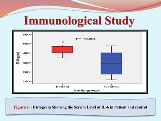
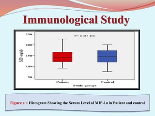
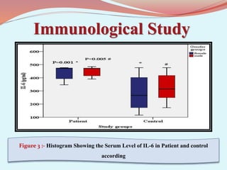
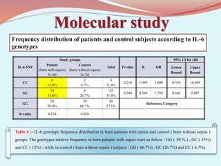
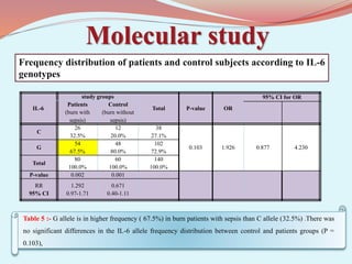
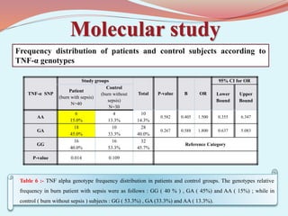
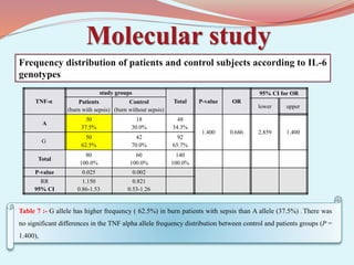
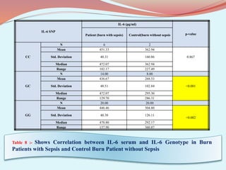
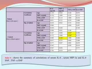
‫بسم‬
‫هللا‬
‫الرحيم‬ ‫الرحمن‬
َ‫ل‬ْ‫ع‬َ‫ت‬ ‫ُن‬‫ك‬َ‫ت‬ ْ‫م‬َ‫ل‬ ‫ا‬َ‫م‬ َ‫ك‬َ‫م‬َّ‫ل‬َ‫ع‬َ‫و‬
ُ‫م‬
ۚ
ُ‫ل‬ْ‫ض‬َ‫ف‬ َ‫ان‬َ‫ك‬َ‫و‬
‫ه‬ َّ
‫اَلل‬
‫ا‬ً‫م‬‫ي‬‫ه‬‫َظ‬‫ع‬ َ‫ك‬ْ‫ي‬َ‫ل‬َ‫ع‬
﴿
113
﴾
‫صدق‬
‫هللا‬
‫العظيم‬ ‫العلي‬
(
‫سورة‬
‫النساء‬
/
‫اية‬
113
)
by
Raghda S. M. alomari
Supervised by
Prof.
Dr. Mahdi H. Al-Ammar
Introduction
Burn injury remains an important cause of
morbidity and mortality worldwide . Infection
complications , including sepsis , septic shock
and sepsis –related organ failure , are common
among patients with moderate to severe burn
injuries .
Sepsis is a life-threatening organ-dysfunction
condition caused by a dysregulated response to
an infectious condition that can cause
complications in patients with major trauma .
Introduction
Burns are one of the most destructive forms of
trauma; despite the improvements in medical
care, infections remain an important cause of
burn injury-related mortality and morbidity , and
complicated sepsis predisposes patients to
diverse complications such as organ failure,
lengthening of hospital stays, and increased costs
Accurate diagnosis and early treatment of sepsis
may have a beneficial impact on clinical
outcome of burn – injured patients
Introduction
 In sepsis , the immune response that is initiated
by an invading pathogen fails to return to
homeostasis, thus culminating in a pathological
syndrome that is characterized by sustained
excessive inflammation and immune suppression
 The increase of cytokine production both in
humans and in experimental models seems to
play important roles in the pathophysiology of
sepsis and septic shock after burn injury.
Introduction
Pro-inflammatory interleukin - 6 is increased
early after burn and secreted by monocytes ,
endothelial cells and fibroblasts , and is able to
stimulate B and T lymphocytes and induce fever
. Some studies have indicated that IL-6 may play
a key role in the inflammatory response to
microbial invasion and also , it is one of the most
important members which may be associated
with sepsis risk and outcome.
Introduction
Macrophage inflammatory protein-1 alpha (MIP-
1α) is a chemotactic chemokine secreted by
macrophages. It performs various biological
functions, such as recruiting inflammatory cells,
wound healing, inhibition of stem cells, and
maintaining effector immune response
Single nucleotide polymorphisms (SNPs) , variations in
a nucleotide at a specific chromosome location, have
been linked to sepsis susceptibility and differences in
prognosis .
Introduction
Among these diverse cytokines , IL-6 and tumor
necrosis factor-α (TNF-α) have attracted
considerable attention . The human IL-6 gene was
mapped to chromosome 7p21 region and
contained several single nucleotide
polymorphisms .
The G to C polymorphism at position-174 of the
IL-6 gene (rs1800795) is associated with an
adverse outcome in a number of inflammatory
diseases, but its association with sepsis remains
unclear.
Introduction
On the other hand , the promoter of single nucleotide
polymorphisms (SNP) at position - 308 (G to A
substitution) of TNF-α gene has been demonstrated to
play a major role in the pathogenesis of sepsis and its
complications after burn injury .
The identified specific Toll-like receptor 4 and TNF-α
SNPs associated with increased risk of sepsis after burn
injury .
TNF-α is pro-inflammatory cytokine produced by
lymphocytes and macrophage.
Aims of the Study
 The current study aimed to investigate the evaluate the correlation of
Single Nucleotide Polymorphisms (SNP) in IL-6 -174 G→C , TNF–α ( -
308 G →A , with serum level of MIP-1 expression in the development of
burn patients with sepsis . The following objectives :-
1.Blood sample collection in a study based on peripheral blood sample .
2.Detection of gene polymorphism in (IL-6 -174 G→C) and TNF–α (-308
G →A) using ARMS-PCR.
3. Screening for IL-6 and MIP-1 level by using ELISA test .
4.The association between gene polymorphism and cytokine levels with the
development of burn patients with sepsis .
Sample collection from burn
patients
Burn patient with
sepsis
5 ml of Blood sample
Serum :- ELISA test for
MIP-1α & IL-6
Whole blood :-Gene
polymorphism for IL-6 &
TNF-α
Data analysis
The diagnosis of sepsis by
PCT ,WBC &
Temperature body
Burn patient without
sepsis
Study design
Diagnostic of Sepsis in Burn Patients
Biomarker Sepsis n=40 Without sepsis n=30 P-value
Procalcitonin (ng/ml) 9.16±5.07 0.57±1.27 0.01*
WBC cells (1*103/cc) 12.87±5.88 7.30±3.46 0.038*
Temperature 38.43±2.43 36.82±0.19 0.47
Blood culture Positive Negative
Table 1 :- Show the result biomarkers levels in sepsis and without sepsis burn
patients
Table 2 :- Shows higher frequency of sepsis in burn patient at age from 24-34 year (47.5%) , there
is no significant difference in mean age between burn with sepsis and burn without sepsis
Chi-Square
Sig.
Control
(burn without sepsis)
Patient
(burn with sepsis)
Age groups
4.618
0.099 NS
13 (43.3%)
8 (20%)
15-24 year
9 (30%)
19 (47.5%)
24-34 year
8 (26.7%)
13 (32.5%)
>35 year
0.051 NS
27.53± 9.03
32.1± 9.62
Mean ± SD
15-50
18-50
Range
Table 3 :- Shows Gender difference between burn with sepsis and burn without sepsis groups ,
sepsis in burn was found to be more prevalent among females(60%) than males (40%)
Chi-Square
p-value.
Control
(burn without sepsis)
Patient
(burn with sepsis)
Gender
0.000
1.000 NS
18 (60%)
24 (60%)
Female
12 (40%)
16 (40%)
Male
Immunological Study
Study
groups
Patients
(burn with sepsis)
n=40
Control
(burn without sepsis)
n=30
healthy
Mean
(SD)
Median
(IQR)
Range
Mean
(SD)
Median
(IQR)
Range
p-
value
Mean
(SD)
Median
(IQR)
Range
IL-6
(pg/ml)
443.77
(42.63)
471.62 137.9
299
(120.11)
295.3 360.87 <0.001
60.3
(32.8)
55.5 130.4
MIP -α
(pg/ml)
148.43
(34.63)
141.9 136.12
140.4
(36.93)
144.49 124.73 0.354
15.3
(2.52)
14.22 7.94
Table 3 :- Shows the serum level and median (IQR) of IL-6 and MIP-1α were significantly higher in
patient group than in control group they were 443.77 pg/ml (471.62) versus 299 pg/ml (295.3) and
148.43 pg/ml (141.9) versus 140.4 pg/ml (144.49) respectively
Immunological Study
Figure 1 :- Histogram Showing the Serum Level of IL-6 in Patient and control
Immunological Study
Figure 2 :- Histogram Showing the Serum Level of MIP-1α in Patient and control
Immunological Study
Figure 3 :- Histogram Showing the Serum Level of IL-6 in Patient and control
according
Immunological Study
Figure4:- Histogram Showing the Serum Level of MIP-1α in Patient and control
according
Molecular study
Figure 5 :- Agarose gel electrophoresis image that show the T . ARMS-PCR product analysis of IL-6 in patient
sample . T . ARMS-PCR product was analysis by 2% agarose gel . Where M: marker (100bp – 1500bp ) , lane
GG wild type homozygote , lane CC mutant type homozygote, and lane GC heterozygote
Detection of interleukin-6 by gel electrophoresis
Molecular study
Figure 6 :- Agarose gel electrophoresis image that show the T . ARMS-PCR product analysis of IL-6 in control
sample . T . ARMS-PCR product was analysis by 2% agarose gel . Where M: marker (100bp – 1500bp ) , lane
GG wild type homozygote , lane CC mutant type homozygote, and lane GC heterozygote
Detection of interleukin-6 by gel electrophoresis
Molecular study
Table 4 :- IL-6 genotype frequency distribution in burn patients with sepsis and control ( burn without sepsis )
groups. The genotypes relative frequency in burn patients with sepsis were as follow : GG ( 50 % ) , GC ( 35%)
and CC ( 15%) ; while in control ( burn without sepsis ) subjects : GG ( 66.7%) , GC (26.7%) and CC ( 6.7%).
Frequency distribution of patients and control subjects according to IL-6
genotypes
IL-6 SNP
Study groups
Total P-value B OR
95% CI for OR
Patient
(burn with sepsis)
N=40
Control
(burn without sepsis)
N=30
Lower
Bound
Upper
Bound
CC
6
15.0%
2
6.7%
8
11.4%
0.210 1.099 3.000 0.539 16.689
GC
14
35.0%
8
26.7%
22
31.4%
0.304 0.560 1.750 0.602 5.087
GG
20
50.0%
20
66.7%
40
57.1%
Reference Category
P-value 0.074 0.058
Molecular study
Table 5 :- G allele is in higher frequency ( 67.5%) in burn patients with sepsis than C allele (32.5%) .There was
no significant differences in the IL-6 allele frequency distribution between control and patients groups (P =
0.103),
Frequency distribution of patients and control subjects according to IL-6
genotypes
IL-6
study groups
Total P-value OR
95% CI for OR
Patients
(burn with
sepsis)
Control
(burn without
sepsis)
C
26
32.5%
12
20.0%
38
27.1%
0.103 1.926 0.877 4.230
G
54
67.5%
48
80.0%
102
72.9%
Total
80
100.0%
60
100.0%
140
100.0%
P-value 0.002 0.001
RR
95% CI
1.292
0.97-1.71
0.671
0.40-1.11
Molecular study
Figure 5 :- Agarose gel electrophoresis image that shows the ARMS-PCR product analysis of TNF–α (-308 G
→A) in patient sample . ARMS-PCR product was analyzed by 2% agarose gel . Where M: marker (100bp –
1500bp ) . GA heterozygous , positive PCR amplification 184 bp product size.
Detection of Tumor necrosis factor alpha by gel electrophoresis
Molecular study
Figure 5 :- Agarose gel electrophoresis image that shows the ARMS-PCR product analysis of TNF–α (-308 G
→A) in control sample . ARMS-PCR product was analyzed by 2% agarose gel . Where M: marker (100bp –
1500bp ) . AA homozygous , GG homozygous and GA heterozygous , positive PCR amplification 184 bp product
size.
Detection of Tumor necrosis factor alpha by gel electrophoresis
Molecular study
Table 6 :- TNF alpha genotype frequency distribution in patients and control groups. The genotypes relative
frequency in burn patient with sepsis were as follows : GG ( 40 % ) , GA ( 45%) and AA ( 15%) ; while in
control ( burn without sepsis ) subjects : GG ( 53.3%) , GA (33.3%) and AA ( 13.3%).
Frequency distribution of patients and control subjects according to
TNF-α genotypes
TNF-α SNP
Study groups
Total P-value B OR
95% CI for OR
Patient
(burn with sepsis)
N=40
Control
(burn without
sepsis)
N=30
Lower
Bound
Upper
Bound
AA
6
15.0%
4
13.3%
10
14.3%
0.582 0.405 1.500 0.355 6.347
GA
18
45.0%
10
33.3%
28
40.0%
0.267 0.588 1.800 0.637 5.083
GG
16
40.0%
16
53.3%
32
45.7%
Reference Category
P-value 0.014 0.109
Molecular study
Table 7 :- G allele has higher frequency ( 62.5%) in burn patients with sepsis than A allele (37.5%) . There was
no significant differences in the TNF alpha allele frequency distribution between control and patients groups (P =
1.400),
Frequency distribution of patients and control subjects according to IL-6
genotypes
TNF-α
study groups
Total P-value OR
95% CI for OR
Patients
(burn with sepsis)
Control
(burn without sepsis)
lower upper
A
30
37.5%
18
30.0%
48
34.3%
1.400 0.686 2.859 1.400
G
50
62.5%
42
70.0%
92
65.7%
Total
80
100.0%
60
100.0%
140
100.0%
P-value 0.025 0.002
RR
95% CI
1.150
0.86-1.53
0.821
0.53-1.26
Table 8 :- Shows Correlation between IL-6 serum and IL-6 Genotype in Burn
Patients with Sepsis and Control Burn Patient without Sepsis
IL-6 SNP
IL-6 (pg/ml)
p-value
Patient (burn with sepsis) Control(burn without sepsis
CC
N 6 2
Mean 451.33 362.94
0.867
Std. Deviation 40.31 160.86
Median 472.07 362.94
Range 102.17 227.49
GC
N 14.00 8.00
Mean 436.67 268.53
<0.001
Std. Deviation 48.51 102.84
Median 472.07 295.30
Range 129.70 286.31
GG
N 20.00 20.00
Mean 446.46 304.80
<0.002
Std. Deviation 40.38 126.11
Median 470.80 292.17
Range 137.90 360.87
Table 8 :- shows the summary of correlations of serum IL-6 , serum MIP-1α and IL-6
SNP , TNF- α SNP
IL-6
(pg/ml)
MIP -α
(pg/ml)
TNF-α SNP IL-6 SNP
Patient
(burn with sepsis )
Correlation
Coefficient
Age 0.134 0.028 0.230 0.076
MIP -α (g/ml) 0.134 1.000 0.114 0.112
TNF-α SNP 0.122 0.114 1.000 0.234
IL-6 SNP 0.011 0.112 0.234 1.000
Sig. (2-tailed) Age 0.410 0.862 0.153 0.642
MIP -α (g/ml) 0.409 . 0.484 0.491
TNF-α SNP 0.454 0.484 . 0.146
IL-6 SNP 0.948 0.491 0.146 .
Control
(burn without sepsis )
Correlation
Coefficient
Age 0.167 0.098 0.547* 0.090
MIP -α (g/ml) 0.172 1.000 0.396* 0.153
TNF-α SNP 0.008 0.396* 1.000 0.006
IL-6 SNP 0.057 0.153 0.006 1.000
Sig. (2-tailed) Age 0.377 0.607 0.002 0.638
MIP -α (g/ml) 0.362 . 0.030 0.419
TNF-α SNP 0.967 0.030 . 0.975
IL-6 SNP 0.766 0.419 0.975 .
Conclusions
1.Sepsis is a medical emergency commonly encountered
during the treatment of burn patients
2.PCT assay can be a valuable complement to the clinical
diagnosis of sepsis and is promising as a method to reduce
antibiotic resistance in critically ill patients.
3.Higher level of serum IL-6 in burn patients with sepsis
compared with burn patients without sepsis individuals .
This plays a role in diagnostic of sepsis .
Conclusions
4. MIP-1α level play an important role in burn patients with
sepsis and without sepsis .
5.The presence of heterozygous GA genotype in the TNF-α-308
was associated with sepsis of burn patients .
6.The present study showed that heterozygous GC genotype was
significantly higher in correlation between IL-6 serum and IL-6
genotype in burn patients with sepsis and burn patients without
sepsis groups (P < 0.001). Also , homozygous GG genotype was
significantly higher in correlation between IL-6 serum and IL-6
genotype in burn patients with sepsis and burn patients without
sepsis groups (P < 0.002) .
Recommendations
1.Studying more gene polymorphism associated with burn patients with
sepsis and burn patients without sepsis .
2.Studying MIP-1α genotype in burn patients with sepsis and compared
with healthy individuals.
3.Studying correlation between TNF-α serum and TNF-α genotype in
burn patients with sepsis and without sepsis .
4.detect other cytokines effect on the development of sepsis .
5.The use of tetra ARMS-PCR to detect genotype single-nucleotide
polymorphisms (SNPs) and allows the study of SNPs in a fast, reliable,
and low-cost way.
Thank you
for listening

presentation of my thesis

  • 1.
    ‫بسم‬ ‫هللا‬ ‫الرحيم‬ ‫الرحمن‬ َ‫ل‬ْ‫ع‬َ‫ت‬ ‫ُن‬‫ك‬َ‫ت‬ْ‫م‬َ‫ل‬ ‫ا‬َ‫م‬ َ‫ك‬َ‫م‬َّ‫ل‬َ‫ع‬َ‫و‬ ُ‫م‬ ۚ ُ‫ل‬ْ‫ض‬َ‫ف‬ َ‫ان‬َ‫ك‬َ‫و‬ ‫ه‬ َّ ‫اَلل‬ ‫ا‬ً‫م‬‫ي‬‫ه‬‫َظ‬‫ع‬ َ‫ك‬ْ‫ي‬َ‫ل‬َ‫ع‬ ﴿ 113 ﴾ ‫صدق‬ ‫هللا‬ ‫العظيم‬ ‫العلي‬ ( ‫سورة‬ ‫النساء‬ / ‫اية‬ 113 )
  • 2.
    by Raghda S. M.alomari Supervised by Prof. Dr. Mahdi H. Al-Ammar
  • 4.
    Introduction Burn injury remainsan important cause of morbidity and mortality worldwide . Infection complications , including sepsis , septic shock and sepsis –related organ failure , are common among patients with moderate to severe burn injuries . Sepsis is a life-threatening organ-dysfunction condition caused by a dysregulated response to an infectious condition that can cause complications in patients with major trauma .
  • 5.
    Introduction Burns are oneof the most destructive forms of trauma; despite the improvements in medical care, infections remain an important cause of burn injury-related mortality and morbidity , and complicated sepsis predisposes patients to diverse complications such as organ failure, lengthening of hospital stays, and increased costs Accurate diagnosis and early treatment of sepsis may have a beneficial impact on clinical outcome of burn – injured patients
  • 6.
    Introduction  In sepsis, the immune response that is initiated by an invading pathogen fails to return to homeostasis, thus culminating in a pathological syndrome that is characterized by sustained excessive inflammation and immune suppression  The increase of cytokine production both in humans and in experimental models seems to play important roles in the pathophysiology of sepsis and septic shock after burn injury.
  • 7.
    Introduction Pro-inflammatory interleukin -6 is increased early after burn and secreted by monocytes , endothelial cells and fibroblasts , and is able to stimulate B and T lymphocytes and induce fever . Some studies have indicated that IL-6 may play a key role in the inflammatory response to microbial invasion and also , it is one of the most important members which may be associated with sepsis risk and outcome.
  • 8.
    Introduction Macrophage inflammatory protein-1alpha (MIP- 1α) is a chemotactic chemokine secreted by macrophages. It performs various biological functions, such as recruiting inflammatory cells, wound healing, inhibition of stem cells, and maintaining effector immune response Single nucleotide polymorphisms (SNPs) , variations in a nucleotide at a specific chromosome location, have been linked to sepsis susceptibility and differences in prognosis .
  • 9.
    Introduction Among these diversecytokines , IL-6 and tumor necrosis factor-α (TNF-α) have attracted considerable attention . The human IL-6 gene was mapped to chromosome 7p21 region and contained several single nucleotide polymorphisms . The G to C polymorphism at position-174 of the IL-6 gene (rs1800795) is associated with an adverse outcome in a number of inflammatory diseases, but its association with sepsis remains unclear.
  • 10.
    Introduction On the otherhand , the promoter of single nucleotide polymorphisms (SNP) at position - 308 (G to A substitution) of TNF-α gene has been demonstrated to play a major role in the pathogenesis of sepsis and its complications after burn injury . The identified specific Toll-like receptor 4 and TNF-α SNPs associated with increased risk of sepsis after burn injury . TNF-α is pro-inflammatory cytokine produced by lymphocytes and macrophage.
  • 11.
    Aims of theStudy  The current study aimed to investigate the evaluate the correlation of Single Nucleotide Polymorphisms (SNP) in IL-6 -174 G→C , TNF–α ( - 308 G →A , with serum level of MIP-1 expression in the development of burn patients with sepsis . The following objectives :- 1.Blood sample collection in a study based on peripheral blood sample . 2.Detection of gene polymorphism in (IL-6 -174 G→C) and TNF–α (-308 G →A) using ARMS-PCR. 3. Screening for IL-6 and MIP-1 level by using ELISA test . 4.The association between gene polymorphism and cytokine levels with the development of burn patients with sepsis .
  • 13.
    Sample collection fromburn patients Burn patient with sepsis 5 ml of Blood sample Serum :- ELISA test for MIP-1α & IL-6 Whole blood :-Gene polymorphism for IL-6 & TNF-α Data analysis The diagnosis of sepsis by PCT ,WBC & Temperature body Burn patient without sepsis Study design
  • 15.
    Diagnostic of Sepsisin Burn Patients Biomarker Sepsis n=40 Without sepsis n=30 P-value Procalcitonin (ng/ml) 9.16±5.07 0.57±1.27 0.01* WBC cells (1*103/cc) 12.87±5.88 7.30±3.46 0.038* Temperature 38.43±2.43 36.82±0.19 0.47 Blood culture Positive Negative Table 1 :- Show the result biomarkers levels in sepsis and without sepsis burn patients
  • 16.
    Table 2 :-Shows higher frequency of sepsis in burn patient at age from 24-34 year (47.5%) , there is no significant difference in mean age between burn with sepsis and burn without sepsis Chi-Square Sig. Control (burn without sepsis) Patient (burn with sepsis) Age groups 4.618 0.099 NS 13 (43.3%) 8 (20%) 15-24 year 9 (30%) 19 (47.5%) 24-34 year 8 (26.7%) 13 (32.5%) >35 year 0.051 NS 27.53± 9.03 32.1± 9.62 Mean ± SD 15-50 18-50 Range
  • 17.
    Table 3 :-Shows Gender difference between burn with sepsis and burn without sepsis groups , sepsis in burn was found to be more prevalent among females(60%) than males (40%) Chi-Square p-value. Control (burn without sepsis) Patient (burn with sepsis) Gender 0.000 1.000 NS 18 (60%) 24 (60%) Female 12 (40%) 16 (40%) Male
  • 18.
    Immunological Study Study groups Patients (burn withsepsis) n=40 Control (burn without sepsis) n=30 healthy Mean (SD) Median (IQR) Range Mean (SD) Median (IQR) Range p- value Mean (SD) Median (IQR) Range IL-6 (pg/ml) 443.77 (42.63) 471.62 137.9 299 (120.11) 295.3 360.87 <0.001 60.3 (32.8) 55.5 130.4 MIP -α (pg/ml) 148.43 (34.63) 141.9 136.12 140.4 (36.93) 144.49 124.73 0.354 15.3 (2.52) 14.22 7.94 Table 3 :- Shows the serum level and median (IQR) of IL-6 and MIP-1α were significantly higher in patient group than in control group they were 443.77 pg/ml (471.62) versus 299 pg/ml (295.3) and 148.43 pg/ml (141.9) versus 140.4 pg/ml (144.49) respectively
  • 19.
    Immunological Study Figure 1:- Histogram Showing the Serum Level of IL-6 in Patient and control
  • 20.
    Immunological Study Figure 2:- Histogram Showing the Serum Level of MIP-1α in Patient and control
  • 21.
    Immunological Study Figure 3:- Histogram Showing the Serum Level of IL-6 in Patient and control according
  • 22.
    Immunological Study Figure4:- HistogramShowing the Serum Level of MIP-1α in Patient and control according
  • 23.
    Molecular study Figure 5:- Agarose gel electrophoresis image that show the T . ARMS-PCR product analysis of IL-6 in patient sample . T . ARMS-PCR product was analysis by 2% agarose gel . Where M: marker (100bp – 1500bp ) , lane GG wild type homozygote , lane CC mutant type homozygote, and lane GC heterozygote Detection of interleukin-6 by gel electrophoresis
  • 24.
    Molecular study Figure 6:- Agarose gel electrophoresis image that show the T . ARMS-PCR product analysis of IL-6 in control sample . T . ARMS-PCR product was analysis by 2% agarose gel . Where M: marker (100bp – 1500bp ) , lane GG wild type homozygote , lane CC mutant type homozygote, and lane GC heterozygote Detection of interleukin-6 by gel electrophoresis
  • 25.
    Molecular study Table 4:- IL-6 genotype frequency distribution in burn patients with sepsis and control ( burn without sepsis ) groups. The genotypes relative frequency in burn patients with sepsis were as follow : GG ( 50 % ) , GC ( 35%) and CC ( 15%) ; while in control ( burn without sepsis ) subjects : GG ( 66.7%) , GC (26.7%) and CC ( 6.7%). Frequency distribution of patients and control subjects according to IL-6 genotypes IL-6 SNP Study groups Total P-value B OR 95% CI for OR Patient (burn with sepsis) N=40 Control (burn without sepsis) N=30 Lower Bound Upper Bound CC 6 15.0% 2 6.7% 8 11.4% 0.210 1.099 3.000 0.539 16.689 GC 14 35.0% 8 26.7% 22 31.4% 0.304 0.560 1.750 0.602 5.087 GG 20 50.0% 20 66.7% 40 57.1% Reference Category P-value 0.074 0.058
  • 26.
    Molecular study Table 5:- G allele is in higher frequency ( 67.5%) in burn patients with sepsis than C allele (32.5%) .There was no significant differences in the IL-6 allele frequency distribution between control and patients groups (P = 0.103), Frequency distribution of patients and control subjects according to IL-6 genotypes IL-6 study groups Total P-value OR 95% CI for OR Patients (burn with sepsis) Control (burn without sepsis) C 26 32.5% 12 20.0% 38 27.1% 0.103 1.926 0.877 4.230 G 54 67.5% 48 80.0% 102 72.9% Total 80 100.0% 60 100.0% 140 100.0% P-value 0.002 0.001 RR 95% CI 1.292 0.97-1.71 0.671 0.40-1.11
  • 27.
    Molecular study Figure 5:- Agarose gel electrophoresis image that shows the ARMS-PCR product analysis of TNF–α (-308 G →A) in patient sample . ARMS-PCR product was analyzed by 2% agarose gel . Where M: marker (100bp – 1500bp ) . GA heterozygous , positive PCR amplification 184 bp product size. Detection of Tumor necrosis factor alpha by gel electrophoresis
  • 28.
    Molecular study Figure 5:- Agarose gel electrophoresis image that shows the ARMS-PCR product analysis of TNF–α (-308 G →A) in control sample . ARMS-PCR product was analyzed by 2% agarose gel . Where M: marker (100bp – 1500bp ) . AA homozygous , GG homozygous and GA heterozygous , positive PCR amplification 184 bp product size. Detection of Tumor necrosis factor alpha by gel electrophoresis
  • 29.
    Molecular study Table 6:- TNF alpha genotype frequency distribution in patients and control groups. The genotypes relative frequency in burn patient with sepsis were as follows : GG ( 40 % ) , GA ( 45%) and AA ( 15%) ; while in control ( burn without sepsis ) subjects : GG ( 53.3%) , GA (33.3%) and AA ( 13.3%). Frequency distribution of patients and control subjects according to TNF-α genotypes TNF-α SNP Study groups Total P-value B OR 95% CI for OR Patient (burn with sepsis) N=40 Control (burn without sepsis) N=30 Lower Bound Upper Bound AA 6 15.0% 4 13.3% 10 14.3% 0.582 0.405 1.500 0.355 6.347 GA 18 45.0% 10 33.3% 28 40.0% 0.267 0.588 1.800 0.637 5.083 GG 16 40.0% 16 53.3% 32 45.7% Reference Category P-value 0.014 0.109
  • 30.
    Molecular study Table 7:- G allele has higher frequency ( 62.5%) in burn patients with sepsis than A allele (37.5%) . There was no significant differences in the TNF alpha allele frequency distribution between control and patients groups (P = 1.400), Frequency distribution of patients and control subjects according to IL-6 genotypes TNF-α study groups Total P-value OR 95% CI for OR Patients (burn with sepsis) Control (burn without sepsis) lower upper A 30 37.5% 18 30.0% 48 34.3% 1.400 0.686 2.859 1.400 G 50 62.5% 42 70.0% 92 65.7% Total 80 100.0% 60 100.0% 140 100.0% P-value 0.025 0.002 RR 95% CI 1.150 0.86-1.53 0.821 0.53-1.26
  • 31.
    Table 8 :-Shows Correlation between IL-6 serum and IL-6 Genotype in Burn Patients with Sepsis and Control Burn Patient without Sepsis IL-6 SNP IL-6 (pg/ml) p-value Patient (burn with sepsis) Control(burn without sepsis CC N 6 2 Mean 451.33 362.94 0.867 Std. Deviation 40.31 160.86 Median 472.07 362.94 Range 102.17 227.49 GC N 14.00 8.00 Mean 436.67 268.53 <0.001 Std. Deviation 48.51 102.84 Median 472.07 295.30 Range 129.70 286.31 GG N 20.00 20.00 Mean 446.46 304.80 <0.002 Std. Deviation 40.38 126.11 Median 470.80 292.17 Range 137.90 360.87
  • 32.
    Table 8 :-shows the summary of correlations of serum IL-6 , serum MIP-1α and IL-6 SNP , TNF- α SNP IL-6 (pg/ml) MIP -α (pg/ml) TNF-α SNP IL-6 SNP Patient (burn with sepsis ) Correlation Coefficient Age 0.134 0.028 0.230 0.076 MIP -α (g/ml) 0.134 1.000 0.114 0.112 TNF-α SNP 0.122 0.114 1.000 0.234 IL-6 SNP 0.011 0.112 0.234 1.000 Sig. (2-tailed) Age 0.410 0.862 0.153 0.642 MIP -α (g/ml) 0.409 . 0.484 0.491 TNF-α SNP 0.454 0.484 . 0.146 IL-6 SNP 0.948 0.491 0.146 . Control (burn without sepsis ) Correlation Coefficient Age 0.167 0.098 0.547* 0.090 MIP -α (g/ml) 0.172 1.000 0.396* 0.153 TNF-α SNP 0.008 0.396* 1.000 0.006 IL-6 SNP 0.057 0.153 0.006 1.000 Sig. (2-tailed) Age 0.377 0.607 0.002 0.638 MIP -α (g/ml) 0.362 . 0.030 0.419 TNF-α SNP 0.967 0.030 . 0.975 IL-6 SNP 0.766 0.419 0.975 .
  • 34.
    Conclusions 1.Sepsis is amedical emergency commonly encountered during the treatment of burn patients 2.PCT assay can be a valuable complement to the clinical diagnosis of sepsis and is promising as a method to reduce antibiotic resistance in critically ill patients. 3.Higher level of serum IL-6 in burn patients with sepsis compared with burn patients without sepsis individuals . This plays a role in diagnostic of sepsis .
  • 35.
    Conclusions 4. MIP-1α levelplay an important role in burn patients with sepsis and without sepsis . 5.The presence of heterozygous GA genotype in the TNF-α-308 was associated with sepsis of burn patients . 6.The present study showed that heterozygous GC genotype was significantly higher in correlation between IL-6 serum and IL-6 genotype in burn patients with sepsis and burn patients without sepsis groups (P < 0.001). Also , homozygous GG genotype was significantly higher in correlation between IL-6 serum and IL-6 genotype in burn patients with sepsis and burn patients without sepsis groups (P < 0.002) .
  • 36.
    Recommendations 1.Studying more genepolymorphism associated with burn patients with sepsis and burn patients without sepsis . 2.Studying MIP-1α genotype in burn patients with sepsis and compared with healthy individuals. 3.Studying correlation between TNF-α serum and TNF-α genotype in burn patients with sepsis and without sepsis . 4.detect other cytokines effect on the development of sepsis . 5.The use of tetra ARMS-PCR to detect genotype single-nucleotide polymorphisms (SNPs) and allows the study of SNPs in a fast, reliable, and low-cost way.
  • 37.