Measurements of immune Functions
Epitope Detection By Antibodies
Epotope quantitation by antibodies
Assessment of immune function
Assessment of hypersensitivity
Introduction
Immune function canbe measured to assess the strength and type of
immune response.
It helps diagnose immunodeficiencies, monitor immune therapy, and
understand immune status.
Measurement is done using serological (antibody-based), cellular, and
molecular techniques.
One key method involves detecting epitopes (antigenic determinants)
using specific antibodies.
Techniques are based on antigen-antibody interactions and can be
qualitative or quantitative.
3.
Epitope Detection byAntibodies
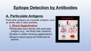
A. Particulate Antigens
Particulate antigens are insoluble antigens, such
as whole cells or latex particles.
1. Direct Agglutination
Antibodies react directly with particulate
antigens (e.g., red blood cells, bacteria).
Leads to visible clumping (agglutination).
Used in blood typing and Widal test for
typhoid.
4.
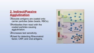
2. Indirect/Passive
Agglutination
Soluble antigensare coated onto
carrier particles (latex beads, RBCs).
Antibodies then react with the
coated particles causing
agglutination.
Increases test sensitivity.
Used for detecting Rheumatoid
factor, CRP, and viral antigens.
5.
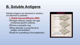
B. Soluble Antigens
Solubleantigens are dissolved in solution,
not attached to particles.
1. Radial Immunodiffusion (RID)
Antigen diffuses radially into agar
containing specific antibody.
Forms a precipitin ring.
Ring diameter is proportional to
antigen concentration.
Used for quantifying immunoglobulins.
6.
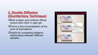
2. Double Diffusion
(OuchterlonyTechnique)
Both antigen and antibody diffuse
toward each other in agar gel.
Forms a line of precipitation at the
zone of equivalence.
Useful for comparing antigenic
relationships between different
samples.
7.
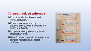
3. Immunoelectrophoresis
Combines electrophoresisand
immunodiffusion.
Proteins are separated by
electrophoresis, then antibodies are
added in a trough.
Antigen-antibody interaction forms
precipitation arcs.
Used for detecting multiple antigens in
complex mixtures (e.g., serum
proteins).
8.
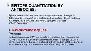
EPITOPE QUANTITATIONBY
ANTIBODIES:
Epitope quantitation involves measuring the number of antigenic
determinants (epitopes) on a protein, cell, or surface. These methods
utilize specific antibodies that bind to epitopes to assess
antigen expression.
1. Radioimmunoassay (RIA)
Principle:
Radioimmunoassay (RIA) is a sensitive technique that measures the
concentration of a specific substance (antigen) in a sample by using
competitive binding between a labeled antigen and an unlabeled antigen
(from the sample) for a limited number of antibody binding sites.
9.
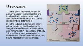
Procedure
1- Inthe direct radioimmuno assay,
primary antibody is radiolabeled and
incubated with antigen. unbound
antibody is washed away, and bound
radioactivity id determined.
2- In the indirect radioimmuno assay,
primary antibody that has bound to
antigen is detected with a radiolabeled,
anti-immunoglobin ( secondary antibody
), the antibody -antigen complex is
washed free of unbound antibodies, and
bound radioactivity is determined.
10.
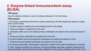
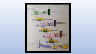
2. Enzyme-linked immunosorbentassay
(ELISA)
Purpose
Enzyme-labeled antibody is used for epitope detection in this technique.
Procedure
1.The assay is generally performed in protein-adsorbing, 96-well polystyrene plates (a single
well is shown here).
2. Soluble antigen is added and noncovalently binds to the plastic.
3. Unbound antigen is washed from the well.
4. Unlabeled (often sera to be tested) primary antibodies are added to the well and allowed to
bind.
5. Unbound primary antibodies are washed from the well.
6. Enzyme-labeled anti-immunoglobulin antibodies are added and allowed to bind.
7. Unbound enzyme-labeled antibodies are washed from the well.
8. An enzyme-cleavable, chromogenic substrate is added to the well and allowed to incubate.
9. Color change indicates the presence of enzyme-labeled secondary antibody. Because the
second antibody only binds to the primary antibody and the primary antibody only binds to the
12.
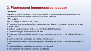
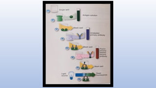
3. Fluorescent immunosorbentassay
Principle
FIA detects specific antigens or antibodies using fluorescent-labeled antibodies or probes,
which emit fluorescence upon binding to the target molecule.
Procedure
The FIA design is similar to the ELISA.
1.The assay may be performed in protein-adsorbing, 96-well polystyrene plates (a single well
is shown here).
2. Soluble antigen is added and noncovalently binds to the plastic.
3. Unbound antigen is washed from the well.
4. Unlabeled (often sera to be tested) primary antibodies are added to the well and allowed to
bind.
5. Unbound primary antibodies are washed from the well.
6. Fluorochrome-labeled anti-immunoglobulin antibodies are added to the well and allowed to
bind.
7. Unbound-labeled antibodies are washed from the well.
8. Fluorescence indicates the presence of epitopes.
14.
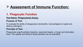
Assesment ofImmune Function:
1. Phagocytic Function
Test Name: Phagocytosis Assay
Purpose of Test: -
To evaluate the ability of phagocytes (neutrophils, macrophages) to ingest and
destroy pathogens.
Principle of Test: -
Phagocytes engulf particles (bacteria, opsonized beads, or fungi) and internalize
them. The uptake and killing of these particles can be quantified.
15.
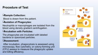
Procedure of Test
SampleCollection:
Blood is drawn from the patient.
Isolation of Phagocytes:
Neutrophils or macrophages are isolated from the
blood using density gradient centrifugation.
Incubation with Particles:
The phagocytes are incubated with labeled
bacteria or opsonized particles.
Assessment:
After incubation, phagocytosis is assessed using
microscopy, flow cytometry, or colony-forming unit
(CFU) assays to measure the phagocytic uptake
and killing efficiency.
16.
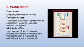
2. Proliferation:
Test Name:
LymphocyteProliferation Assay
Purpose of Test:
To evaluate the ability of lymphocytes to
proliferate in response to specific
antigens or mitogens.
Principle of Test:
Lymphocytes (T and B cells) are
exposed to mitogens or antigens, and
their proliferation is measured by
radioactive thymidine incorporation.
17.
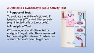
3.Cytotoxic T Lymphocyte(CTL) Activity Test
Purpose of Test:
To evaluate the ability of cytotoxic T
lymphocytes (CTLs) to kill target cells
(e.g., infected cells or tumor cells).
Principle of Test:
CTLs recognize and kill infected or
malignant target cells. This is assessed
by measuring the release of radioactive
sodium chromate lysed target cells.
18.
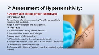
Assessment ofHypersensitivity:
1.Allergy Skin Testing (Type 1 Sensitivity) :
Purpose of Test
To identify specific allergens causing Type I hypersensitivity
reactions (IgE mediated).
Helps in allergy diagnosis and management.
Procedure
1. Clean skin area (usually forearm or back).
2. Mark and label sites for each allergen.
3. Apply a drop of allergen extract.
4. Prick skin through the drop using a sterile lancet.
5. Observe for reaction (wheal and redness) after 15–20 minutes.
6. Measure and record reaction size.
7. Compare with histamine (positive control) and saline (negative
control).
19.
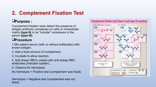
2. Complement FixationTest
Purpose :
Complement fixation tests detect the presence of
antigen-antibody complexes on cells or intracellular
matrix (type II) or as "soluble" complexes in the
serum (type III).
Procedure
1.Mix patient serum (with or without antibodies) with
known antigen.
2. Add a fixed amount of complement.
3. Incubate to allow reaction.
4. Add sheep RBCs coated with anti-sheep RBC
antibodies (indicator system).
5. Observe for hemolysis:
No hemolysis = Positive test (complement was fixed).
Hemolysis = Negative test (complement was not
fixed).
20.
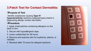
3.Patch Test forContact Dermatitis:
Purpose of Test
Identify substances causing Type IV
hypersensitivity reactions (delayed-type).Useful in
diagnosing allergic contact dermatitis.
Procedure
1. Apply small patches containing allergens on the
back or arm.
2. Secure with hypoallergenic tape.
3. Leave undisturbed for 48 hours.
4. Remove and examine skin for erythema, edema, or
vesicles.
5. Recheck after 72 hours for delayed reactions.
.


![HIV MLS Presentation [Autosaved]-1.pptxx](https://cdn.slidesharecdn.com/ss_thumbnails/hivmlspresentationautosaved-1-251126074336-0b1c62b0-thumbnail.jpg?width=640&height=640&fit=bounds)

![LAB INFORMATION SYSTEM presentation [Autosaved].pptx](https://cdn.slidesharecdn.com/ss_thumbnails/labinformationsystempresentationautosaved-251126072037-cfeb429f-thumbnail.jpg?width=640&height=640&fit=bounds)


