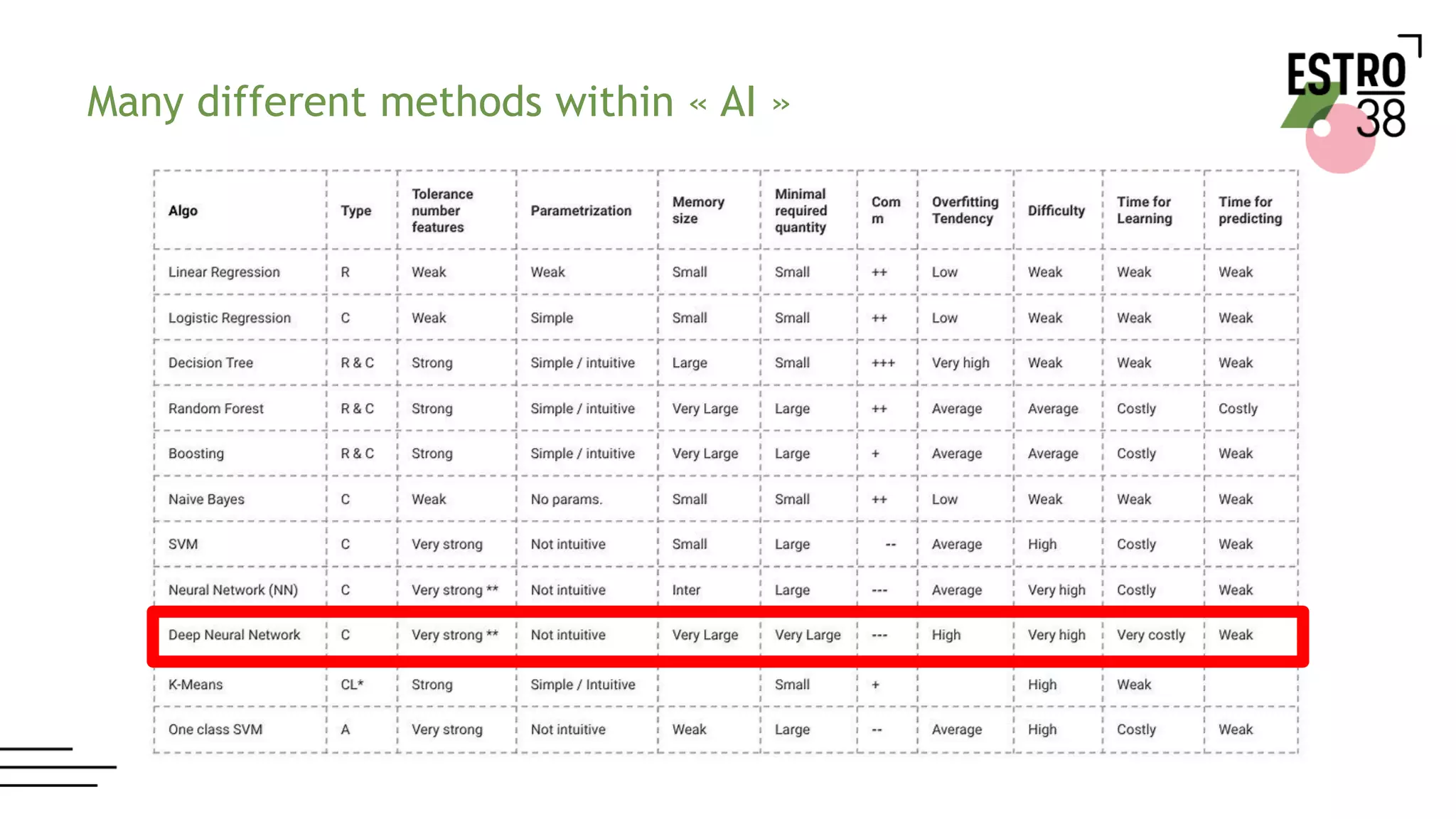
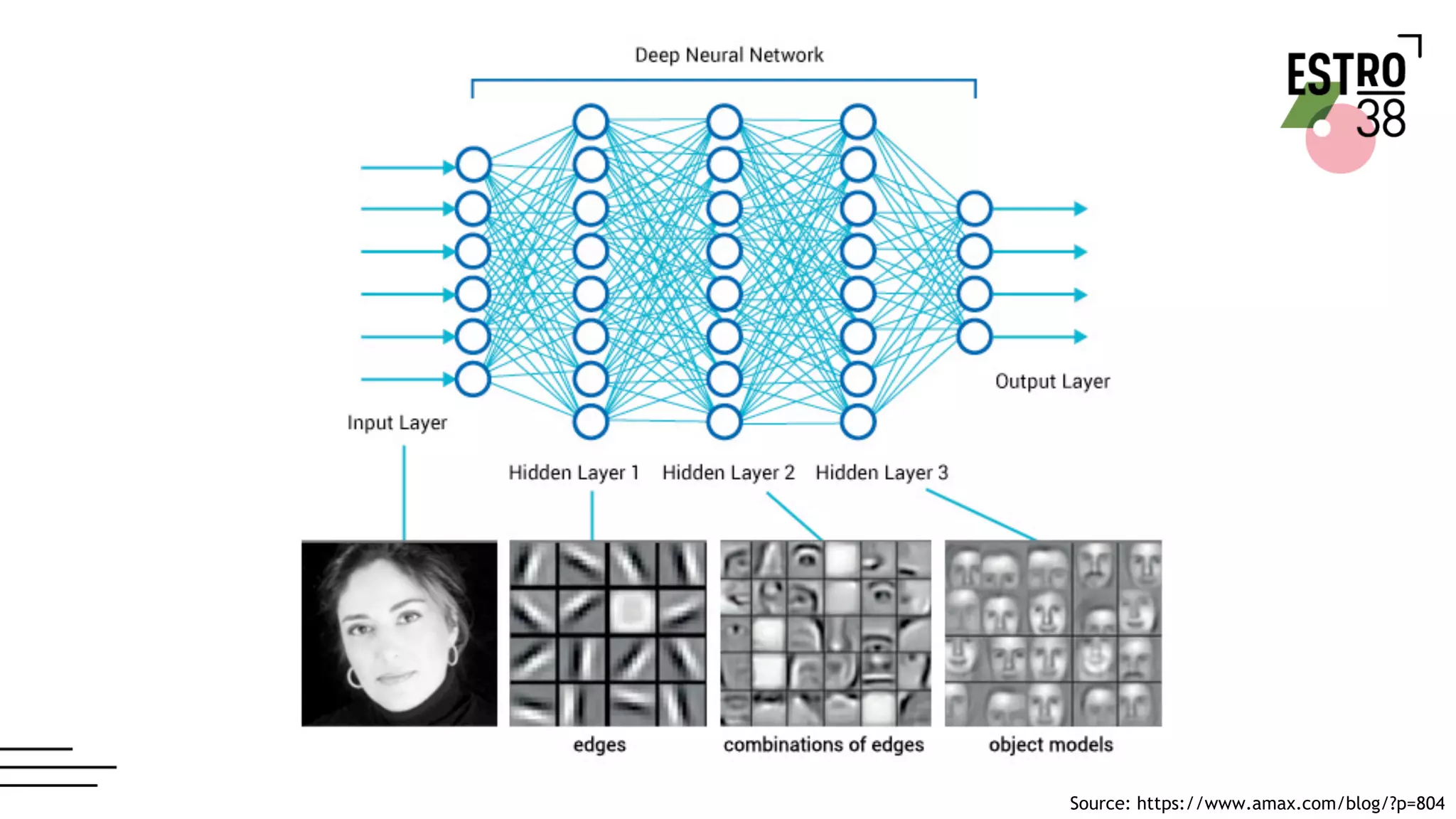
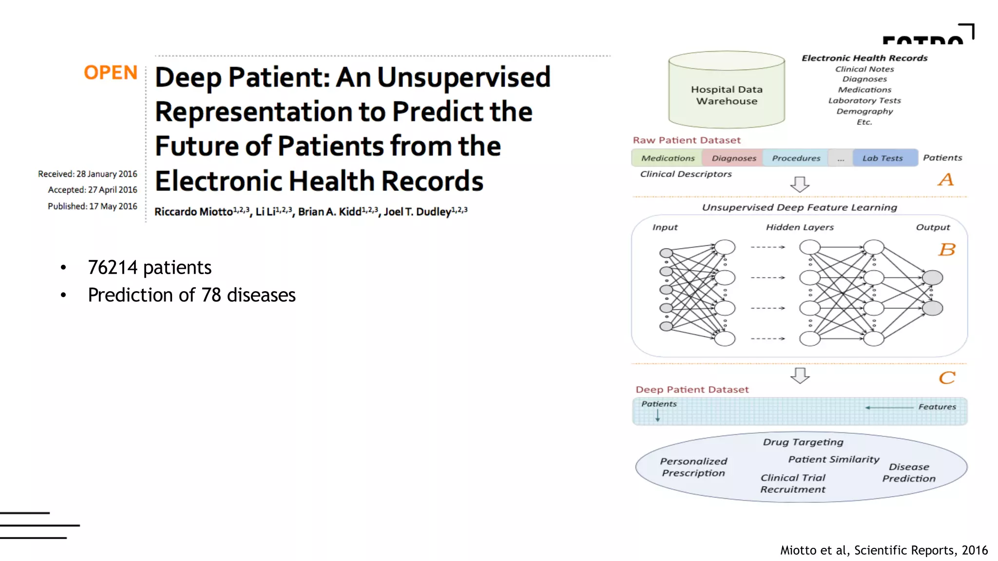
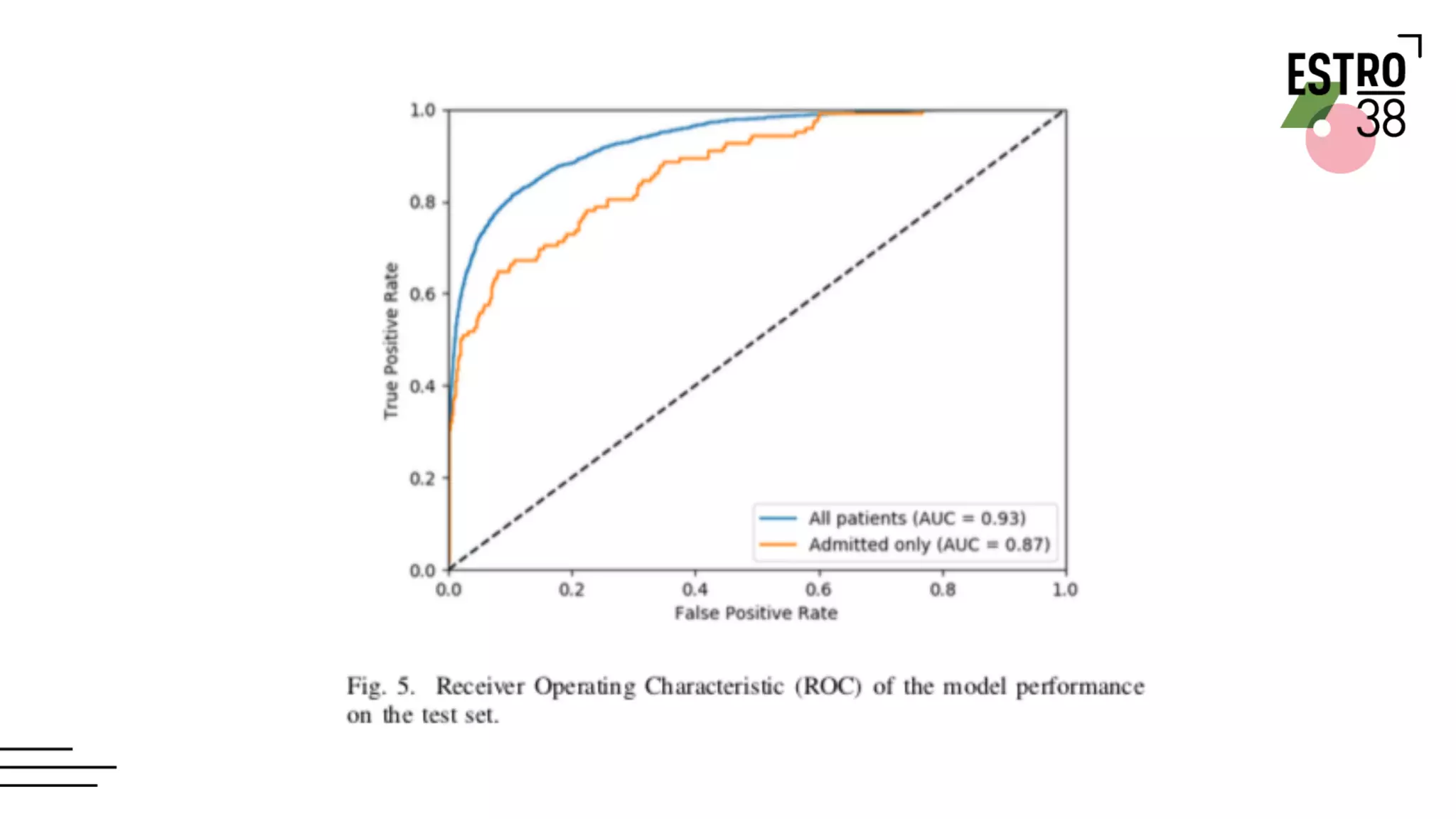
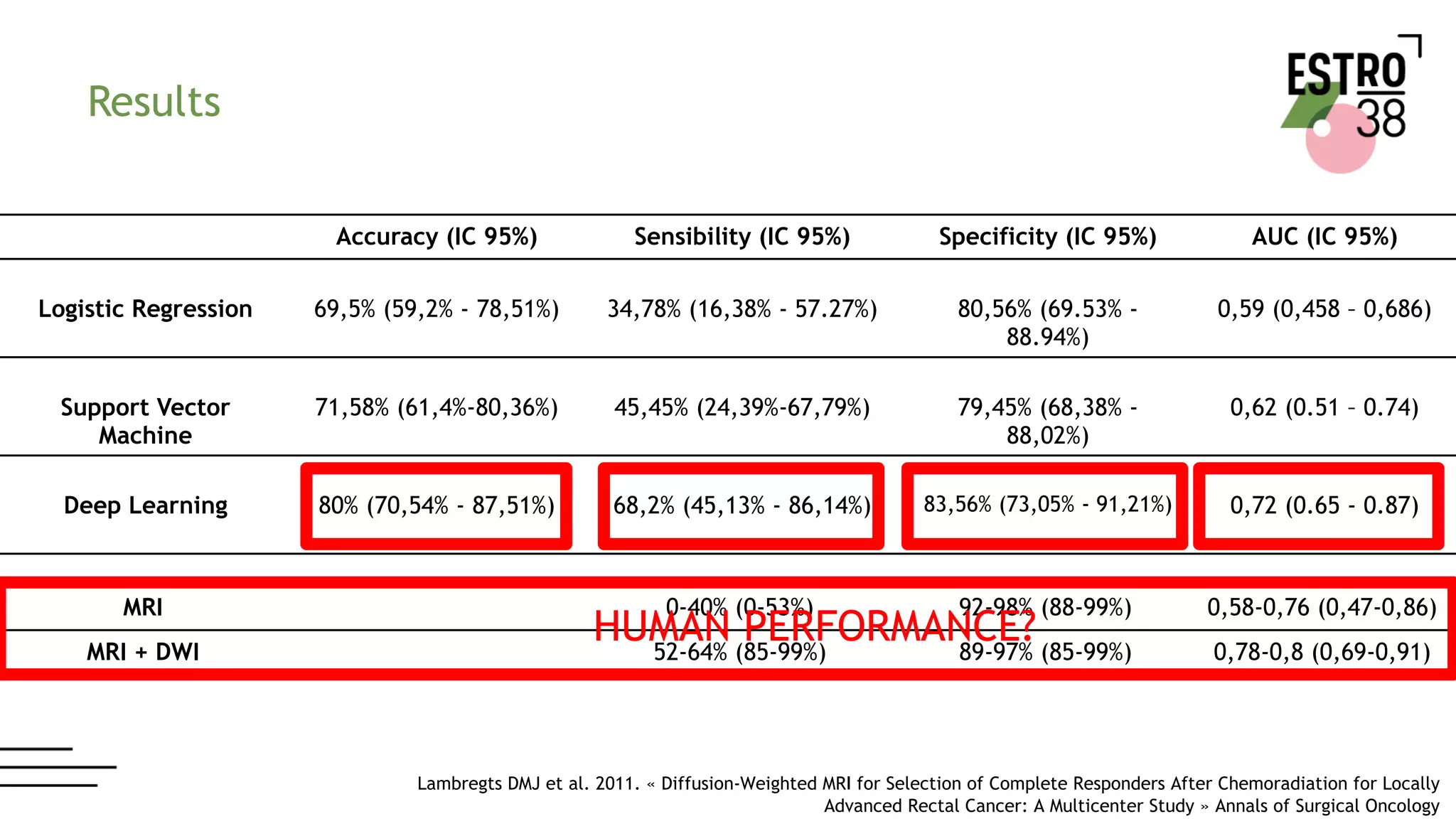
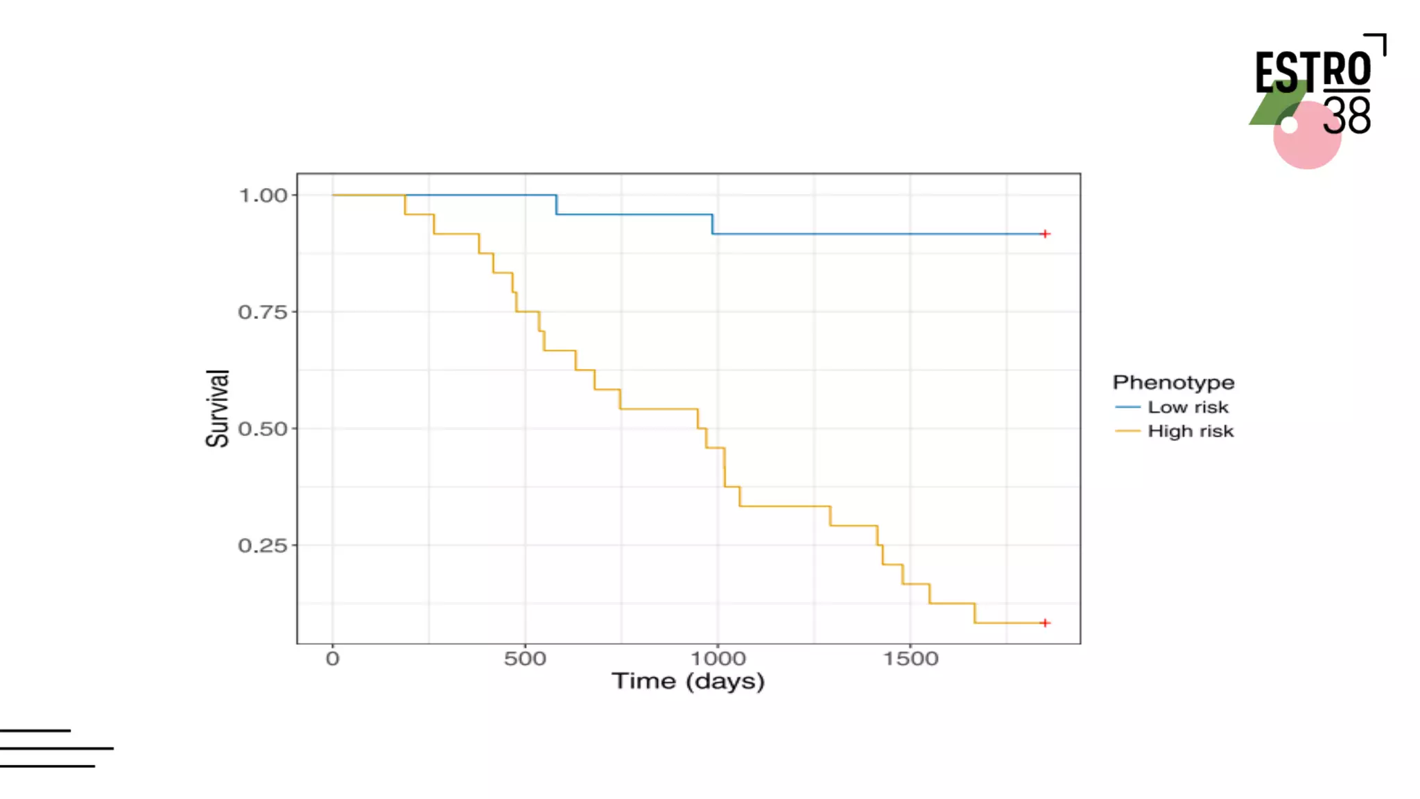
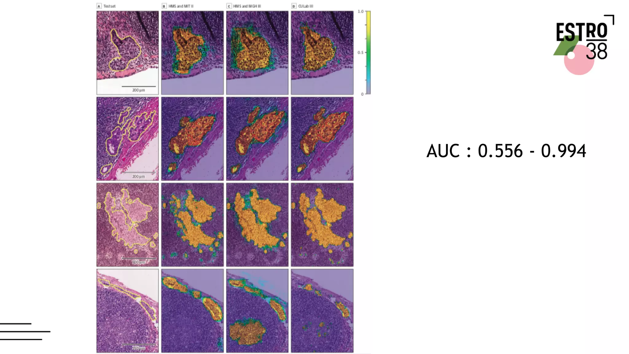
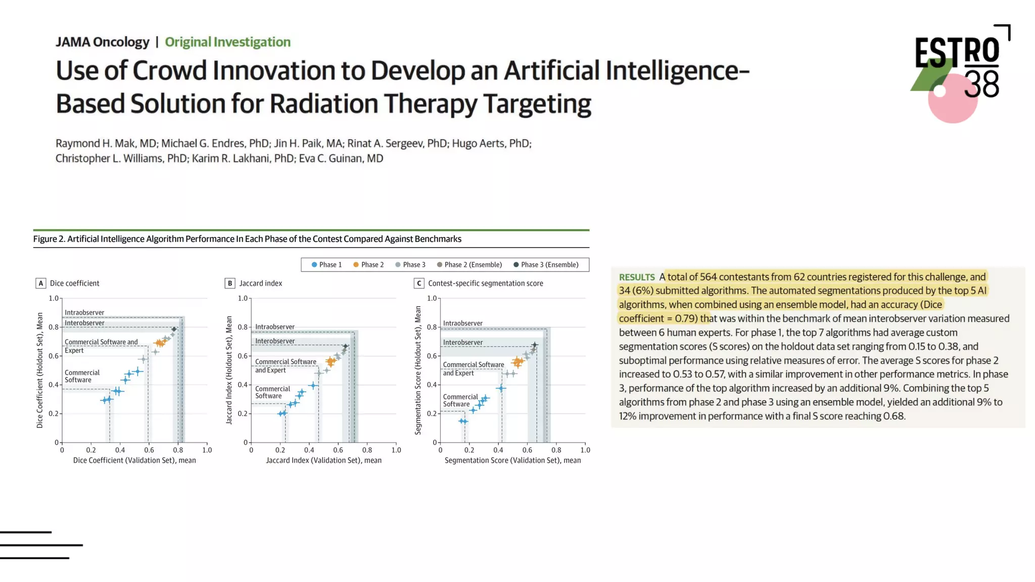
Artificial Intelligence in Radiation OncologyWookjin Choi
- The document discusses the use of artificial intelligence and radiomics in radiation oncology. It presents frameworks for radiomics analysis involving image registration, tumor segmentation, feature extraction, and predictive modeling.
- Specific applications discussed include using radiomics for lung cancer screening and prediction of tumor response. Radiomics features combined with machine learning models show improved performance over clinical guidelines for assessing lung nodule malignancy.
- Methods are also presented for quantifying tumor characteristics like spiculation through image analysis and extracting interpretable radiomics features. This can provide semantic information to radiologists for assessment.
Artificial Intelligence in Radiation Oncology.pptxWookjin Choi
The document discusses artificial intelligence applications in radiation oncology, including automatic delineation of organs-at-risk using deep learning models like OARNet. It also discusses radiomics approaches for clinical decision support and outcomes prediction using features extracted from medical images with techniques like spiculation quantification for lung cancer screening.
Artificial Intelligence in Radiation OncologyWookjin Choi
This document discusses artificial intelligence applications in radiation oncology. It begins with acknowledgements and then outlines several AI applications including radiomics tools for lung cancer screening, tumor response prediction, and predicting aggressive lung adenocarcinoma subtypes. It also discusses using AI for automatic tumor delineation and quantification of delineation variability as well as local tumor morphological changes prediction and metabolic tumor volume changes. The document provides details on methods and results for several of these AI applications in radiation oncology.
This document provides an overview of image-guided radiation therapy (IGRT) for lung cancer. It discusses the role of IGRT in managing tumor motion through techniques like breath hold methods, free breathing with gating or tracking, and 4D imaging. Segmentation of the tumor and organs at risk on 4D CT scans is covered. Dose fractionation schedules and biological effective dose calculations for hypofractionated stereotactic body radiation therapy are reviewed. Toxicities, outcomes, and challenges of IGRT in lung cancer are also mentioned.
Final ICRU 62 ( International commission on radiation units and measurements)DrAyush Garg
The document discusses recommendations from reports by the International Commission on Radiation Units and Measurements (ICRU) for defining volumes used in radiation therapy planning and reporting. ICRU Report 62 provides additional details on volumes such as the internal target volume (ITV) and planning organ at risk volume (PRV), and introduces metrics like the conformity index. It also further classifies organs at risk as serial, parallel or serial-parallel based on their radiosensitivity.
TBI is the radiotherapy technique to irradiate whole body before doing stem cell transplant. The main purpose of doing TBIB is to condition the immune system of body so that there will be maximum chance of transplant acceptance.
This document provides an overview of planning systems in radiotherapy and discusses various topics related to clinical treatment planning using computerized treatment planning systems. It begins with an introduction to the author and their experience with different treatment planning systems. It then covers definitions and concepts important for clinical treatment planning such as volumes, dose specifications, patient data acquisition, beam combinations, and dose statistics. The document also discusses virtual simulation, image fusion, treatment aids, oblique incidence corrections, and portal imaging. It provides details on the hardware, calculations algorithms, and commissioning of computerized treatment planning systems. In summary, the document offers a comprehensive review of clinical treatment planning processes and considerations for computerized treatment planning systems.
Motion management strategies in radiation therapy aim to account for tumor movement during treatment. Key strategies include gating methods that deliver radiation only during specific respiratory phases, breath hold methods that immobilize tumors during deep inhalation or exhalation, tracking methods that follow tumor motion in real-time and adjust beam targeting accordingly, and encompassing methods that define larger target volumes to cover full respiratory excursion. No single approach is clearly superior, as appropriate management depends on tumor location, motion extent, and available technology. The goal of all motion management is to safely escalate dose to tumors while reducing dose to surrounding healthy tissues.
Artificial Intelligence in Radiation OncologyWookjin Choi
- The document discusses the use of artificial intelligence and radiomics in radiation oncology. It presents frameworks for radiomics analysis involving image registration, tumor segmentation, feature extraction, and predictive modeling.
- Specific applications discussed include using radiomics for lung cancer screening and prediction of tumor response. Radiomics features combined with machine learning models show improved performance over clinical guidelines for assessing lung nodule malignancy.
- Methods are also presented for quantifying tumor characteristics like spiculation through image analysis and extracting interpretable radiomics features. This can provide semantic information to radiologists for assessment.
Artificial Intelligence in Radiation Oncology.pptxWookjin Choi
The document discusses artificial intelligence applications in radiation oncology, including automatic delineation of organs-at-risk using deep learning models like OARNet. It also discusses radiomics approaches for clinical decision support and outcomes prediction using features extracted from medical images with techniques like spiculation quantification for lung cancer screening.
Artificial Intelligence in Radiation OncologyWookjin Choi
This document discusses artificial intelligence applications in radiation oncology. It begins with acknowledgements and then outlines several AI applications including radiomics tools for lung cancer screening, tumor response prediction, and predicting aggressive lung adenocarcinoma subtypes. It also discusses using AI for automatic tumor delineation and quantification of delineation variability as well as local tumor morphological changes prediction and metabolic tumor volume changes. The document provides details on methods and results for several of these AI applications in radiation oncology.
This document provides an overview of image-guided radiation therapy (IGRT) for lung cancer. It discusses the role of IGRT in managing tumor motion through techniques like breath hold methods, free breathing with gating or tracking, and 4D imaging. Segmentation of the tumor and organs at risk on 4D CT scans is covered. Dose fractionation schedules and biological effective dose calculations for hypofractionated stereotactic body radiation therapy are reviewed. Toxicities, outcomes, and challenges of IGRT in lung cancer are also mentioned.
Final ICRU 62 ( International commission on radiation units and measurements)DrAyush Garg
The document discusses recommendations from reports by the International Commission on Radiation Units and Measurements (ICRU) for defining volumes used in radiation therapy planning and reporting. ICRU Report 62 provides additional details on volumes such as the internal target volume (ITV) and planning organ at risk volume (PRV), and introduces metrics like the conformity index. It also further classifies organs at risk as serial, parallel or serial-parallel based on their radiosensitivity.
TBI is the radiotherapy technique to irradiate whole body before doing stem cell transplant. The main purpose of doing TBIB is to condition the immune system of body so that there will be maximum chance of transplant acceptance.
This document provides an overview of planning systems in radiotherapy and discusses various topics related to clinical treatment planning using computerized treatment planning systems. It begins with an introduction to the author and their experience with different treatment planning systems. It then covers definitions and concepts important for clinical treatment planning such as volumes, dose specifications, patient data acquisition, beam combinations, and dose statistics. The document also discusses virtual simulation, image fusion, treatment aids, oblique incidence corrections, and portal imaging. It provides details on the hardware, calculations algorithms, and commissioning of computerized treatment planning systems. In summary, the document offers a comprehensive review of clinical treatment planning processes and considerations for computerized treatment planning systems.
Motion management strategies in radiation therapy aim to account for tumor movement during treatment. Key strategies include gating methods that deliver radiation only during specific respiratory phases, breath hold methods that immobilize tumors during deep inhalation or exhalation, tracking methods that follow tumor motion in real-time and adjust beam targeting accordingly, and encompassing methods that define larger target volumes to cover full respiratory excursion. No single approach is clearly superior, as appropriate management depends on tumor location, motion extent, and available technology. The goal of all motion management is to safely escalate dose to tumors while reducing dose to surrounding healthy tissues.
This document discusses lung stereotactic body radiotherapy (SBRT) for the treatment of early stage non-small cell lung cancer (NSCLC). It covers treatment indications for SBRT, methods used to account for tumor motion including 4DCT planning and respiratory gating, treatment planning guidelines, evidence from studies showing high rates of local control and survival, and results from RTOG trials of SBRT for lung cancer. In particular, it highlights that SBRT achieves local control rates of 85-95% and overall survival rates of 50-95% at 3-5 years for early stage NSCLC.
Stereotactic body radiation therapy (SBRT) uses advanced technology to deliver high ablative doses of radiation to tumors in a precise manner. SBRT has been shown to be effective in treating various tumor types with acceptable toxicity. However, long term toxicity requires further study. New techniques aim to reduce treatment margins and account for organ motion to minimize dose to surrounding healthy tissues while ensuring accurate dose delivery to the tumor. SBRT shows promise but further prospective clinical trials are needed to fully evaluate efficacy and safety.
RADIOTHERAPY IN CARCINOMA BREAST (EARLY AND LOCALLY ADVANCED)DrAnkitaPatel
This document discusses radiation therapy for breast cancer. It begins by outlining the important role of radiation therapy at various stages of breast cancer, including as part of breast conservation and after mastectomy. It then discusses indications for adjuvant radiation therapy based on factors like tumor size and lymph node involvement. The document reviews evidence from clinical trials demonstrating the benefits of radiation therapy after breast-conserving surgery in reducing recurrence rates and improving survival. It also discusses techniques, dosing, and toxicity considerations for radiation therapy delivery.
Treatment verification systems in radiation therapyanju k.v.
This document discusses various techniques used for treatment verification in radiation therapy. It describes electronic portal imaging devices (EPID) which can be used for daily treatment localization and verification through portal images with little additional dose. Cone beam computed tomography (CBCT) is also discussed, which provides volumetric CT images with submillimeter resolution, allowing verification of patient positioning before treatment. Both EPID and CBCT help ensure the correct radiation dose is delivered to the intended target volume.
This document discusses hemi body irradiation (HBI) technique used to treat metastatic cancer. HBI involves irradiating only the upper or lower half of the body using parallel opposed radiation fields. It has advantages over total body irradiation like smaller field size and less side effects. HBI is used to palliate widely metastatic disease and as adjuvant therapy for certain cancers. Potential complications include nausea, diarrhea, pneumonitis and hematological effects. The document also provides an overview of cancer registries in India, which systematically collect cancer data to help understand cancer patterns and guide control programs. Population-based and hospital-based registries use active and passive methods to collect data on cancer incidence, stages and survival.
This document discusses the use of stereotactic body radiation therapy (SBRT) for liver tumors. It provides details on common liver tumors including hepatocellular carcinoma and metastases. It describes SBRT as a treatment option for inoperable early stage tumors, as a bridge to transplant, and for intermediate or locally advanced stages. Key factors for patient selection and treatment planning such as tumor size, number and location, as well as liver function are summarized. The document also briefly discusses proton beam therapy and current clinical trials investigating SBRT for liver cancer.
This document discusses the history and techniques of stereotactic radiosurgery (SRS) and stereotactic body radiation therapy (SBRT). It begins by outlining the early development of SRS by Lars Leksell in the 1950s. It then defines key terms like SRS, SBRT, and fractionated stereotactic radiosurgery. The document goes on to discuss the rationale and advantages of SRS/SBRT, including its ability to deliver high radiation doses with steep dose gradients using multiple beams and image guidance. It also covers topics like tumor oxygenation, cell kill mechanisms, and recent technological advances in the field like VMAT, flattening filter free beams, and 4D
This document discusses motion management techniques for lung cancer radiotherapy. It begins by explaining why motion management is important, as standard CT scans do not fully capture lung tumor motion. It then describes 4DCT and other methods for assessing tumor motion, as well as techniques like ITV, gating, tracking and breath-holding to control for motion. Specific examples of tracking systems like ExacTrac and Cyberknife are provided. Overall, the document provides an overview of the challenges of lung tumor motion and different strategies used to manage it in radiation treatment planning and delivery.
This document discusses Image Guided Radiation Therapy (IGRT). It begins by explaining that radiotherapy has traditionally used imaging for treatment planning and execution when the target is not on the surface. It then describes various IGRT technologies, dividing them into non-radiation based systems like ultrasound, cameras, electromagnetic tracking and MRI; and radiation based systems like EPID, CBCT, fan beam KVCT and MVCT. These systems provide improved target localization and allow for corrections. IGRT aims to reduce errors and improve precision of radiotherapy.
This document discusses various modern radiation therapy techniques including IMRT, IGRT, MVCBCT, and KVCBCT. It provides background on 2D and 3D conformal radiation therapy. IMRT uses intensity modulated beams and inverse planning to improve dose distribution. IGRT uses imaging before and during treatment for precise targeting. MVCBCT and KVCBCT provide volumetric imaging using megavoltage and kilovoltage sources, with KVCBCT offering better soft tissue contrast. Errors in patient positioning can be detected and corrected using these image-guided techniques.
1) Radiotherapy is the primary treatment for nasopharyngeal carcinoma due to the excellent response to radiation and proximity to critical structures making surgery difficult.
2) Concurrent chemoradiation provides the greatest survival benefits compared to radiotherapy alone or adjuvant chemotherapy alone based on multiple studies. A network meta-analysis found the treatments with the highest probability of benefit were chemoradiation followed by adjuvant chemotherapy.
3) Positioning and immobilization techniques such as thermoplastic masks are used to precisely deliver high radiation doses to target volumes while sparing surrounding healthy tissues. Intensity-modulated radiotherapy has improved the conformality and reduced side effects compared to 3D conformal radiotherapy.
Prophylactic cranial irradiation (PCI) is used to prevent brain metastases in cancers with a high risk of spreading to the brain. It is indicated for small cell lung cancer and certain leukemias. PCI significantly reduces the rate of brain metastases in small cell lung cancer, especially when administered early at higher doses. For extensive stage small cell lung cancer, MRI surveillance may be an alternative to PCI. While PCI reduces brain metastases in leukemia, the risk of brain involvement is low for some types such as AML. The standard dose for PCI is 1200-1800 cGy in fractions, with timing and volumes depending on the cancer type. Potential toxicities include neurocognitive effects, endocrine disorders, and secondary cancers.
Smart radiotherapy aims to precisely target tumor cells while sparing healthy cells. New techniques described in the document include using hypoxic cell sensitizers to target hypoxic tumor regions, anti-angiogenic agents to inhibit tumor blood vessels, and nanoparticles to enhance radiation dose and selectively deliver drugs. Molecular imaging helps optimize treatment by identifying tumor characteristics. Combining radiotherapy with immunotherapy or targeted depletion of host cells may also improve outcomes. Overall, the document discusses developing more precise radiation approaches through better understanding of tumor biology and microenvironment.
An overview of imrt, vmat optimization algorithmsRahim Gohar
This document discusses intensity-modulated radiation therapy (IMRT) and volumetric modulated arc therapy (VMAT) optimization algorithms, including direct machine parameter optimization (DVO), leaf motion calculation (LMC), progressive gradient optimization (PGO), and multi-resolution dose calculation (MRDC). It emphasizes that theoretical physicists developing these algorithms have revolutionized the field of radiation oncology by enabling more conformal radiation treatments.
This document discusses image-guided radiation therapy (IGRT) and its evolution and applications. It begins by defining IGRT as external beam radiation therapy using imaging prior to each treatment fraction to verify patient positioning. IGRT allows for reduction of safety margins by compensating for set-up errors and organ motion. The document then reviews the history of IGRT from early portal imaging to modern cone-beam CT and other volumetric imaging techniques. It provides examples of IGRT protocols and clinical outcomes for sites such as prostate, lung, liver, and central nervous system tumors.
Learn about the process of radiation therapy to treat soft tissue sarcoma, and how new radiation technology has improved treatment of the disease.
This presentation was given by Elizabeth H. Baldini, MD, MPH, radiation oncology director for the Center for Sarcoma and Bone Oncology at Dana-Farber Cancer Institute. It was originally presented as part of the "15 Years of GIST/Soft Tissue Sarcoma Symposium," held on Sept. 12, 2015 at Dana-Farber in Boston, Mass.
PENTEC GUIDELINES FOR PAEDIATRIC RADIOTHERAPYKanhu Charan
This document outlines guidelines from the PENTEC group for minimizing late effects from radiation therapy in pediatric patients. It discusses the importance of considering both physical and cognitive development differences between adult and pediatric patients. Key recommendations include prioritizing the tumor target volume while avoiding excessive constraints on other organs. Dose constraints and gradients are discussed for specific organs like the vertebrae, where growth is ongoing. The goals of the PENTEC group are outlined as developing evidence-based dose guidelines to improve survivor outcomes and propose standardized dose-volume reporting.
1) The document discusses various radiation techniques for treating cancer of the esophagus including 2D, 3D conformal radiation therapy, IMRT, and IGRT.
2) It covers topics like target volume delineation, field design considerations for different esophageal subsites, and evolution from 2D to 3D treatment planning.
3) While there is no consensus, most contemporary trials use margins of 3-5cm cranially and caudally on the gross tumor with approximately a 2cm radial margin.
AI techniques are being explored to derive real-world evidence from routine medical imaging and reports. Image segmentation algorithms can identify tumors and organs in medical images. Natural language processing of radiology reports containing over 700,000 structured records dating back to 2009 has mapped patterns of metastatic disease and generated real-time survival curves for different cancers using only the uncurated data. Further development aims to uncover true response rates, map cancers of unknown primary back in time, and generate hypotheses for clinical trials to potentially expedite research. Addressing issues around data biases, identity, and social justice will be important to responsibly develop these techniques.
This document discusses how echocardiography can be used as both a diagnostic and hemodynamic tool in intensive care units (ICUs) to help save lives. A 1-day study observed echocardiography being used in 140 ICUs to assess 36% of patients. A prospective study of 540 ICU patients found that changes in echocardiography dynamic parameters such as delta pulse pressure and delta superior vena cava collapsibility had high sensitivity and specificity for predicting fluid responsiveness. Echocardiography can accurately record hemodynamic data relevant for improving patient prognosis and guiding treatment in the ICU.
At the 35th AICC-RCOG Annual Conference in association with FOGSI and MOGS, Dr. Niranjan Chavan, President of MOGS, gave an address on Artificial Intelligence in Gynaecologic Oncology at Taj Lands' End, Bandra, Mumbai on the 6th November 2022
Interpretable Spiculation Quantification for Lung Cancer ScreeningWookjin Choi
Spiculations are spikes on the surface of pulmonary nodule and are important predictors of malignancy in lung cancer. In this work, we introduced an interpretable, parameter-free technique for quantifying this critical feature using the area distortion metric from the spherical conformal (angle-preserving) parameterization. The conformal factor in the spherical mapping formulation provides a direct measure of spiculation which can be used to detect spikes and compute spike heights for geometrically-complex spiculations. The use of the area distortion metric from conformal mapping has never been exploited before in this context. Based on the area distortion metric and the spiculation height, we introduced a novel spiculation score. A combination of our spiculation measures was found to be highly correlated (Spearman's rank correlation coefficient ρ = 0.48) with the radiologist's spiculation score. These measures were also used in the radiomics framework to achieve state-of-the-art malignancy prediction accuracy of 88.9% on a publicly available dataset.
Multivariable model development and internal validation for prostate cancer s...Max Peters
A multivariable model was developed to predict prostate cancer specific survival (PCaSS) and overall survival (OS) after salvage Iodine-125 brachytherapy based on the largest cohort to date of 62 patients. PSA doubling time (PSADT) remained the only statistically significant predictor of both PCaSS and OS in the multivariable analysis. A PSADT of >24 months resulted in an >80% probability of 8-year PCaSS, and a PSADT of >33 months resulted in an >80% probability of 8-year OS. Calibration of the model was accurate up to 8 years of follow-up. Larger validation studies are needed to confirm these
This document provides an overview of neoadjuvant and adjuvant therapy strategies for patients with resectable pancreatic cancer. It summarizes results from several key clinical trials evaluating different chemotherapy regimens in the neoadjuvant and adjuvant settings. It also discusses ongoing trials investigating newer treatment approaches for resectable and borderline resectable disease.
[Review] High-performance medicine: the convergence of human and artificial i...Dongmin Choi
The document discusses the convergence of human and artificial intelligence in medicine. It outlines two major trends: 1) rising healthcare expenditures with no productivity growth, and 2) the generation of massive amounts of medical data that exceeds human abilities to analyze. While the integration of human and AI has barely begun, AI has the potential to solve problems in healthcare like diagnostic errors. The paper aims to summarize existing evidence for using AI in various medical fields like radiology, pathology, dermatology, ophthalmology, and cardiology. It provides examples of studies applying AI to tasks like detecting diseases in medical images and reports performance that matches or exceeds human experts. However, limitations and challenges for clinical adoption are also noted.
Data Science in Healthcare -The University Malaya Medical Centre Breast Cance...University of Malaya
The document discusses an MRI report for a patient with a history of metastatic breast cancer. The MRI showed abnormal high signal intensity lesions in multiple vertebral bodies, sacrum, ilium and sternum, consistent with known metastatic disease. Correlation was made with a previous CT from two months prior.
iHT2 Health IT Summit Atlanta 2013 – John Doulis, MD , CIO, MedCare Investment Funds, Former Assistant Vice Chancellor – Health Affairs Chief Operations Officer – Informatics, Vanderbilt University - Case Study: "The Chaos in Health Care Today and the Need for an Analytics Platform"
MediaEval 2017 - Medical Multimedia Task: Multimedia for Medicine: The Medico...multimediaeval
Presenter: Konstantin Pogorelov, Simula Research Laboratory, University of Oslo, Norway
Paper: http://ceur-ws.org/Vol-1984/Mediaeval_2017_paper_3.pdf
Video: https://youtu.be/V2vFNXKSFrM
Authors: Michael Riegler, Konstantin Pogorelov, Pål Halvorsen, Carsten Griwodz, Thomas de Lange, Kristin Ranheim Randel, Sigrun Losada Eskeland, Duc-Tien Dang-Nguyen, Mathias Lux, Concetto Spampinato
Abstract: The Multimedia for Medicine Medico Task, running for the first time as part of MediaEval 2017, focuses on detecting abnormalities, diseases and anatomical landmarks in images captured by medical devices in the gastrointestinal tract. The task characteristics are described, including the use case and its challenges, the dataset with ground truth, the required participant runs and the evaluation metrics.
The document summarizes findings from a study on the appropriateness of upper endoscopy referrals in Italy. Key findings include:
1) 22% of over 13,000 endoscopies were deemed inappropriate based on guidelines. Relevant findings were found in 51% of appropriate referrals and 32% of inappropriate ones.
2) Common relevant findings included esophagitis, gastric erosions, esophageal varices, and ulcers. New cancers were found in 1.6% of patients.
3) Guidelines had high sensitivity but low specificity for relevant findings and cancer. A simple rule using age and alarm symptoms had similar accuracy to guidelines.
4) Over 20% of endoscopy
Christopher A. Longhurst, MD, MS
Chief Medical Information Officer
Lucile Packard Children’s Hospital
Clinical Associate Professor of Pediatrics
Stanford School of Medicine
Case Study "Using Big Data to Shift from Evidence-based Practice to Practice-based Evidence"
Packard Children's Hospital at Stanford has been on a journey towards a comprehensive electronic medical record (EMR) since 2004. These efforts resulted in national attention in 2010 with the publication of the first-ever correlation between implementation of computerized physician order entry (CPOE) and a decrease in hospital-wide mortality. This landmark was followed in 2011 with a paper in the NEJM describing the first documented use of aggregate EMR data to make a real-time patient care decision. In this talk, Dr. Chris Longhurst will share the "story behind the story" of these accomplishments and highlight opportunities for using applied clinical informatics to improve the value of healthcare we deliver to our patients.
Challenges and opportunities for machine learning in biomedical researchFranciscoJAzuajeG
1. Machine learning faces challenges in biomedical research due to data heterogeneity, lack of labeled data, and complexity in biological patterns and networks.
2. Combining machine learning and biological network models can help address these challenges by encoding data in biologically meaningful networks and extracting network-based features for prediction.
3. Examples applying this approach to cancer datasets showed that models based on network centrality features outperformed other methods, and deep learning using these features achieved the best prediction performance across multiple neuroblastoma datasets.
Michael DeBrota et al. - Assessment of Computational Histopathology in Thorac...Michael DeBrota
Background and Hypothesis:
Thoracic aortic aneurysm (TAA) histopathology includes elastic fiber (EF) abnormalities, mucoid extracellular matrix (MECM) accumulation, and smooth muscle derangement in the aortic medial layer. While semi-quantitative grading of these characteristics is a standard practice, computational characterization of medial layer components may facilitate novel quantitative analyses at higher throughput. We hypothesized that computational results would correlate with results of semi-quantitative grading of aortic histopathology.
Experimental Design:
Formalin-fixed, paraffin-embedded human aortic tissue sections were stained with Movat’s pentachrome to characterize aortic microstructure. Sections were also immunostained for nitrotyrosine residues to assess oxidative stress. Samples were initially graded semi-quantitatively by two independent blinded readers. Next, computational histopathology software was used a) to quantify the proportions of EF, MECM, and cellular area in the medial layer of pentachrome-stained sections and b) to quantify the distribution and intensity of positive nitrotyrosine staining in immunostained sections. Association between semi-quantitative grading and computed values was tested with ANOVA.
Results:
The cohort included 74 participants who underwent prophylactic aortic replacement for TAA and 23 healthy controls. The mean age was 54±17 years. On average, EFs accounted for 49% (range 6-90%) of medial tissue area, whereas MECM accounted for 25% (1-73%). The overall semi-quantitative grade of medial degeneration severity was associated with decrease in EF fraction (p=0.02). The grade of EF thinning also strongly correlated with decrease in EF fraction (p=1x10⁻⁶). Meanwhile, grade for accumulation of MECM was associated with increase in MECM (p=0.004). Increased semi-quantitative grading for nitrotyrosine levels was associated with increased nuclear signal optical density (p=9x10⁻¹⁰) and greater percentage of cells labeled as strongly positive (p=8x10⁻¹⁰).
Conclusion and Potential Impact:
We observed significant correlations between computed quantitative values and semi-quantitative grading. This suggests that computational histopathology is a valid method for investigation of human TAA tissues.
WEBINAR: The Four Ws of Preclinical Small Animal Imaging; What, When, Where &...Scintica Instrumentation
We will begin this 3-part preclinical imaging learning mini-series with this 60-minute webinar in which we will highlight five important preclinical imaging modalities including optical imaging, high frequency ultrasound, MRI, PET/CT, and fluorescence in vivo endomicroscopy. We will give a brief technical overview of each modality and discuss how an image is acquired. We will then review the strengths and weaknesses of each technology and provide some insight on when to use one over the other by highlighting some example images acquired on preclinical systems offered by Scintica Instrumentation.
Topics discussed in this webinar include:
The need for preclinical imaging
Technical overview: Optical, High Frequency Ultrasound, MRI, PET/CT, and Fluorescence in vivo Endomicroscopy
When each modality should be used
Different types of preclinical imaging applications
Multiplex imaging
Taiwan has achieved universal health coverage with a service coverage index of 85, on par with other high coverage countries like Canada, South Korea, and Japan. Key health indicators for Taiwan include a 97.7% antenatal care coverage rate, 70% tuberculosis treatment effectiveness, and 97.8% coverage for childhood immunizations. Taiwan also has strong programs for non-communicable diseases like diabetes, with quality targets met by over 90% of patients and extensive primary and secondary prevention programs for cancer.
Intensity-modulated radiotherapy with simultaneous modulated accelerated boos...Enrique Moreno Gonzalez
To present our experience of intensity-modulated radiotherapy (IMRT) with simultaneous modulated accelerated radiotherapy (SMART) boost technique in patients with nasopharyngeal carcinoma (NPC).
Similar to Presentation clinical applications of Artificial Intelligence for radiation oncology (20)
NVBDCP.pptx Nation vector borne disease control programSapna Thakur
NVBDCP was launched in 2003-2004 . Vector-Borne Disease: Disease that results from an infection transmitted to humans and other animals by blood-feeding arthropods, such as mosquitoes, ticks, and fleas. Examples of vector-borne diseases include Dengue fever, West Nile Virus, Lyme disease, and malaria.
share - Lions, tigers, AI and health misinformation, oh my!.pptxTina Purnat
• Pitfalls and pivots needed to use AI effectively in public health
• Evidence-based strategies to address health misinformation effectively
• Building trust with communities online and offline
• Equipping health professionals to address questions, concerns and health misinformation
• Assessing risk and mitigating harm from adverse health narratives in communities, health workforce and health system
Muktapishti is a traditional Ayurvedic preparation made from Shoditha Mukta (Purified Pearl), is believed to help regulate thyroid function and reduce symptoms of hyperthyroidism due to its cooling and balancing properties. Clinical evidence on its efficacy remains limited, necessitating further research to validate its therapeutic benefits.
Recomendações da OMS sobre cuidados maternos e neonatais para uma experiência pós-natal positiva.
Em consonância com os ODS – Objetivos do Desenvolvimento Sustentável e a Estratégia Global para a Saúde das Mulheres, Crianças e Adolescentes, e aplicando uma abordagem baseada nos direitos humanos, os esforços de cuidados pós-natais devem expandir-se para além da cobertura e da simples sobrevivência, de modo a incluir cuidados de qualidade.
Estas diretrizes visam melhorar a qualidade dos cuidados pós-natais essenciais e de rotina prestados às mulheres e aos recém-nascidos, com o objetivo final de melhorar a saúde e o bem-estar materno e neonatal.
Uma “experiência pós-natal positiva” é um resultado importante para todas as mulheres que dão à luz e para os seus recém-nascidos, estabelecendo as bases para a melhoria da saúde e do bem-estar a curto e longo prazo. Uma experiência pós-natal positiva é definida como aquela em que as mulheres, pessoas que gestam, os recém-nascidos, os casais, os pais, os cuidadores e as famílias recebem informação consistente, garantia e apoio de profissionais de saúde motivados; e onde um sistema de saúde flexível e com recursos reconheça as necessidades das mulheres e dos bebês e respeite o seu contexto cultural.
Estas diretrizes consolidadas apresentam algumas recomendações novas e já bem fundamentadas sobre cuidados pós-natais de rotina para mulheres e neonatos que recebem cuidados no pós-parto em unidades de saúde ou na comunidade, independentemente dos recursos disponíveis.
É fornecido um conjunto abrangente de recomendações para cuidados durante o período puerperal, com ênfase nos cuidados essenciais que todas as mulheres e recém-nascidos devem receber, e com a devida atenção à qualidade dos cuidados; isto é, a entrega e a experiência do cuidado recebido. Estas diretrizes atualizam e ampliam as recomendações da OMS de 2014 sobre cuidados pós-natais da mãe e do recém-nascido e complementam as atuais diretrizes da OMS sobre a gestão de complicações pós-natais.
O estabelecimento da amamentação e o manejo das principais intercorrências é contemplada.
Recomendamos muito.
Vamos discutir essas recomendações no nosso curso de pós-graduação em Aleitamento no Instituto Ciclos.
Esta publicação só está disponível em inglês até o momento.
Prof. Marcus Renato de Carvalho
www.agostodourado.com
Histololgy of Female Reproductive System.pptxAyeshaZaid1
Dive into an in-depth exploration of the histological structure of female reproductive system with this comprehensive lecture. Presented by Dr. Ayesha Irfan, Assistant Professor of Anatomy, this presentation covers the Gross anatomy and functional histology of the female reproductive organs. Ideal for students, educators, and anyone interested in medical science, this lecture provides clear explanations, detailed diagrams, and valuable insights into female reproductive system. Enhance your knowledge and understanding of this essential aspect of human biology.
Integrating Ayurveda into Parkinson’s Management: A Holistic ApproachAyurveda ForAll
Explore the benefits of combining Ayurveda with conventional Parkinson's treatments. Learn how a holistic approach can manage symptoms, enhance well-being, and balance body energies. Discover the steps to safely integrate Ayurvedic practices into your Parkinson’s care plan, including expert guidance on diet, herbal remedies, and lifestyle modifications.
Adhd Medication Shortage Uk - trinexpharmacy.comreignlana06
The UK is currently facing a Adhd Medication Shortage Uk, which has left many patients and their families grappling with uncertainty and frustration. ADHD, or Attention Deficit Hyperactivity Disorder, is a chronic condition that requires consistent medication to manage effectively. This shortage has highlighted the critical role these medications play in the daily lives of those affected by ADHD. Contact : +1 (747) 209 – 3649 E-mail : sales@trinexpharmacy.com
ABDOMINAL TRAUMA in pediatrics part one.drhasanrajab
Abdominal trauma in pediatrics refers to injuries or damage to the abdominal organs in children. It can occur due to various causes such as falls, motor vehicle accidents, sports-related injuries, and physical abuse. Children are more vulnerable to abdominal trauma due to their unique anatomical and physiological characteristics. Signs and symptoms include abdominal pain, tenderness, distension, vomiting, and signs of shock. Diagnosis involves physical examination, imaging studies, and laboratory tests. Management depends on the severity and may involve conservative treatment or surgical intervention. Prevention is crucial in reducing the incidence of abdominal trauma in children.
Cell Therapy Expansion and Challenges in Autoimmune DiseaseHealth Advances
There is increasing confidence that cell therapies will soon play a role in the treatment of autoimmune disorders, but the extent of this impact remains to be seen. Early readouts on autologous CAR-Ts in lupus are encouraging, but manufacturing and cost limitations are likely to restrict access to highly refractory patients. Allogeneic CAR-Ts have the potential to broaden access to earlier lines of treatment due to their inherent cost benefits, however they will need to demonstrate comparable or improved efficacy to established modalities.
In addition to infrastructure and capacity constraints, CAR-Ts face a very different risk-benefit dynamic in autoimmune compared to oncology, highlighting the need for tolerable therapies with low adverse event risk. CAR-NK and Treg-based therapies are also being developed in certain autoimmune disorders and may demonstrate favorable safety profiles. Several novel non-cell therapies such as bispecific antibodies, nanobodies, and RNAi drugs, may also offer future alternative competitive solutions with variable value propositions.
Widespread adoption of cell therapies will not only require strong efficacy and safety data, but also adapted pricing and access strategies. At oncology-based price points, CAR-Ts are unlikely to achieve broad market access in autoimmune disorders, with eligible patient populations that are potentially orders of magnitude greater than the number of currently addressable cancer patients. Developers have made strides towards reducing cell therapy COGS while improving manufacturing efficiency, but payors will inevitably restrict access until more sustainable pricing is achieved.
Despite these headwinds, industry leaders and investors remain confident that cell therapies are poised to address significant unmet need in patients suffering from autoimmune disorders. However, the extent of this impact on the treatment landscape remains to be seen, as the industry rapidly approaches an inflection point.
TEST BANK For Community Health Nursing A Canadian Perspective, 5th Edition by...Donc Test
TEST BANK For Community Health Nursing A Canadian Perspective, 5th Edition by Stamler, Verified Chapters 1 - 33, Complete Newest Version Community Health Nursing A Canadian Perspective, 5th Edition by Stamler, Verified Chapters 1 - 33, Complete Newest Version Community Health Nursing A Canadian Perspective, 5th Edition by Stamler Community Health Nursing A Canadian Perspective, 5th Edition TEST BANK by Stamler Test Bank For Community Health Nursing A Canadian Perspective, 5th Edition Pdf Chapters Download Test Bank For Community Health Nursing A Canadian Perspective, 5th Edition Pdf Download Stuvia Test Bank For Community Health Nursing A Canadian Perspective, 5th Edition Study Guide Test Bank For Community Health Nursing A Canadian Perspective, 5th Edition Ebook Download Stuvia Test Bank For Community Health Nursing A Canadian Perspective, 5th Edition Questions and Answers Quizlet Test Bank For Community Health Nursing A Canadian Perspective, 5th Edition Studocu Test Bank For Community Health Nursing A Canadian Perspective, 5th Edition Quizlet Test Bank For Community Health Nursing A Canadian Perspective, 5th Edition Stuvia Community Health Nursing A Canadian Perspective, 5th Edition Pdf Chapters Download Community Health Nursing A Canadian Perspective, 5th Edition Pdf Download Course Hero Community Health Nursing A Canadian Perspective, 5th Edition Answers Quizlet Community Health Nursing A Canadian Perspective, 5th Edition Ebook Download Course hero Community Health Nursing A Canadian Perspective, 5th Edition Questions and Answers Community Health Nursing A Canadian Perspective, 5th Edition Studocu Community Health Nursing A Canadian Perspective, 5th Edition Quizlet Community Health Nursing A Canadian Perspective, 5th Edition Stuvia Community Health Nursing A Canadian Perspective, 5th Edition Test Bank Pdf Chapters Download Community Health Nursing A Canadian Perspective, 5th Edition Test Bank Pdf Download Stuvia Community Health Nursing A Canadian Perspective, 5th Edition Test Bank Study Guide Questions and Answers Community Health Nursing A Canadian Perspective, 5th Edition Test Bank Ebook Download Stuvia Community Health Nursing A Canadian Perspective, 5th Edition Test Bank Questions Quizlet Community Health Nursing A Canadian Perspective, 5th Edition Test Bank Studocu Community Health Nursing A Canadian Perspective, 5th Edition Test Bank Quizlet Community Health Nursing A Canadian Perspective, 5th Edition Test Bank Stuvia
TEST BANK For Community Health Nursing A Canadian Perspective, 5th Edition by...
Presentation clinical applications of Artificial Intelligence for radiation oncology
1. Clinical applications of AI for
Radiation Oncology
Jean-Emmanuel Bibault, MD, PhD
Service d’oncologie radiothérapie - HEGP
INSERM UMRS 872, Equipe 22: Information Sciences to support
Personalized Medicine
14. 2 million adult and pediatric patients cared for at either the Stanford Hospital or the
Lucile Packard Children’s hospital between 1995 and 2014
Prediction of death (3-12 months)