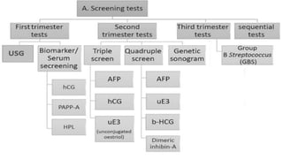
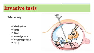
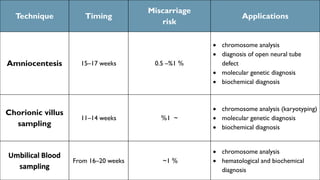
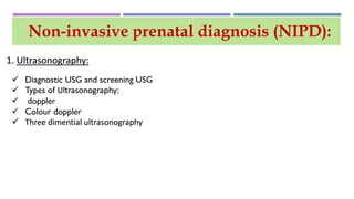
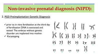
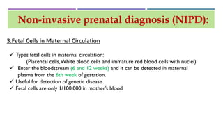
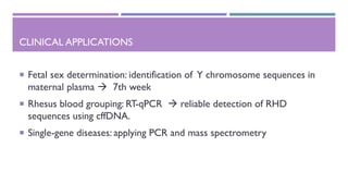
PRENATAL DIAGNOSIS
PRESENTED BY :
THARAA MOHAMMAD RAND SLEIMAN
RUBA MAHMOUD QUSSAI ABBAS
SUPERVISED BY: DR . RENWAH AL-SAYYED
Prenatal diagnosis
When ? Why? And how ?
What’s the difference ??
SCREENING TESTS
SCREENING TESTS
ULTRASONOGRAPHY (USG)
Started at 6 WEEKS 16-20WEEKS looking for anomalies
NuchalTranslucency (NT) (2.5ml)
BLOOD TEST
 maternal serum human chorionic
gonadotropin [βhCG],
 pregnancy-associated plasma
protein [PAPP-A]
 Human placental lactogen [HPL]
 Rh factor
 HIV
 HBV
 Rubella
 Syphlis
Biomarkers and blood test
MATERNAL SERUM HUMAN CHORIONIC
GONADOTROPIN [ΒHCG]
Quantitative?? Qualitative??
immunometric assay
PREGNANCY-ASSOCIATED PLASMA PROTEIN (PAPP-A)
PAPP-A concentrations early in pregnancy have
been associated with:
 Down syndrome
a high rate of fetal loss
poor fetal growth
 premature delivery
SCREENING TESTS
ALPHA FETOPROTEIN (AFP)
 Yolk sac in first trimester
 Fetal liver in second and third trimester
 MSAFP normally increase from 12-32 weeks and it is most sensitive between 16-18 weeks
Maternal Serum Estriol(uE3)
 urinary and serum estriol
 in trisomies and in neural tube defects associated with adrenal hypoplasia
Dimeric inhibin-A
 negative feedback regulator of FSH secretion
 Inhibin is secreted by the placenta and corpus luteum
SCREENING TESTS
DIAGNOSTIC TESTS
1- Amniocentesis
Invasive tests
Mechanism
Time
Risks
Investigations
Hydroamnios
2-Chorionic Villus Sampling
Invasive tests
Mechanism
Time
Risks
Investigations
3-Umbilical Blood sampling :
Invasive tests
Mechanism
Time
Risks
Investigations
Rh incompatibility
4-Fetoscopy
Invasive tests
Mechanism
Time
Risks
Investigations
Hydronephrosis
FFTS
Technique Timing
Miscarriage
risk
Applications
Amniocentesis 15–17 weeks 0.5 %– 1 %
 chromosome analysis
 diagnosis of open neural tube
defect
 molecular genetic diagnosis
 biochemical diagnosis
Chorionic villus
sampling
11–14 weeks ~1%
 chromosome analysis (karyotyping)
 molecular genetic diagnosis
 biochemical diagnosis
Umbilical Blood
sampling
From 16–20 weeks ~1 %
 chromosome analysis
 hematological and biochemical
diagnosis
Non-invasive prenatal diagnosis (NIPD):
1. Ultrasonography:
 Diagnostic USG and screening USG
 Types of Ultrasonography:
 doppler
 Colour doppler
 Three dimential ultrasonography
 Diagnostic USG assess fetal proportions:
sex, position, growth; placenta, amniotic fluid
 Conditions detected by ultrasound:
 Neural tube defects
 Dawn syndrome
 Body wall defects
 Growth disturbances
 Major limb abnormalities
 Oligo- or polyhydramnios
 Major organ abnormalities
Non-invasive prenatal diagnosis (NIPD):
1. Ultrasonography:
Non-invasive prenatal diagnosis (NIPD):
 prior to in vitro fertilization at the third day
of fertilization DNA is extracted and
tested The embryo without genetic
disorder are implanted into mother.
 Karyotyping
2. PGD Preimplantation Genetic Diagnosis
Non-invasive prenatal diagnosis (NIPD):
3.Fetal Cells in Maternal Circulation
 Types fetal cells in maternal circulation:
(Placental cells,White blood cells and immature red blood cells with nuclei)
 Enter the bloodstream (6 and 12 weeks) and it can be detected in maternal
plasma from the 6th week of gestation.
 Useful for detection of genetic disease.
 Fetal cells are only 1/100,000 in mother’s blood
Non-invasive prenatal diagnosis (NIPD):
4.Cff DNA/RNA in Maternal Circulation
• Circulating cell-free DNA 
apoptotic cells, necrotic cells
• 10%
• Placental microparticles
• 28th
• 143bp
• mRNA-Y chromosome
SECOND: verify the presence of fetal material in the
maternal circulation (detection of paternally inherited
sequences e.g., sequences located on the Y
chromosome, paternally inherited single-nucleotide
polymorphisms (SNPs), epigenetic markers)
FIRST: cffDNA and cffRNA have to be
isolated and purified from maternal plasma
SAMPLE COLLECTION
AND PROCESSING
• EDTA
• Formaldehyde to prevent
lysis of maternal cells
• Centrifugation
EXTRACTION OF CELL
FREE FETAL DNA / RNA
• Size fractionation
Plasma DNA isolation is
performed manually with
commercially available kits
EpiQuik™ Circulating Cell-Free DNA Isolation Kit
Quick-cfDNA™ Serum & Plasma Kit
NextPrep-Mag™ cfDNA Isolation Kit
The EpiQuik™ Circulating Cell-Free DNA Isolation Kit utilizes magnetic beads
based size-fractionation technology to isolate circulating cell-free DNA
(ccfDNA) from mono-nucleosomal and di-nucleosomal complexes in
plasma/serum samples.The isolated ccfDNA can be directly used for real
time-PCR and DNA library preparation suitable for next generation
sequencing.The kit has the following advantages:
 Uses innovative magnetic bead based size-fractionation technology for
selective isolation of circulating cell-free DNA from plasma/serum that is
mainly 170 bps in size.The isolated DNA can be directly used for both
qPCR and NGS DNA library preparation.
 Fast and straightforward procedure can be finished within 2 hours. No
gels, columns or centrifugation is needed.
 Efficient removal of proteins, salts, nucleases, PCR inhibiting substances,
and other impurities such as polysaccharides, polyphenols and lipids.
 Sensitive and efficient DNA capture enables successful isolation with high
recovery (>80% of input monoucleosomal DNA), even when the
quantities of starting material are limited (as low as 0.1 ml).
 Manual and automation friendly – Scalable for single tube or 96-well plate
formats.
CLINICAL APPLICATIONS
 Fetal sex determination: identification of Y chromosome sequences in
maternal plasma  7th week
 Rhesus blood grouping: RT-qPCR  reliable detection of RHD
sequences using cffDNA.
 Single-gene diseases: applying PCR and mass spectrometry

Prenatal diagnosis test

  • 1.
    PRENATAL DIAGNOSIS PRESENTED BY: THARAA MOHAMMAD RAND SLEIMAN RUBA MAHMOUD QUSSAI ABBAS SUPERVISED BY: DR . RENWAH AL-SAYYED
  • 2.
  • 3.
  • 4.
  • 5.
  • 6.
    ULTRASONOGRAPHY (USG) Started at6 WEEKS 16-20WEEKS looking for anomalies NuchalTranslucency (NT) (2.5ml)
  • 7.
    BLOOD TEST  maternalserum human chorionic gonadotropin [βhCG],  pregnancy-associated plasma protein [PAPP-A]  Human placental lactogen [HPL]  Rh factor  HIV  HBV  Rubella  Syphlis Biomarkers and blood test
  • 8.
    MATERNAL SERUM HUMANCHORIONIC GONADOTROPIN [ΒHCG] Quantitative?? Qualitative?? immunometric assay
  • 9.
    PREGNANCY-ASSOCIATED PLASMA PROTEIN(PAPP-A) PAPP-A concentrations early in pregnancy have been associated with:  Down syndrome a high rate of fetal loss poor fetal growth  premature delivery
  • 10.
  • 11.
    ALPHA FETOPROTEIN (AFP) Yolk sac in first trimester  Fetal liver in second and third trimester  MSAFP normally increase from 12-32 weeks and it is most sensitive between 16-18 weeks
  • 12.
    Maternal Serum Estriol(uE3) urinary and serum estriol  in trisomies and in neural tube defects associated with adrenal hypoplasia
  • 14.
    Dimeric inhibin-A  negativefeedback regulator of FSH secretion  Inhibin is secreted by the placenta and corpus luteum
  • 15.
  • 16.
  • 18.
  • 19.
    2-Chorionic Villus Sampling Invasivetests Mechanism Time Risks Investigations
  • 20.
    3-Umbilical Blood sampling: Invasive tests Mechanism Time Risks Investigations Rh incompatibility
  • 21.
  • 22.
    Technique Timing Miscarriage risk Applications Amniocentesis 15–17weeks 0.5 %– 1 %  chromosome analysis  diagnosis of open neural tube defect  molecular genetic diagnosis  biochemical diagnosis Chorionic villus sampling 11–14 weeks ~1%  chromosome analysis (karyotyping)  molecular genetic diagnosis  biochemical diagnosis Umbilical Blood sampling From 16–20 weeks ~1 %  chromosome analysis  hematological and biochemical diagnosis
  • 23.
    Non-invasive prenatal diagnosis(NIPD): 1. Ultrasonography:  Diagnostic USG and screening USG  Types of Ultrasonography:  doppler  Colour doppler  Three dimential ultrasonography
  • 24.
     Diagnostic USGassess fetal proportions: sex, position, growth; placenta, amniotic fluid  Conditions detected by ultrasound:  Neural tube defects  Dawn syndrome  Body wall defects  Growth disturbances  Major limb abnormalities  Oligo- or polyhydramnios  Major organ abnormalities Non-invasive prenatal diagnosis (NIPD): 1. Ultrasonography:
  • 25.
    Non-invasive prenatal diagnosis(NIPD):  prior to in vitro fertilization at the third day of fertilization DNA is extracted and tested The embryo without genetic disorder are implanted into mother.  Karyotyping 2. PGD Preimplantation Genetic Diagnosis
  • 26.
    Non-invasive prenatal diagnosis(NIPD): 3.Fetal Cells in Maternal Circulation  Types fetal cells in maternal circulation: (Placental cells,White blood cells and immature red blood cells with nuclei)  Enter the bloodstream (6 and 12 weeks) and it can be detected in maternal plasma from the 6th week of gestation.  Useful for detection of genetic disease.  Fetal cells are only 1/100,000 in mother’s blood
  • 27.
    Non-invasive prenatal diagnosis(NIPD): 4.Cff DNA/RNA in Maternal Circulation • Circulating cell-free DNA  apoptotic cells, necrotic cells • 10% • Placental microparticles • 28th • 143bp • mRNA-Y chromosome
  • 28.
    SECOND: verify thepresence of fetal material in the maternal circulation (detection of paternally inherited sequences e.g., sequences located on the Y chromosome, paternally inherited single-nucleotide polymorphisms (SNPs), epigenetic markers) FIRST: cffDNA and cffRNA have to be isolated and purified from maternal plasma
  • 29.
    SAMPLE COLLECTION AND PROCESSING •EDTA • Formaldehyde to prevent lysis of maternal cells • Centrifugation EXTRACTION OF CELL FREE FETAL DNA / RNA • Size fractionation
  • 30.
    Plasma DNA isolationis performed manually with commercially available kits EpiQuik™ Circulating Cell-Free DNA Isolation Kit Quick-cfDNA™ Serum & Plasma Kit NextPrep-Mag™ cfDNA Isolation Kit
  • 31.
    The EpiQuik™ CirculatingCell-Free DNA Isolation Kit utilizes magnetic beads based size-fractionation technology to isolate circulating cell-free DNA (ccfDNA) from mono-nucleosomal and di-nucleosomal complexes in plasma/serum samples.The isolated ccfDNA can be directly used for real time-PCR and DNA library preparation suitable for next generation sequencing.The kit has the following advantages:  Uses innovative magnetic bead based size-fractionation technology for selective isolation of circulating cell-free DNA from plasma/serum that is mainly 170 bps in size.The isolated DNA can be directly used for both qPCR and NGS DNA library preparation.  Fast and straightforward procedure can be finished within 2 hours. No gels, columns or centrifugation is needed.  Efficient removal of proteins, salts, nucleases, PCR inhibiting substances, and other impurities such as polysaccharides, polyphenols and lipids.  Sensitive and efficient DNA capture enables successful isolation with high recovery (>80% of input monoucleosomal DNA), even when the quantities of starting material are limited (as low as 0.1 ml).  Manual and automation friendly – Scalable for single tube or 96-well plate formats.
  • 32.
    CLINICAL APPLICATIONS  Fetalsex determination: identification of Y chromosome sequences in maternal plasma  7th week  Rhesus blood grouping: RT-qPCR  reliable detection of RHD sequences using cffDNA.  Single-gene diseases: applying PCR and mass spectrometry