Cox et al., lab on a chip poster 7.26.12 final with apyrase
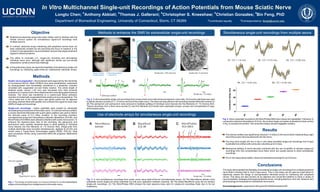
Simultaneous single-unit recordings from multiple peripheral nerve fibers
1. Fig. 1. The design and fabrication of a tissue chamber for In vitro extracellular
single-unit recordings from multiple axons in the sciatic nerve.
Objective
Multichannel electrode arrays have been widely used to interface with the
central nervous system for simultaneous signal-unit recordings from
multiple neurons.
In contrast, electrode arrays interfacing with peripheral nerves have not
been extensively studied but are becoming the focus of research in the
context of next-generation neuromodulation devices that target peripheral
organs to relieve symptoms.
The ability to modulate (i.e., single-unit recording and stimulating)
individual nerve axon, although with significant clinical and pre-clinical
implications, remains a technical challenge.
In this preliminary study, we report the feasibility of simultaneous single-unit
recordings by interfacing state-of-the-art multichannel electrode arrays.
& & & & #
Longtu Chen; Anthony Abbiati; Thomas J. Gallerani; Christopher S. Kneeshaw; Christian Gonzales; Bin Feng, PhD
The stimulus artifact was significantly reduced (< 6 mSec) in the record when implementing a tight
seal of the suction stimulus electrode onto the nerve.
The long nerve length (25 mm) in the in vitro setup permitted single-unit recordings from A-type
myelinated nerve fibers with conduction velocities up to 4 m/sec.
Mechanical splitting of nerve fascicles combined with the use of paraffin oil allowed single-unit
recordings from fine unmyelinated nerve fibers which are usually elusive to direct extracellular
recordings.
Our in vitro setup allows stable, robust and repeated recordings for up to 6 hours.
Sciatic nerve preparation - All procedures were approved by the University
of Connecticut IACUC. Male C57BL/6 mice were anesthetized, euthanized
by exsanguination and immediately transferred to a dissection chamber
circulated with oxygenated ice-cold Krebs solution. The whole length of
bilateral sciatic nerves (~30 mm) was harvested from their proximal
projection to L4 spinal cord to their distal branches innervating gastrocnemius
muscles. The nerve was transferred to a custom-built tissue perfusion
chamber circulated with oxygenated Krebs solution at room temperature. The
~5mm distal end of the sciatic nerve was gently pulled into an adjacent
recording chamber filled with paraffin oil to enhance the signal-to-noise ratio
(SNR) of single-unit recordings.
In Vitro Multichannel Single-unit Recordings of Action Potentials from Mouse Sciatic Nerve
Fig. 3. In vitro simultaneous recordings from sciatic nerve using state-of-the-art microelectrode arrays. (A) The NeuroNexus tetrode could
potentially record single-unit action potentials but with poor separation between channels. (B) The tested BlackRock array did not achieve
single-unit recordings. (C) The MicroProbes MEA showed the best signal-to-noise ratio for single-unit recordings likely due to its low
impedance.
Fig. 2. In vitro extracellular single-unit recordings from mouse sciatic nerve with enhanced signal-to-noise ratio. (A)Asuction electrode was used
to deliver stimulus currents (0.1-1.5 mA) to one end of the sciatic nerve. The other end was placed in the recording chamber filled with mineral oil.
(B) The epineurium and perineurium were removed to facilitate splitting of individual nerve fascicle into fine filaments of ~10 microns thick.
Typical recordings from a thick nerve bundle (C) and thin filaments (D) include both myelinated (A-type, arrows) and unmyelinated (C-type,
arrow heads) axons.
Department of Biomedical Engineering, University of Connecticut, Storrs, CT 06269
Methods
Results
This study demonstrated the feasibility of simultaneous single-unit recordings from multiple peripheral
nerve fibers including both A- and C-type axons. This in vitro setup can be used as a test bench to
objectively assess the design of next-generation electrode arrays for interfacing with peripheral
nerves. This setup can also function as an objective and convenient platform to study a variety of
neuromodulation strategies that target peripheral nerves, including electrical, infra-red, ultrasonic and
pharmacological manipulations of peripheral nerve axons.
Acknowledgements: supported by NIH grant DK100460 awarded to BF.
Conclusions
Single-unit recordings - Action potentials were evoked by electrically
stimulating the proximal end of the sciatic nerve. To minimize stimulus artifact,
a suction electrode fabricated with quartz glass capillary was used to deliver
the stimulus pulse of 0.2 mSec duration. In the recording chamber,
microelectrode arrays from NeuroNexus (tetrode), BlackRock (ICS 96), and
MicroProbes (4Ch MEA) were utilized to interface with sciatic nerve axons. To
enhance the signal-to-noise ratio of the recording, the epineurium and
perineurium were carefully dissected away to allow splitting of individual
nerve fascicle into fine filaments of ~10 microns thick. Single-units from
multiple electrodes were recorded simultaneously, digitized at 24 kHz and
stored using a Tucker-Davis Technologies system (RZ5D, PZ5-32). Data
were processed off-line using customized MATLAB programs (Mathworks
R2016b).
#
Correspondance: fengb@uconn.edu
Use of electrode arrays for simultaneous single-unit recordings
Simultaneous single-unit recordings from multiple axonsMethods to enhance the SNR for extracellular single-unit recordings
Vacuum grease
Opening for nerve
Scale bar: 5 micronsTissue chamber
(Krebs solution)
Recording chamber
(mineral oil)
Stimulus
electrode
Stereoscope
Recording
electrode
Tissue chamber
(Krebs solution)
Recording chamber
(mineral oil)
Sciatic
nerve
Nerve
fascicle
A B
C D
Scale bar: 10 mSec
Stimulus artifact
A NeuroNexus
tetrode
B BlackRock
ICS 96
C MicroProbes
4Ch MEA
Scale bar: 10 mSec
#1 CV = 1.6 m/s #2 CV = 0.72 m/s #3 CV = 0.60 m/s
#4 CV = 0.44 m/s #5 CV = 0.40 m/s
#1
#2
#3
#4
#5
0
20
40
60
Recording
electrode
ConductionDelay(ms)
Fig. 4. Action potentials recorded by the MicroProbes MEA were robust and repeatable. Following 10
consecutive electrical stimulations (0.5 Hz), the evoked single-unit spikes (indicated by black arrows
in Fig. 3) overlaid one another.The conduction delays showed negligible variation.
1
2 3 4
5
Scale bar: 100 microns
&
Contributed equally