2007 roma, campus biomedico, università di ingegnieria. quale raporto tra bioingegniere e medico
•Download as PPT, PDF•
0 likes•320 views

2007 roma, campus biomedico, università di ingegnieria. quale raporto tra bioingegniere e medico

Recommended
More Related Content
What's hot
What's hot (12)
Similar to 2007 roma, campus biomedico, università di ingegnieria. quale raporto tra bioingegniere e medico
Similar to 2007 roma, campus biomedico, università di ingegnieria. quale raporto tra bioingegniere e medico (20)
More from Centro Diagnostico Nardi
More from Centro Diagnostico Nardi (20)
Recently uploaded
Recently uploaded (20)
2007 roma, campus biomedico, università di ingegnieria. quale raporto tra bioingegniere e medico
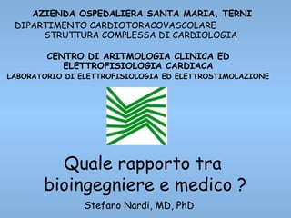
- 1. Quale rapporto tra bioingegniere e medico ? Stefano Nardi, MD, PhD AZIENDA OSPEDALIERA SANTA MARIA, TERNI DIPARTIMENTO CARDIOTORACOVASCOLARE STRUTTURA COMPLESSA DI CARDIOLOGIA CENTRO DI ARITMOLOGIA CLINICA ED ELETTROFISIOLOGIA CARDIACA LABORATORIO DI ELETTROFISIOLOGIA ED ELETTROSTIMOLAZIONE
- 2. .....chiara.... Parlare di tutto....... ......in maniera approfondita, ...ed in poco tempo!!!! Mission: Impossibile
- 3. Cell-Membrane Resting Potential + - 0 mV ….a “resting” potential of -90 mV is observed inside the cell with respect to outside the cell Advance needle electrode across the cell membrane….
- 4. Cell-Membrane Resting Potential + The resting potential is maintained by an ATP powered sodium-potassium “pump” within the membrane that transports Na+ ions outward and K+ ions inward (3 Na+ per 2 K+ ). Na+ K+ Na+ Na+ The gradient of ion-concentration separates charge across the membrane with an equal and opposite electrical gradient of -90 mV. - K+ Advance needle electrode across the cell membrane…. - - -- -- --- -- - - --- - --- -- - - -- - + + + + +++ + + + + +++ + + + + + + + + + +
- 5. Cell Membrane Action Potential (AP) + - Stimulate the cell…. 0 mV ….a transmembrane “AP” is observed with 5 characteristic phases (Φ)
- 6. Cell Membrane Action Potential (AP) + - Stimulate the cell…. Φ0 – Upstroke Φ2 – Plateau (absolute refractory) Φ3 – Recovery (relative refractory) Φ4 – Resting Φ1 – Initial Recovery 0 mV
- 8. C’era una volta la Paleo- Cardiologia 1887 First Electrical recording of the Heart
- 18. Infero mediale Infero-laterale VPIL VPSL Limitation of 2D ApproachLimitation of 2D Approach
- 19. • Surface-based – 5.6 kHz current signal emitted from 3 pairs of surface electrodes –Each catheter electrode located 93 times per second – Visualize all catheters in 3D space for cardiac navigation Virtual Reality Methodology
- 20. STANDARD FLUOROSTANDARD FLUOROVIRTUAL GEOMETRYVIRTUAL GEOMETRY
- 21. Virtual Geometry ReconstructionVirtual Geometry Reconstruction
- 22. Real Time reconstructionReal Time reconstruction
- 25. virtual geometry reconstructionvirtual geometry reconstruction
- 26. virtual geometry reconstructionvirtual geometry reconstruction
- 27. virtual geometry reconstructionvirtual geometry reconstruction
- 29. virtual geometry reconstructionvirtual geometry reconstruction
- 30. virtual geometry reconstructionvirtual geometry reconstruction
- 32. Geometry Color
- 33. Multipoint Mapping with A Regional Mapping Catheter
- 37. Begin Segmentation • Slice data: – Transverse – Coronal – Sagittal
- 38. Create a Model • Select a structure • Click an area of interest • Adjust threshold and boundary emphasis • Apply
- 39. Create a Model • The resulting 3D model appears
- 41. Separate the Structures • Select a structure • Click in slice or 3D • Repeat • Apply
- 43. Display the LA • Remove extra structures
- 44. Using Manual Tools • Draw the desired result
- 45. Save the Model • Tile outer surface • Ready for use in EnSite System Study
- 46. virtual geometry reconstructionvirtual geometry reconstruction
- 47. Atrial Fibrillation ablationAtrial Fibrillation ablation virtual geometry reconstructionvirtual geometry reconstruction
- 48. virtual geometry reconstructionvirtual geometry reconstruction
Editor's Notes
- <number>
- <number>
- <number>
- <number>
- <number>
- <number> Accesso dalla vena femorale. HRA = registra l’impulso così come si genera dal nodo SA HBE = Registrazione del fascio di His. Non acquisisce il segnale dal nodo AV, ma lo acquisisce subito sotto sul fascio di His (vicino la tricuspide). Questo rivela all’elettrofisiologo quanto tempo impiega l’impulso per passare dal nodo SA al nodo AV. RVA = segnale dal ventricolo destro Coronary Sinus = riceve il segnale da LA e LV.
- Any type of catheter (electrode based) Up to 12 catheters, 64 electrodes
- EnSite NavX enables physicians to visualize and navigate a myriad of intracardiac catheters in any chamber of the heart. It also simultaneously displays more electrodes and catheters than any other 3-D mapping system currently available. EnSite NavX is compatible with catheters from all manufacturers. The non-proprietary nature of our technology gives you freedom of choice for diagnostic and ablation catheters.
- <number> AVI movie Here’s a quick illustration to show you how the chamber maps are built. At the start of an EnSite procedure, the catheter is inserted in the chamber and validated by the system. (click on map image) As a catheter is moved within the chamber, the system records three-dimensional points. The operator can also give certain points special emphasis (indicated by white squares)—these are called locked points, to help define key areas of the anatomy, such as the isthmus or crista. As seen in the published literature, chamber maps or geometries can be built in as little as five minutes. Thereafter, there is no need for fluoroscopy, since the system provides superior orientation to fluoroscopy through the 3D model and superior catheter orientation through the 3D catheter display. When the geometry is finished, event data can then be recorded.
- (SLIDE 5) At this purpose nowadays, novel and different technologies for mapping, tracking and ablation are available for approaching AF and in this view the technologic progress continuous to evolving over the time.
- In this example we will be segmenting a left atrium
- Subregioning crops away information that is not pertinent to the segmentation
- Segmentation in EnSite Verismo is a point-and-click interface. Select a tool for a specific task, click in the slice views to create a 3D model in the fourth panel. Controls in the panel views allow navigation to any slice in the model in each direction. The 2D views also feature zoom, pan, and window/level (brightness and contrast) adjustments.
- The first tool is Region Grow. Region Grow is used to quickly create the majority of the model. Subsequent tools will refine the model. In Region Grow, the user defines an area of interest by placing a “seed point” in a structure, in this example, the left atrium. After placing the seed, the user will adjust the min and max threshold using the sliders; thresholding defines what intensity of greyscale values (light vs dark) will be used to create the model. The area of the model covered by the current threshold setting is colored translucent red. Boundary emphasis identifies sharp transitions in intensity; those areas are colored grey. The effect of these controls: The seed point identifies where the algorithm should search for connected intensities. Thresholding controls how well the model is filled in. Boundary emphasis identifies the edges between structures, important for the following step
- Correlating edges of the model appear as lines in the slice views
- Note that the created model includes partial models of the RA, RV, and LV. The partial creation of these structures is relative to their distance from the point that we placed during region grow, and the relative contrast and connection of the blood pool. The 3D model can be rotated, zoomed, and panned The worldview torso will rotate with the model Clicking the buttons at the top of the screen will access familiar angles The current model includes all of the data we need, and more
- The list controls which structure is currently being segmented.
- Here is the model as it appears with separated structures. If a break between structures is not at the desired location, the separator tool can be repeated on the structures in question.
- To review the LA, simply disable the display of the other structures in the list.
- Optionally, manual segmentation tools can be used to trim away extraneous information.
- In the final step, Verismo converts the solid model of the blood pool to a tiled surface comprised of thousands of triangles. This model is similar to the geometries on the EnSite System and allows the use of familiar clinical functions such as translucency, clipping, and labeling. The completed model may be saved to the system hard drive or exported to CD.
- Distal PV branches can serve as useful spatial landmarks, and can be readily delineated by the placement of 3D spherical markers during catheter pullback.