The restorations ofedentulous areas with
fixed partial dentures (FPDs) present a
particular challenge for the clinician. Because
of their ease of use and favorable long term
results, conventional FPDs represent the most
popular treatment measure today. In these
restorations, the pontic must fulfill the complex
roles of replacing the function of the lost tooth,
achieving an esthetic appearance, enabling
adequate oral hygiene, and preventing tissue
irritation. In addition the pontic must meet
certain structural requirements to ensure the
mechanical stability of the restorations.
INTRODUCTION
3.
The histories offixed and of
removable partial prosthetic appliances
go more or less in hand and it is difficult
at times to tell just where to draw the
line between these two types from the
available data. Since the use of
prosthodontics, the most old dental
prosthesis is believed to be a fixed
type.
HISTORY
4.
Replaced tooth wassewed in place
by using ligatures made from gold or
silver. Egyptians and Phoeniceans
were the pioneers in the field of
pontics and were the first to construct
dental bridge work. These were
mostly made of calf bone or ivory. It is
suggested that teeth of ivory and
bone secured by copper wire or
catgut string were used in China for
ages before they were introduced in
Europe.
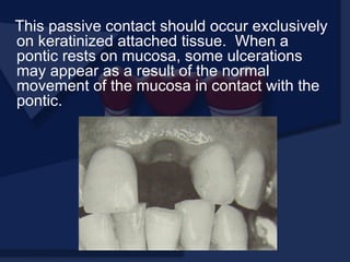
5.
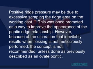
It was Mancyin 1928 who laid the
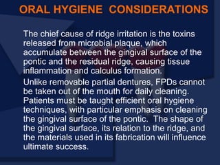
foundation to present day FPD design,
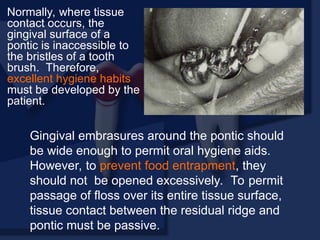
however Pierre Fauchard (1923) has often
been referred to as the ‘Father of Modern
Dental Prosthesis’. In his work in the field of
FPD he used what he called ‘tenons’ which
were in reality dowels or pivots screwed
into the roots to retain some of the bridges
and it is possible that he may have been
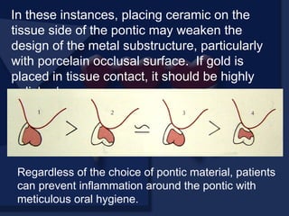
the first to attach dental bridges to tooth
roots by this method.
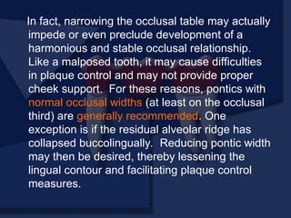
6.
Selberg (1936) pointedout that basic
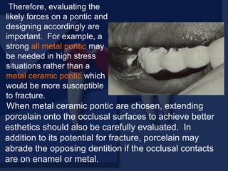
materials had changed but little in the past
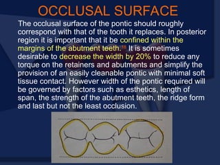
few years. These materials were gold or
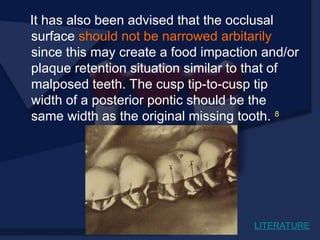
porcelain or a combination of the two. He
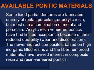
summed up by saying that the restoration
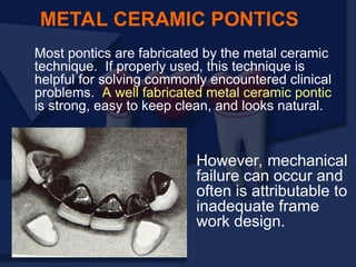
must meet the following requirements:
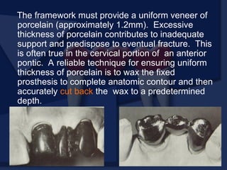
Protection, comfort,
esthetics, durability and
utility.
7.
The Glossary ofprosthodontic terms 5
defines Pontics as - An artificial teeth on a
fixed partial denture that replaces missing
natural teeth, restores it’s function and
usually fills the space previously filled by
the natural teeth.
Tylman 4
defines Pontics as the
suspended member of a fixed partial
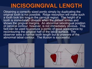
denture which replaces the lost natural
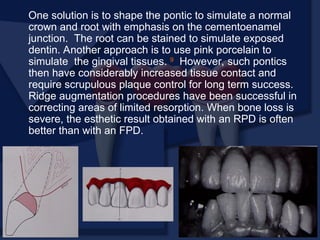
tooth, restores function and occupies the
space of the missing tooth.
DEFINITION
8.
It is nota simple replacement, because placing an
exact anatomic replica of the tooth in the space would
be hygienically unmanageable. They must be
compatible with continued oral health and comfort.
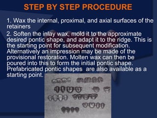
The edentulous areas where a fixed prosthesis is to be
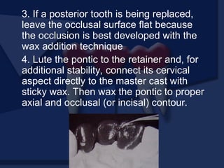
provided may be overlooked during the treatment-
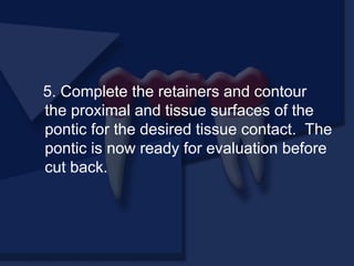
planning phase. Unfortunately, any deficiency or
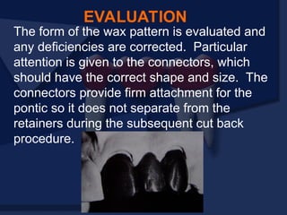
potential problem that may arise during the fabrication
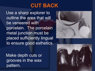
of a pontic is often identified only after the teeth have
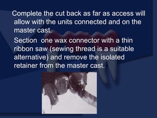
been prepared or even when the master cast is ready
to be sent to the laboratory. Proper preparation
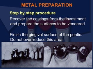
includes a careful analysis of the critical dimensions of
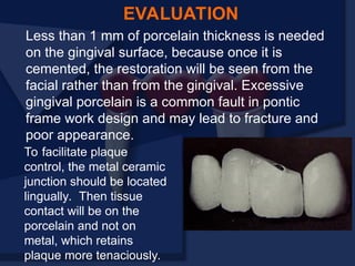
the edentulous areas: mesiodistal width,
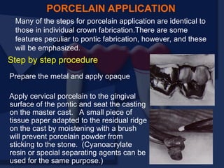
occlusocervical distance, buccolingual diameter, and
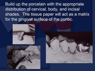
location of the residual ridge.
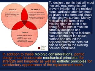
9.
To design apontic that will meet
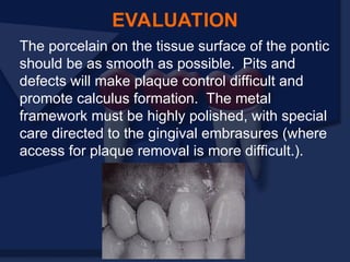
hygienic requirements and
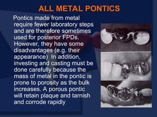
prevent irritation of the residual
ridge, particular attention must
be given to the form and shape
of the gingival surface. Merely
replicating the form of the
missing tooth or teeth is not
enough. The pontic must be
carefully designed and
fabricated not only to facilitate
plaque control of the tissue
surface and around the
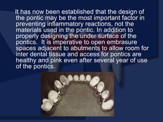
adjacent abutment teeth but
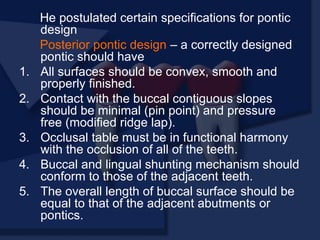
also to adjust to the existing
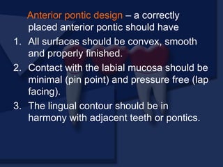
occlusal conditions.
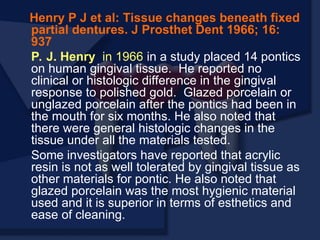
In addition to these biologic considerations, pontic
design must incorporate mechanical principles for
strength and longevity as well as esthetic principles for
satisfactory appearance of the replacement teeth.
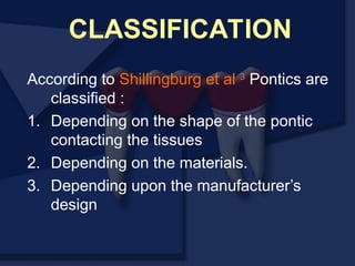
10.
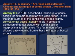
According to Shillingburget al 3
Pontics are
classified :
1. Depending on the shape of the pontic
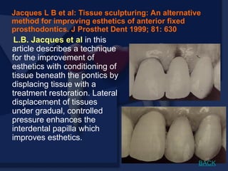
contacting the tissues
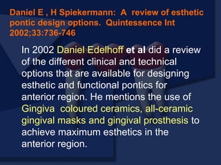
2. Depending on the materials.
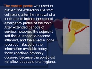
3. Depending upon the manufacturer’s
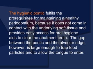
design
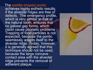
CLASSIFICATION
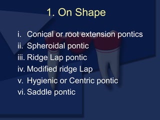
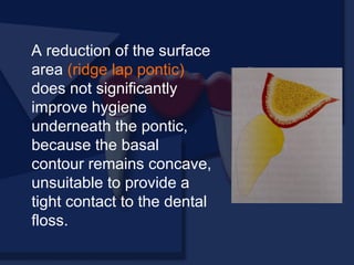
11.
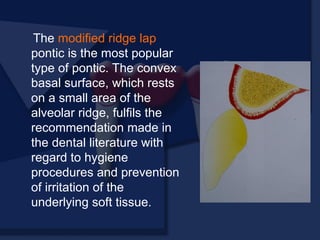
1. On Shape
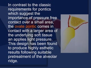
i.Conical or root extension pontics
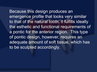
ii. Spheroidal pontic
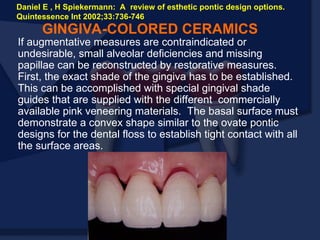
iii. Ridge Lap pontic
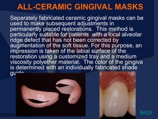
iv. Modified ridge Lap
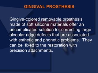
v. Hygienic or Centric pontic
vi. Saddle pontic
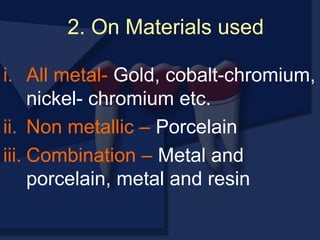
12.
i. All metal-Gold, cobalt-chromium,
nickel- chromium etc.
ii. Non metallic – Porcelain
iii. Combination – Metal and
porcelain, metal and resin
2. On Materials used
13.
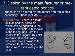
3. Design bythe manufacturer or pre-
fabricated pontics
a. Trupontic – There is a large
bulk of gingival porcelain
which can be adapted to the
ridge. A horizontal tubular slot
in the facing runs from the
center to the lingual. This slot
in combination with wide
proximal bevels provides the
retention for the facings.
These were used widely in the
past.
These can be altered by the dentist and reglazed if
necessary. These include:
14.
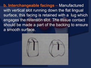
b. Interchangeable facings– Manufactured
with vertical slot running down the flat lingual
surface, this facing is retained with a lug which
engages the retention slot. The tissue contact
should be made a part of the backing to ensure
a smooth surface.
15.
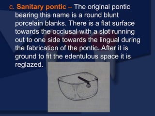
c. Sanitary pontic– The original pontic
bearing this name is a round blunt
porcelain blanks. There is a flat surface
towards the occlusal with a slot running
out to one side towards the lingual during
the fabrication of the pontic. After it is
ground to fit the edentulous space it is
reglazed.
16.
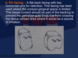
d. Pin facing– A flat back facing with two
horizontal pins for retention. This facing has been
used where the occluso-gingival space is limited.
This tissue contact should be part of the backing to
prevent the porcelain-gold finish line from crossing
the tissue contact area where it would be a source
of irritation.
17.
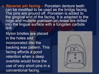
e. Reverse pinfacing – Porcelain denture teeth
can be modified to be used as the bridge facing.
The pins are ground off. Porcelain is added to
the gingival end of the facing. It is adapted to the
ridge and multiple precision pin holes are drilled
into the lingual surface with a tungsten carbide
drill.
Nylon bristles are placed
in the holes and
incorporated into the
backing wax pattern. This
facing affords a good
retention when a deep
overbite would force the
use of very short pins in a
conventional facing.
18.
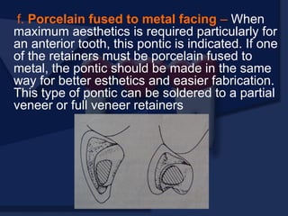
f. Porcelain fusedto metal facing – When
maximum aesthetics is required particularly for
an anterior tooth, this pontic is indicated. If one
of the retainers must be porcelain fused to
metal, the pontic should be made in the same
way for better esthetics and easier fabrication.
This type of pontic can be soldered to a partial
veneer or full veneer retainers
19.
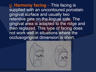
g. Harmony facing– This facing is
supplied with an uncontoured porcelain
gingival surface and usually two
retentive pins on the lingual side. The
gingival area is adapted to the ridge and
then reglazed. This type of facing does
not work well in situations where the
occlusogingival dimension is short.
20.
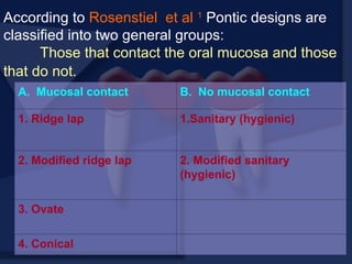
According to Rosenstielet al 1
Pontic designs are
classified into two general groups:
Those that contact the oral mucosa and those
that do not.
A. Mucosal contact B. No mucosal contact
1. Ridge lap 1.Sanitary (hygienic)
2. Modified ridge lap 2. Modified sanitary
(hygienic)
3. Ovate
4. Conical
21.
Pontic selection dependsprimarily on
esthetics and oral hygiene. In the
anterior region, where esthetics is a
concern, the pontic should be well
adapted to the tissue to make it
appear that it emerges from the
gingival. Conversely, in the posterior
regions (mandibular premolar and
molar areas), esthetics can be
compromised in the interest of
designs that are more amenable to
oral hygiene
PONTIC SELECTION
22.
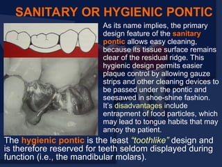
As its nameimplies, the primary
design feature of the sanitary
pontic allows easy cleaning,
because its tissue surface remains
clear of the residual ridge. This
hygienic design permits easier
plaque control by allowing gauze
strips and other cleaning devices to
be passed under the pontic and
seesawed in shoe-shine fashion.
It’s disadvantages include
entrapment of food particles, which
may lead to tongue habits that may
annoy the patient.
SANITARY OR HYGIENIC PONTIC
The hygienic pontic is the least “toothlike” design and
is therefore reserved for teeth seldom displayed during
function (i.e., the mandibular molars).
23.
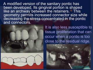
A modified versionof the sanitary pontic has
been developed. Its gingival portion is shaped
like an archway between the retainers.15
This
geometry permits increased connector size while
decreasing the stress concentrated in the pontic
and connectors.
It is also less susceptible to
tissue proliferation that can
occur when a pontic is too
close to the residual ridge.
LITERATURE
24.
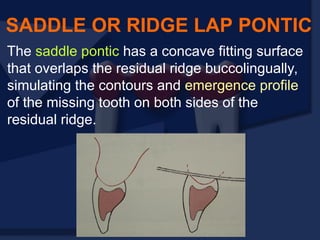
The saddle pontichas a concave fitting surface
that overlaps the residual ridge buccolingually,
simulating the contours and emergence profile
of the missing tooth on both sides of the
residual ridge.
SADDLE OR RIDGE LAP PONTIC
25.
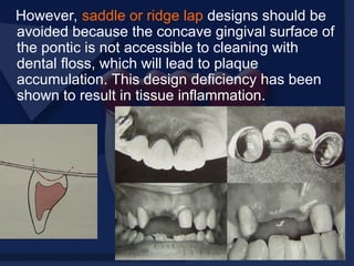
However, saddle orridge lap designs should be
avoided because the concave gingival surface of
the pontic is not accessible to cleaning with
dental floss, which will lead to plaque
accumulation. This design deficiency has been
shown to result in tissue inflammation.
26.
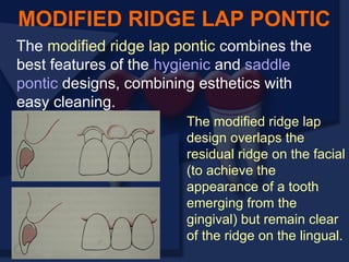
The modified ridgelap pontic combines the
best features of the hygienic and saddle
pontic designs, combining esthetics with
easy cleaning.
MODIFIED RIDGE LAP PONTIC
The modified ridge lap
design overlaps the
residual ridge on the facial
(to achieve the
appearance of a tooth
emerging from the
gingival) but remain clear
of the ridge on the lingual.
27.
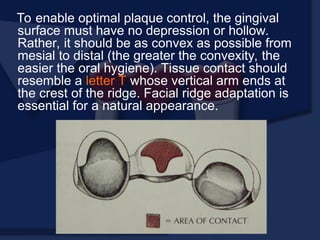
To enable optimalplaque control, the gingival
surface must have no depression or hollow.
Rather, it should be as convex as possible from
mesial to distal (the greater the convexity, the
easier the oral hygiene). Tissue contact should
resemble a letter T whose vertical arm ends at
the crest of the ridge. Facial ridge adaptation is
essential for a natural appearance.
28.
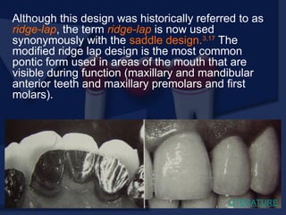
Although this designwas historically referred to as
ridge-lap, the term ridge-lap is now used
synonymously with the saddle design.3,17
The
modified ridge lap design is the most common
pontic form used in areas of the mouth that are
visible during function (maxillary and mandibular
anterior teeth and maxillary premolars and first
molars).
LITERATURE
29.
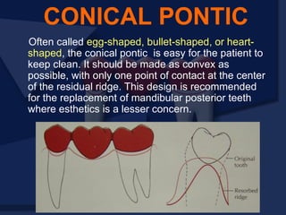
Often called egg-shaped,bullet-shaped, or heart-
shaped, the conical pontic is easy for the patient to
keep clean. It should be made as convex as
possible, with only one point of contact at the center
of the residual ridge. This design is recommended
for the replacement of mandibular posterior teeth
where esthetics is a lesser concern.
CONICAL PONTIC
30.
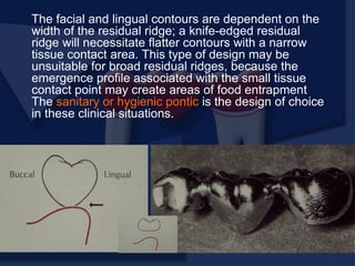
The facial andlingual contours are dependent on the
width of the residual ridge; a knife-edged residual
ridge will necessitate flatter contours with a narrow
tissue contact area. This type of design may be
unsuitable for broad residual ridges, because the
emergence profile associated with the small tissue
contact point may create areas of food entrapment
The sanitary or hygienic pontic is the design of choice
in these clinical situations.
31.
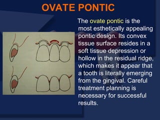
The ovate ponticis the
most esthetically appealing
pontic design. Its convex
tissue surface resides in a
soft tissue depression or
hollow in the residual ridge,
which makes it appear that
a tooth is literally emerging
from the gingival. Careful
treatment planning is
necessary for successful
results.
OVATE PONTIC
32.
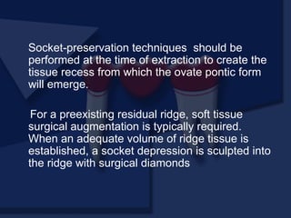
Socket-preservation techniques shouldbe
performed at the time of extraction to create the
tissue recess from which the ovate pontic form
will emerge.
For a preexisting residual ridge, soft tissue
surgical augmentation is typically required.
When an adequate volume of ridge tissue is
established, a socket depression is sculpted into
the ridge with surgical diamonds
33.
The ovate pontic’sadvantages include it’s
pleasing appearance and it’s strength, when
used successfully with ridge augmentation, it’s
emergence from the ridge appears identical to
that of a natural tooth. This type of pontic design,
however, requires an adequate amount of soft
tissue, which has to be sculpted accordingly.13
Various techniques are available for this
purpose, ranging from controlled regeneration
directly after the extraction of the tooth
(immediate pontic technique) to plastic surgery
(gingival grafting), which is accompanied by
tissue conditioning in the course of the
subsequent prosthodontic treatment.
LITERATURE
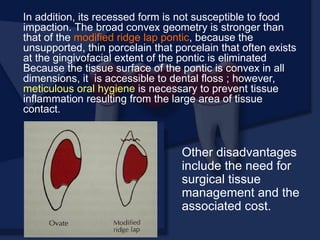
34.
In addition, itsrecessed form is not susceptible to food
impaction. The broad convex geometry is stronger than
that of the modified ridge lap pontic, because the
unsupported, thin porcelain that porcelain that often exists
at the gingivofacial extent of the pontic is eliminated
Because the tissue surface of the pontic is convex in all
dimensions, it is accessible to dental floss ; however,
meticulous oral hygiene is necessary to prevent tissue
inflammation resulting from the large area of tissue
contact.
Other disadvantages
include the need for
surgical tissue
management and the
associated cost.
35.
In addition tothese other modifications
of pontics like soft tissue conditioning 13
,
gingival- coloured ceramics, all-ceramic
gingival masks and gingival masks have
been discussed. 9
LITERATURE
36.
The biologic principlesof pontic design
pertain to the maintenance and
preservation of the residual ridge,
abutment and opposing teeth, and
supporting tissue. Factors of specific
influence are pontic ridge contact,
amenability to oral hygiene, and the
direction of occlusal forces.
BIOLOGIC CONSIDERATIONS
37.
Pressure free contactbetween the pontic
and the underlying tissue is indicated to
prevented ulceration and inflammation of
the soft tissues. If any blanching of the
soft tissues is observed in try-in, the
pressure area should be identified with a
disclosing medium (i.e, pressure indicating
paste) and the pontic recontoured until
tissue contact is entirely passive.
RIDGE CONTACT
38.
This passive contactshould occur exclusively
on keratinized attached tissue. When a
pontic rests on mucosa, some ulcerations
may appear as a result of the normal
movement of the mucosa in contact with the
pontic.
39.
Positive ridge pressuremay be due to
excessive scraping the ridge area on the
working cast. 7
This was once promoted
as a way to improve the appearance of the
pontic ridge relationship. However,
because of the ulceration that inevitably
results when flossing is not meticulously
performed, the concept is not
recommended, unless done as previously
described as an ovate pontic.
LITERATURE
40.
The chief causeof ridge irritation is the toxins
released from microbial plaque, which
accumulate between the gingival surface of the
pontic and the residual ridge, causing tissue
inflammation and calculus formation.
Unlike removable partial dentures, FPDs cannot
be taken out of the mouth for daily cleaning.
Patients must be taught efficient oral hygiene
techniques, with particular emphasis on cleaning
the gingival surface of the pontic. The shape of
the gingival surface, its relation to the ridge, and
the materials used in its fabrication will influence
ultimate success.
ORAL HYGIENE CONSIDERATIONS
41.
Normally, where tissue
contactoccurs, the
gingival surface of a
pontic is inaccessible to
the bristles of a tooth
brush. Therefore,
excellent hygiene habits
must be developed by the
patient.
Gingival embrasures around the pontic should
be wide enough to permit oral hygiene aids.
However, to prevent food entrapment, they
should not be opened excessively. To permit
passage of floss over its entire tissue surface,
tissue contact between the residual ridge and
pontic must be passive.
42.
If the pontichas a depression or
concavity in its gingival surface,
plaque will accumulate, because the
floss cannot clean this area, and
tissue irritation will follow. This is
usually reversible; when the surface
is subsequently modified to eliminate
the concavity, inflammation
disappears.
43.
Therefore, an accuratedescription of
pontic design should be known to the
laboratory, and the prosthesis should be
checked and corrected if necessary before
cementation. Prevention is the best
solution for controlling tissue irritation.
44.
Any material chosento fabricate the pontic
should provide good esthetic results where
needed; biocompatibility, rigidity, and strength to
withstand occlusal forces; and longevity. FPDs
should be made as rigid as possible, because
any flexure during mastication or parafunction
may cause pressure on the gingiva and cause
fractures of the veneering material. Occlusal
contacts should not fall on the junction between
metal and porcelain during centric or eccentric
tooth contracts, nor should a metal ceramic
junction occur in contact with the residual ridge
on the gingival surface of the pontic.
PONTIC MATERIAL
45.
Investigations into thebiocompatibility of
materials used to fabricate pontics have
centered on two factors :
1. The effect of the materials and
2. The effects of surface adherence.
46.
Glazed porcelain isgenerally considered the
most biocompatible of the available pontic
materials, 11
and clinical data tend to support this
opinion 7
, although the critical factor seems to be
the material’s ability to resist plaque
accumulation (rather than the material itself).
Well polished gold is smoother, less prone to
corrosion, and less retentive of plaque than an
unpolished or porous casting.
However, even highly polished surfaces will
accumulate plaque if oral hygiene measures are
ignored.
LITERATURE
47.
Although glazed porcelainlooks very smooth,
when viewed under a microscope, its surface
shows many voids and is rougher than either
polished gold or acrylic resin. Nevertheless,
highly glazed porcelain is easier to clean than
other materials. For easier plaque removal and
biocompatibility, the tissue surface of the pontic
should be made in glazed porcelain. However,
ceramic tissue contact may be contra indicated
in edentulous areas where there is minimal
distance between the residual ridge and the
occlusal table.
48.
In these instances,placing ceramic on the
tissue side of the pontic may weaken the
design of the metal substructure, particularly
with porcelain occlusal surface. If gold is
placed in tissue contact, it should be highly
polished.
Regardless of the choice of pontic material, patients
can prevent inflammation around the pontic with
meticulous oral hygiene.
49.
Reducing the buccolingualwidth of the pontic by as
much as 30% has been suggested as a way to lessen
occlusal forces on, and thus the loading of, abutment
teeth. This practice continues today, although it has
little scientific basis. Critical analysis reveals that
forces are lessened only when chewing food of
uniform consistency and that a mere 12% increase in
chewing efficiency can be expected from a one third
reduction of pontic width. Potentially harmful forces
are more likely to be encountered if an FPD is loaded
by the accidental bitting on a hard object or by
parafunctional activities like bruxism rather than by
chewing foods of uniform consistency. These forces
are not reduced by narrowing the occlusal table.
OCCLUSAL FORCES
50.
In fact, narrowingthe occlusal table may actually
impede or even preclude development of a
harmonious and stable occlusal relationship.
Like a malposed tooth, it may cause difficulties
in plaque control and may not provide proper
cheek support. For these reasons, pontics with
normal occlusal widths (at least on the occlusal
third) are generally recommended. One
exception is if the residual alveolar ridge has
collapsed buccolingually. Reducing pontic width
may then be desired, thereby lessening the
lingual contour and facilitating plaque control
measures.
51.
The prognosis offixed partial denture
pontics will be compromised if mechanical
principles are not followed closely.
Mechanical problems may be caused by
improper choice of materials, poor frame
work design, poor tooth preparation, or
poor occlusion. These factors can lead to
fracture of the prosthesis or displacement
of the retainers. Long span posterior FPDs
are particularly susceptible to mechanical
problems.
MECHANICAL CONSIDERATIONS
52.
When metal ceramicpontic are chosen, extending
porcelain onto the occlusal surfaces to achieve better
esthetics should also be carefully evaluated. In
addition to its potential for fracture, porcelain may
abrade the opposing dentition if the occlusal contacts
are on enamel or metal.
Therefore, evaluating the
likely forces on a pontic and
designing accordingly are
important. For example, a
strong all metal pontic may
be needed in high stress
situations rather than a
metal ceramic pontic which
would be more susceptible
to fracture.
53.
OCCLUSAL SURFACE
The occlusalsurface of the pontic should roughly
correspond with that of the tooth it replaces. In posterior
region it is important that it be confined within the
margins of the abutment teeth.18
It is sometimes
desirable to decrease the width by 20% to reduce any
torque on the retainers and abutments and simplify the
provision of an easily cleanable pontic with minimal soft
tissue contact. However width of the pontic required will
be governed by factors such as esthetics, length of
span, the strength of the abutment teeth, the ridge form
and last but not the least occlusion.
54.
It has alsobeen advised that the occlusal
surface should not be narrowed arbitarily
since this may create a food impaction and/or
plaque retention situation similar to that of
malposed teeth. The cusp tip-to-cusp tip
width of a posterior pontic should be the
same width as the original missing tooth. 8
LITERATURE
55.
Some fixed partialdentures are fabricated
entirely of metal, porcelain, or acrylic resin,
but most use a combination of metal and
porcelain. Acrylic resin veneered pontics
have had limited acceptance because of their
reduced durability (wear and discoloration).
The newer indirect composites, based on high
inorganic filled resins and the fiber reinforced
materials, have revived interest in composite
resin and resin-veneered pontics.
AVAILABLE PONTIC MATERIALS
56.
Most pontics arefabricated by the metal ceramic
technique. If properly used, this technique is
helpful for solving commonly encountered clinical
problems. A well fabricated metal ceramic pontic
is strong, easy to keep clean, and looks natural.
METAL CERAMIC PONTICS
However, mechanical
failure can occur and
often is attributable to
inadequate frame
work design.
57.
The framework mustprovide a uniform veneer of
porcelain (approximately 1.2mm). Excessive
thickness of porcelain contributes to inadequate
support and predispose to eventual fracture. This
is often true in the cervical portion of an anterior
pontic. A reliable technique for ensuring uniform
thickness of porcelain is to wax the fixed
prosthesis to complete anatomic contour and then
accurately cut back the wax to a predetermined
depth.
58.
The metal surfacesto be veneered must be
smooth and free of pits. Surface irregularities
will cause incomplete wetting by the porcelain
slurry, leading to voids at the porcelain metal
interface that reduce bond strength and
increase the possibility of mechanical failure
Sharp angles on the veneering area should be
rounded. They produce increased stress
concentrations that can cause mechanical
failure.
59.
The location anddesign of the external
metal porcelain junction require particular
attention. Any deformation of the metal
frame work at the junction can lead to
chipping of the porcelain. For this
reason, occlusal centric contacts must
be placed at least 1.5mm away from the
junction. Excursive eccentric contacts
that might deform the metal ceramic
interface must be watched carefully.
60.
Historically, acrylic resin-veneeredrestorations had
deficiencies that made them acceptable only as longer
term provisionals. Their resistance to abrasion was
lower then enamel or porcelain, and noticeable wear
occurred with normal tooth-brushing. Furthermore,
the relatively high surface area/volume ratio of a thin
resin veneer made dimensional change from water
absorption and thermal fluctuations (thermo cycling) a
problem. Because no chemical bond existed between
the resin and the metal framework, the resin was
retained by mechanical means (eg., undercuts).
Continuous dimensional change of the veneers often
caused leakage at the metal-resin interface, with
subsequent discoloration of the restoration.
RESIN-VENEERED PONTICS
61.
Nevertheless, there arecertain advantages to
using polymeric materials instead of ceramics;
they are easy to manipulate and repair and do
not require the high melting range alloys needed
for metal ceramic techniques. Recently
introduced indirect composite resin systems
have resolved some of the problems inherent in
previous indirect resin veneers. These new
generation indirect resins have a higher density
of inorganic ceramic filler than traditional direct
and indirect composite resins. Most use a post
curing process that results in high flexural
strength, minimal polymerization shrinkage, and
wear rates comparable to those of tooth enamel.
In addition, improvements in the bond between
the composite resin and metal may lead to a
reappraisal of resin veneers.
62.
Composite resins canbe used in fixed
partial dentures without a metal
substructure. A substructure matrix of
impregnated glass or polymer fiber
provides structural strength. The physical
properties of this system, combined with its
excellent marginal adaptation and
esthetics, make it a possible metal free
alternative for FPDs, although long term
clinical performance is not yet known.
FIBER-REINFORCED COMPOSITE RESIN
PONTICS
63.
No matter howwell biologic and
mechanical principles have been followed
during fabrication, the patient will evaluate
the result by how it looks, especially when
anterior teeth have been replaced. Many
esthetic considerations that pertain to
single crowns also apply to the pontic.
Several problems unique to the pontic may
be encountered when attempting to
achieve a natural appearance.
ESTHETIC CONSIDERATIONS
64.
As esthetically successfulpontic will
replicate the form, contours, incisal edge,
gingival and incisal embrasures, and color
of adjacent teeth. The pontic’s simulation
of a natural tooth is most often betrayed at
the tissue pontic interface. The greatest
challenge here is to compensate for
anatomic changes that occur after
extraction. Special attention should be
paid to the contour of the labial surface as
it approaches the pontic-tissue junction to
achieve a “natural” appearance.
THE GINGIVAL INTERFACE
65.
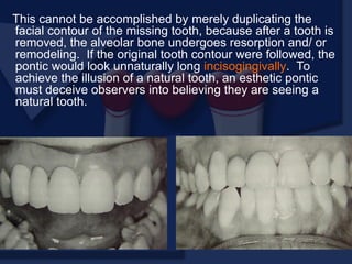
This cannot beaccomplished by merely duplicating the
facial contour of the missing tooth, because after a tooth is
removed, the alveolar bone undergoes resorption and/ or
remodeling. If the original tooth contour were followed, the
pontic would look unnaturally long incisogingivally. To
achieve the illusion of a natural tooth, an esthetic pontic
must deceive observers into believing they are seeing a
natural tooth.
66.
The modified ridge-lappontic is recommended for
most anterior situations; it compensates for lost
buccolingual width in the residual ridge by
overlapping what remains. Rather than emerging
from the crest of the ridge as a natural tooth would,
the cervical aspect of the pontic sits in front of the
ridge, covering any abnormal ridge morphology
resulting from tooth loss. Fortunately, because
most teeth are viewed from only two dimensions,
this relationship remains undetected. A properly
designed, modified ridge lap provides the required
convexity on the tissue side, with smooth and open
embrasures on the lingual side for ease of cleaning.
This is difficult to accomplish.
67.
Clinically, many ponticsare seen with less than
optimal contour, many pontics are seen with less
than optimal contour, resulting in an unnatural
appearance. This can be avoided with careful
preparation at the diagnostic waxing stage.
In normal situations, light falls from above and
an object’s shadow is below it. Unexpected
lighting or unexpectedly placed shadows can be
confusing to the brain. Because of past
experience, the brain “knows” that a tooth grows
out of the gingiva, and it therefore “sees” a
pontic as a tooth unless telltale shadows
suggest otherwise.
68.
Special care mustbe taken when studying where
shadows fall around natural teeth, particularly around
the gingival margin. If a pontic is poorly adapted to the
residual ridge, there will be an unnatural shadow in the
cervical area that looks odd and spoils the illusion of a
natural tooth. In additional, recesses occurring at the
gingival interface will collect food debris, further
betraying the illusion of a natural tooth.
69.
When appearance isof utmost concern,
the ovate pontic, used in conjunction with
alveolar preservation or soft tissue ridge
augmentation, can provide an appearance
at the gingival interface that it virtually
indistinguishable from a natural tooth.
Because it emerges from a soft tissue
recess, this pontic is not susceptible to
many of the esthetic pitfalls previously
described for the modified ridge lap pontic.
However, in most cases, the patient must
be willing to undergo the additional
surgical procedures that an ovate pontic
requires.
70.
Obtaining a correctlysized pontic simply by duplicating the
original tooth is not possible. Ridge resorption will make such
a tooth look too long in the cervical region. The height of a
tooth is immediately obvious when the patient smiles and
shows the gingival margins. An abnormal labiolingual position
or cervical contour, however, is not immediately obvious. This
fact can be used to produce a pontic of good appearance by
recontouring the gingival half of the labial surface. The
observer sees a normal tooth length but is unaware of the
abnormal labial contour. The illusion is successful.
INCISOGINGIVAL LENGTH
71.
One solution isto shape the pontic to simulate a normal
crown and root with emphasis on the cementoenamel
junction. The root can be stained to simulate exposed
dentin. Another approach is to use pink porcelain to
simulate the gingival tissues. 9
However, such pontics
then have considerably increased tissue contact and
require scrupulous plaque control for long term success.
Ridge augmentation procedures have been successful in
correcting areas of limited resorption. When bone loss is
severe, the esthetic result obtained with an RPD is often
better than with an FPD.
72.
Frequently, the spaceavailable for a pontic will be greater or
smaller than the width of the contra lateral tooth. This is
usually due to uncontrolled tooth movement that occurred
when a tooth was removed and not replaced.
If possible, such a discrepancy should be corrected by
orthodontic treatment. If this is not possible, an acceptable
appearance may be obtained by incorporating visual
perception principles into the pontic design.
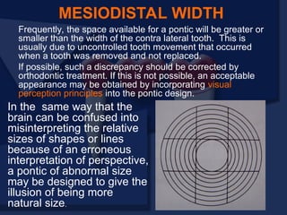
MESIODISTAL WIDTH
In the same way that the
brain can be confused into
misinterpreting the relative
sizes of shapes or lines
because of an erroneous
interpretation of perspective,
a pontic of abnormal size
may be designed to give the
illusion of being more
natural size.
73.
The width ofan anterior tooth is usually identified by the
relative positions of the mesiofacial and distofacial line
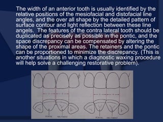
angles, and the over all shape by the detailed pattern of
surface contour and light reflection between these line
angels. The features of the contra lateral tooth should be
duplicated as precisely as possible in the pontic, and the
space discrepancy can be compensated by altering the
shape of the proximal areas. The retainers and the pontic
can be proportioned to minimize the discrepancy. (This is
another situations in which a diagnostic waxing procedure
will help solve a challenging restorative problem).
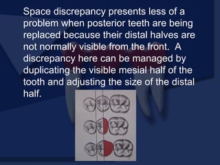
74.
Space discrepancy presentsless of a
problem when posterior teeth are being
replaced because their distal halves are
not normally visible from the front. A
discrepancy here can be managed by
duplicating the visible mesial half of the
tooth and adjusting the size of the distal
half.
75.
Available materials
Over time,several techniques for pontic
fabrication evolved. Prefabricated
porcelain facings were very popular for
use with conventional gold alloys. As use
of the metal ceramic technique increased
during the 1970s, prefabricated facings
lost their popularity and essentially
disappeared. Although an acceptable
substitute, custom made metal ceramic
facings never gained widespread
acceptance.
PONTIC FABRICATION
76.
Most pontics arenow made with a
metal ceramic technique, which
provides the best solution to the
biologic, mechanical, and esthetic
challenges encountered in pontic
design. Their fabrication, however,
differs slightly from the fabrication of
individual crowns.
77.
METAL CERAMIC PONTICS
Awell designed metal ceramic pontic
provides easy plaque removal, strength,
wear resistance, and esthetics. It
fabrication is relatively simple. The metal
frame work for the pontic and one or both
of its retainers is cast in one piece. This
facilitates pontic manipulation during the
successive laboratory and clinical phases.
78.
For strength andesthetics, an accurately
controlled thickness of porcelain is needed
in the finished restoration. To ensure this,
a wax pattern is made to the final
anatomic contour. This also permits an
assessment of connector design adequacy
and the relationship between the
connectors and the proposed configuration
of the ceramic veneer.
ANATOMIC CONTOUR WAXING
79.
1. Wax theinternal, proximal, and axial surfaces of the
retainers
2. Soften the inlay wax, mold it to the approximate
desired pontic shape, and adapt it to the ridge. This is
the starting point for subsequent modification.
Alternatively an impression may be made of the
provisional restoration. Molten wax can then be
poured into this to form the initial pontic shape.
Prefabricated pontic shapes are also available as a
starting point.
STEP BY STEP PROCEDURE
80.
3. If aposterior tooth is being replaced,
leave the occlusal surface flat because
the occlusion is best developed with the
wax addition technique
4. Lute the pontic to the retainer and, for
additional stability, connect its cervical
aspect directly to the master cast with
sticky wax. Then wax the pontic to proper
axial and occlusal (or incisal) contour.
81.
5. Complete theretainers and contour
the proximal and tissue surfaces of the
pontic for the desired tissue contact. The
pontic is now ready for evaluation before
cut back.
82.
The form ofthe wax pattern is evaluated and
any deficiencies are corrected. Particular
attention is given to the connectors, which
should have the correct shape and size. The
connectors provide firm attachment for the
pontic so it does not separate from the
retainers during the subsequent cut back
procedure.
EVALUATION
83.
Use a sharpexplorer to
outline the area that will
be veneered with
porcelain. The porcelain
metal junction must be
placed sufficiently lingual
to ensure good esthetics.
Make depth cuts or
grooves in the wax
pattern.
CUT BACK
84.
Complete the cutback as far as access will
allow with the units connected and on the
master cast.
Section one wax connector with a thin
ribbon saw (sewing thread is a suitable
alternative) and remove the isolated
retainer from the master cast.
85.
Finish the cut-backof this retainer, making
sure there is a distinct 90-degree porcelain
metal junction.
Reflow and finalize the margins. The pontic is
held in position by the other retainer during this
procedure.
Refined the pontic cut back where access is
improved by removal of the first retainer.
Reseat the first retainer, reattach it to the
pontic, section the other connector, and repeat
the process.
Sprue the units and do any final reshaping as
needed.
Invest and cast
86.
Step by stepprocedure
Recover the castings from the investment
and prepare the surfaces to be veneered
Finish the gingival surface of the pontic.
Do not over-reduce this area.
METAL PREPARATION
87.
EVALUATION
Less than 1mm of porcelain thickness is needed
on the gingival surface, because once it is
cemented, the restoration will be seen from the
facial rather than from the gingival. Excessive
gingival porcelain is a common fault in pontic
frame work design and may lead to fracture and
poor appearance.
To facilitate plaque
control, the metal ceramic
junction should be located
lingually. Then tissue
contact will be on the
porcelain and not on
metal, which retains
plaque more tenaciously.
88.
Prepare the metaland apply opaque
Apply cervical porcelain to the gingival
surface of the pontic and seat the casting
on the master cast. A small piece of
tissue paper adapted to the residual ridge
on the cast by moistening with a brush
will prevent porcelain powder from
sticking to the stone. (Cyanoacrylate
resin or special separating agents can be
used for the same purpose.)
PORCELAIN APPLICATION
Many of the steps for porcelain application are identical to
those in individual crown fabrication.There are some
features peculiar to pontic fabrication, however, and these
will be emphasized.
Step by step procedure
89.
Build up theporcelain with the appropriate
distribution of cervical, body, and incisal
shades. The tissue paper will act as a matrix
for the gingival surface of the pontic.
90.
When the porcelainhas been condensed,
section between the units with a thin
razor blade. This will prevent the
porcelain from puling away from the
framework as a result of firing shrinkage.
A second application of porcelain will be
needed to correct any deficiencies
caused by firing shrinkage. Such
additions usually are needed proximally
and gingivally on the pontic.
Apply a porcelain separating liquid to the
stone ridge so that the additional gingival
porcelain can be lifted directly from the
cast
91.
Mark the desiredtissue contact and
contour the gingival surface to
provide as convex a surface as
possible. The pontic is now ready for
clinical evaluation and soldering
procedures, characterization,
glazing, finishing and polishing.
92.
EVALUATION
The porcelain onthe tissue surface of the pontic
should be as smooth as possible. Pits and
defects will make plaque control difficult and
promote calculus formation. The metal
framework must be highly polished, with special
care directed to the gingival embrasures (where
access for plaque removal is more difficult.).
93.
Pontics made frommetal
require fewer laboratory steps
and are therefore sometimes
used for posterior FPDs.
However, they have some
disadvantages (e.g. their
appearance) In addition,
investing and casting must be
done carefully because the
mass of metal in the pontic is
prone to porosity as the bulk
increases. A porous pontic
will retain plaque and tarnish
and corrode rapidly
ALL METAL PONTICS
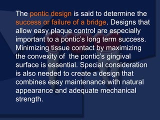
The pontic designis said to determine the
success or failure of a bridge. Designs that
allow easy plaque control are especially
important to a pontic’s long term success.
Minimizing tissue contact by maximizing
the convexity of the pontic’s gingival
surface is essential. Special consideration
is also needed to create a design that
combines easy maintenance with natural
appearance and adequate mechanical
strength.
96.
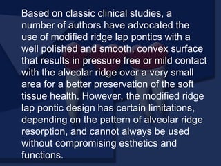
Based on classicclinical studies, a
number of authors have advocated the
use of modified ridge lap pontics with a
well polished and smooth, convex surface
that results in pressure free or mild contact
with the alveolar ridge over a very small
area for a better preservation of the soft
tissue health. However, the modified ridge
lap pontic design has certain limitations,
depending on the pattern of alveolar ridge
resorption, and cannot always be used
without compromising esthetics and
functions.
97.
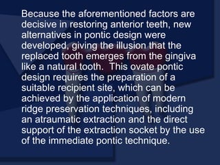
Because the aforementionedfactors are
decisive in restoring anterior teeth, new
alternatives in pontic design were
developed, giving the illusion that the
replaced tooth emerges from the gingiva
like a natural tooth. This ovate pontic
design requires the preparation of a
suitable recipient site, which can be
achieved by the application of modern
ridge preservation techniques, including
an atraumatic extraction and the direct
support of the extraction socket by the use
of the immediate pontic technique.
98.
Metal ceramic ponticfabrication is
straightforward and practical. However, it
requires careful execution for maximum
strength, appearance, and effective plaque
control. Alternatively procedures may
some times be helpful, particularly when
gold alloys are used for the retainers.
Resin veneered pontics should be
restricted to use as longer term provisional
restorations, and all metal pontics may be
the restoration of choice in non-esthetics
situations, particularly where forces are
high.
99.
Thus the designof the pontic is
probably the most important
factor in determining the
success of the restoration of the
patient. If the patient is unable to
clean effectively and maintain
the pontic the restoration will be
unsuccessful.
Harmon C B:Pontic design. J Prosthet
Dent 1958; 8: 496
Carlos B Harmon in 1958 doing a study on the
pontic design said that the success of a bridge can
be attained only when correct form and materials
are combined in a well engineering pontic design
capable of meeting the exact factors of durability
and the maintenance of health and cleanliness.
According to him porcelain, despite certain
unfortunate properties, was the standard as a
component part of pontic construction. Colour form
and texture of natural teeth are readily reproduced
in porcelain. Also its remarkable tissue tolerance,
when contacting the gingival has played an
important part in advanced fixed bridge work. High
fusing porcelain when correctly glazed will display
surface traits remarkably close to those of a natural
tooth.
BACK
102.
Stein RS: Pontic-residual ridge
relationship: A research report. J Prosthet
Dent 1966; 16: 251
Shaldon Stein in 1966 did a study on the
pontic residual ridge relationship. The purpose
of his study were :
To determine the frequency and the nature of
tissue reaction of underlying the residual ridge
mucosa to specific pontic designs.
To compare the frequency and the nature of
tissue reactions of the residual ridge mucosa
to various materials used in pontic
constructions.
103.
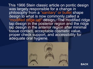
This 1966 Steinclassic article on pontic design
was largely responsible for a change in
philosophy from a “sanitary” or bullet shape
design to what is now commonly called a
“modified ridge lap” design. The modified ridge
lap design in the posterior region and the ridge
lap design in the anterior region after minimal
tissue contact, acceptable cosmetic value,
proper check support, and accessibility for
adequate oral hygiene.
BACK
104.
It has nowbeen established that the design of
the pontic may be the most important factor in
preventing inflammatory reactions, not the
materials used in the pontic. In addition to
properly designing the under surface of the
pontics. It is imperative to open embrasure
spaces adjacent to abutments to allow room for
inter dental tissue and access for pontics are
healthy and pink even after several year of use
of the pontics.
105.
He postulated certainspecifications for pontic
design
Posterior pontic design – a correctly designed
pontic should have
1. All surfaces should be convex, smooth and
properly finished.
2. Contact with the buccal contiguous slopes
should be minimal (pin point) and pressure
free (modified ridge lap).
3. Occlusal table must be in functional harmony
with the occlusion of all of the teeth.
4. Buccal and lingual shunting mechanism should
conform to those of the adjacent teeth.
5. The overall length of buccal surface should be
equal to that of the adjacent abutments or
pontics.
106.
Anterior pontic design– a correctly
placed anterior pontic should have
1. All surfaces should be convex, smooth
and properly finished.
2. Contact with the labial mucosa should be
minimal (pin point) and pressure free (lap
facing).
3. The lingual contour should be in
harmony with adjacent teeth or pontics.
107.
Henry P Jet al: Tissue changes beneath fixed
partial dentures. J Prosthet Dent 1966; 16:
937
P. J. Henry in 1966 in a study placed 14 pontics
on human gingival tissue. He reported no
clinical or histologic difference in the gingival
response to polished gold. Glazed porcelain or
unglazed porcelain after the pontics had been in
the mouth for six months. He also noted that
there were general histologic changes in the
tissue under all the materials tested.
Some investigators have reported that acrylic
resin is not as well tolerated by gingival tissue as
other materials for pontic. He also noted that
glazed porcelain was the most hygienic material
used and it is superior in terms of esthetics and
ease of cleaning.
108.
Cavozos E :Tissue response to fixed partial
denture pontics. J Prosthet Dent 1968; 20:
143
Cavazos in 1968 did a study to
demonstrate that the adaptations of
pontic to the ridge or the amount of
“relief” (scraping of the cast
provided) on the cast is highly
significant and directly proportional
to the amount of unfavourable
tissue change. Absolute minimal
(0.0 to 0.25mm of cast scraping)
produced no tissue changes. When
the cast scraping was increased to
1mm, tissue changes were produced
varying from mild inflammation to
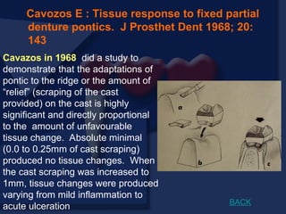
acute ulceration BACK
109.
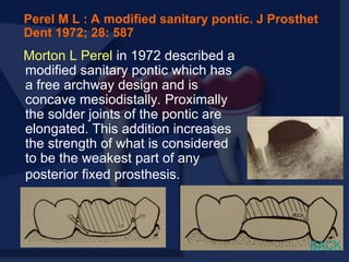
Morton L Perelin 1972 described a
modified sanitary pontic which has
a free archway design and is
concave mesiodistally. Proximally
the solder joints of the pontic are
elongated. This addition increases
the strength of what is considered
to be the weakest part of any
posterior fixed prosthesis.
Perel M L : A modified sanitary pontic. J Prosthet
Dent 1972; 28: 587
BACK
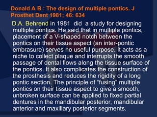
110.
D.A. Behrend in1981 did a study for designing
multiple pontics. He said that in multiple pontics,
placement of a V-shaped notch between the
pontics on their tissue aspect (an inter-pontic
embrasure) serves no useful purpose. It acts as a
niche to collect plaque and interrupts the smooth
passage of dental flows along the tissue surface of
the pontics. It also complicates the construction of
the prosthesis and reduces the rigidity of a long
pontic section. The principle of “fusing” multiple
pontics on their tissue aspect to give a smooth,
unbroken surface can be applied to fixed partial
dentures in the mandibular posterior, mandibular
anterior and maxillary posterior segments.
Donald A B : The design of multiple pontics. J
Prosthet Dent 1981; 46: 634
111.
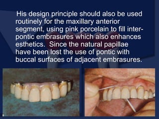
His design principleshould also be used
routinely for the maxillary anterior
segment, using pink porcelain to fill inter-
pontic embrasures which also enhances
esthetics. Since the natural papillae
have been lost the use of pontic with
buccal surfaces of adjacent embrasures.
112.
Antony H Lin 1983 described a technique of pontic
design for extreme resorption of alveolar ridge. In this
the undersurface of the pontic was shaped slightly
convex or flat bucco-lingually to aid in complete
disruption of dental plaque with dental floss or
interproximal toothbrushes. The flat undersurface
allowed easy cleaning from either the lingual or buccal
aspect.
Antony H L: A sanitary “ Arc- fixed partial denture” :
Concept and technique of pontic design. J Prosthet Dent
1983; 50: 338
113.
Porter CB: Anteriorpontic design; a
logical progression. J Prosthet Dent 1984;
51; 774-776.
Carles. B. Porter in 1984 carried out a study
on the anterior pontic design. He stated that
until Stein described his pontic modification
in 1966, only limited deviations have been
noted on traditional pontic design. With minor
exceptions steins pontic design has replaced
the “Saddle Type” pontic, but it seems limited
when multiple pontics must be used.
114.
Parkinson C.F: Ponticdesign of posterior fixed
partial prosthesis; is it a microbial misadventure?
J Prosthet Dent 1984; 51; 51-54
In 1984 Parkinson and Schoberg did a study on the
pontic design of posterior fixed partial prosthesis.
Present designs are commonly based on tooth
replacement without replacement of basic or soft
tissue. The designs cause patients dissatisfaction
because of “whistling” during speech and cause
patient complaints such as, “food always get stuck
under the bridge”. Calculus build up on fixed partial
denture pontics, which is difficult to remove can be
due to poor oral hygiene caused by manipulation
difficulties.
115.
By the restrictionof pontic embrasures,
plaque accumulation and calculus
deposition are eliminated.The number of
surfaces the patient must clean are
reduced and oral hygiene is simplified. In
addition, more of the missing natural
structures are replaced.
116.
L.B. Jacques etal in this
article describes a technique
for the improvement of
esthetics with conditioning of
tissue beneath the pontics by
displacing tissue with a
treatment restoration. Lateral
displacement of tissues
under gradual, controlled
pressure enhances the
interdental papilla which
improves esthetics.
Jacques L B et al: Tissue sculpturing: An alternative
method for improving esthetics of anterior fixed
prosthodontics. J Prosthet Dent 1999; 81: 630
BACK
117.
In 2002 DanielEdelhoff et al did a review
of the different clinical and technical
options that are available for designing
esthetic and functional pontics for
anterior region. He mentions the use of
Gingiva coloured ceramics, all-ceramic
gingival masks and gingival prosthesis to
achieve maximum esthetics in the
anterior region.
Daniel E , H Spiekermann: A review of esthetic
pontic design options. Quintessence Int
2002;33:736-746
118.
The conical ponticwas used to
prevent the extraction site from
collapsing after the removal of a
tooth and to imitate the natural
emergency profile of the tooth.
After extended periods of
service, however, the adjacent
soft tissue tended to become
inflamed, and the alveolar bone
resorbed. Based on the
information available today,
these reactions probably
occurred because the pontic did
not allow adequate oral hygiene.
119.
The hygienic ponticfulfils the
prerequisites for maintaining a healthy
periodontium, because it does not come in
contact with the underlying soft tissue and
provides easy access for oral hygiene
aids to clear the abutment teeth. The gap
between the pontic and the alveolar ridge,
however, is large enough to trap food
particles and to allow the tongue to enter.
120.
The saddle-shaped pontic
achieveshighly esthetic results,
if the alveolar ridges are free of
defects. The emergency profile,
which is very similar to that of
the natural tooth, ensures that
no palatal gap forms, which
could cause phonetic problems
Trapping of food particles is not
expected, because the pontic
seamlessly adapts itself to the
alveolar ridge. Today, however,
it is generally agreed that this
technique should not be used,
because the large concave
contact area with the alveolar
ridge prevents the removal of
adherent plaque.
121.
A reduction ofthe surface
area (ridge lap pontic)
does not significantly
improve hygiene
underneath the pontic,
because the basal
contour remains concave,
unsuitable to provide a
tight contact to the dental
floss.
122.
The modified ridgelap
pontic is the most popular
type of pontic. The convex
basal surface, which rests
on a small area of the
alveolar ridge, fulfils the
recommendation made in
the dental literature with
regard to hygiene
procedures and prevention
of irritation of the
underlying soft tissue.
123.
In contrast tothe classic
requirements for pontics
which suggest the
importance of pressure free
contact over a small area,
the ovate pontic comes in
contact with a larger area of
the underlying soft tissue
an applies light pressure.
This design has been found
to produce highly esthetic
results following suitable
pretreatment of the alveolar
ridge.
124.
Because this designproduces an
emergence profile that looks very similar
to that of the natural tooth, it fulfills ideally
the esthetic and functional requirements of
a pontic for the anterior region. This type
of pontic design, however, requires an
adequate amount of soft tissue, which has
to be sculpted accordingly.
125.
If augmentative measuresare contraindicated or
undesirable, small alveolar deficiencies and missing
papillae can be reconstructed by restorative measures.
First, the exact shade of the gingiva has to be established.
This can be accomplished with special gingival shade
guides that are supplied with the different commercially
available pink veneering materials. The basal surface must
demonstrate a convex shape similar to the ovate pontic
designs for the dental floss to establish tight contact with all
the surface areas.
GINGIVA-COLORED CERAMICS
Daniel E , H Spiekermann: A review of esthetic pontic design options.
Quintessence Int 2002;33:736-746
126.
Separately fabricated ceramicgingival masks can be
used to make subsequent adjustments in
permanently placed restorations. This method is
particularly suitable for patients with a local alveolar
ridge defect that has not been corrected by
augmentation of the soft tissue. For this purpose, an
impression is taken of the labial surface of the
restoration using a customized tray and a medium
viscosity polyether material. The color of the gingiva
is determined with an individually fabricated shade
guide.
ALL-CERAMIC GINGIVAL MASKS
BACK
127.
GINGIVAL PROSTHESIS
Gingiva-colored removableprosthesis
made of soft silicone materials offer an
uncomplicated solution for correcting large
alveolar ridge defects that are associated
with esthetic and phonetic problems. They
can be fixed to the restoration with
precision attachments.
128.
1. Rosenstiel SF et al : Contemporary Fixed Prosthodontics, ed
3, Missouri, Mosby Inc, pg 513
2. Shillingburg H T et al : Fundamentals of fixed prosthodontics,
ed 3, Chicago , Quintessence Publishing, pg 485
3. Shillingburg H T et al : Fundamentals of fixed prosthodontics,
ed 2, Chicago , Quintessence Publishing, pg 387
4. Malone F P et al : Theory and practice of fixed prosthodontics,
Eight Edition , Ishiyaku Euro America, Inc
5. The glossary of prosthodontic terms : J Prosthet Dent 1999; 81
6. Antony H L: A sanitary “ Arc- fixed partial denture” : Concept
and technique of pontic design. J Prosthet Dent 1983; 50: 338
7. Cavozos E : Tissue response to fixed partial denture pontics.
J Prosthet Dent 1968; 20: 143
8. Curtis M B: Current theories of crown contour, margin
placement and pontic design. J Prosthet Dent 1981; 45: 268
9. Daniel Edelhoff, H Spiekermann: A review of esthetic pontic
design options. Quintessence Int 2002;33:736-746
REFERENCES
129.
10. Donald AB : The design of multiple pontics. J Prosthet Dent 1981;
46: 634
11. Harmon C B: Pontic design. J Prosthet Dent 1958; 8: 496
12. Henry P J et al: Tissue changes beneath fixed partial dentures. J
Prosthet Dent 1966; 16: 937
13. Jacques L B et al: Tissue sculpturing: An alternative method for
improving esthetics of anterior fixed prosthodontics. J Prosthet
Dent 1999; 81: 630
14. Parkinson C.F: Pontic design of posterior fixed partial prosthesis; is
it a microbial misadventure? J Prosthet Dent 1984; 51; 51-54.
15. Perel M L : A modified sanitary pontic. J Prosthet Dent 1972; 28:
587
16. Porter CB: Anterior pontic design; a logical progression. J Prosthet
Dent 1984; 51; 774-776.
17. Stein RS: Pontic- residual ridge relationship: A research report. J
Prosthet Dent 1966; 16: 251
18. Roberts DH : Fixed Bridge Prostheses ; John Wright and Sons,
Bristol 1980, pg 68


![/'.,mklppontics final year.pptx m,l;;[']\](https://cdn.slidesharecdn.com/ss_thumbnails/ponticsfinalyear-251031103915-60ae6a8b-thumbnail.jpg?width=640&height=640&fit=bounds)


![5)pontic and pontic designs [Autosaved].pptx](https://cdn.slidesharecdn.com/ss_thumbnails/5ponticandponticdesignsautosaved-250715095630-2c1631ed-thumbnail.jpg?width=640&height=640&fit=bounds)


![PERI-PROSTHETIC FRACTURE NAIL-PLATE CONSTRUCT [NPC].pptx](https://cdn.slidesharecdn.com/ss_thumbnails/drarunkumardrmohamedashrafperiprostheticfrasturenail-plateconstructnpc-260209164459-7e9d15a1-thumbnail.jpg?width=640&height=640&fit=bounds)


![ONFH[AVN HIP] -TRIPLE REGIME -A NOVAL SURGICAL CONCEPT .pptx](https://cdn.slidesharecdn.com/ss_thumbnails/onfhavnhip2026koaconcalicutdrgokuldevdrmashraf-260210064517-213ec005-thumbnail.jpg?width=640&height=640&fit=bounds)


