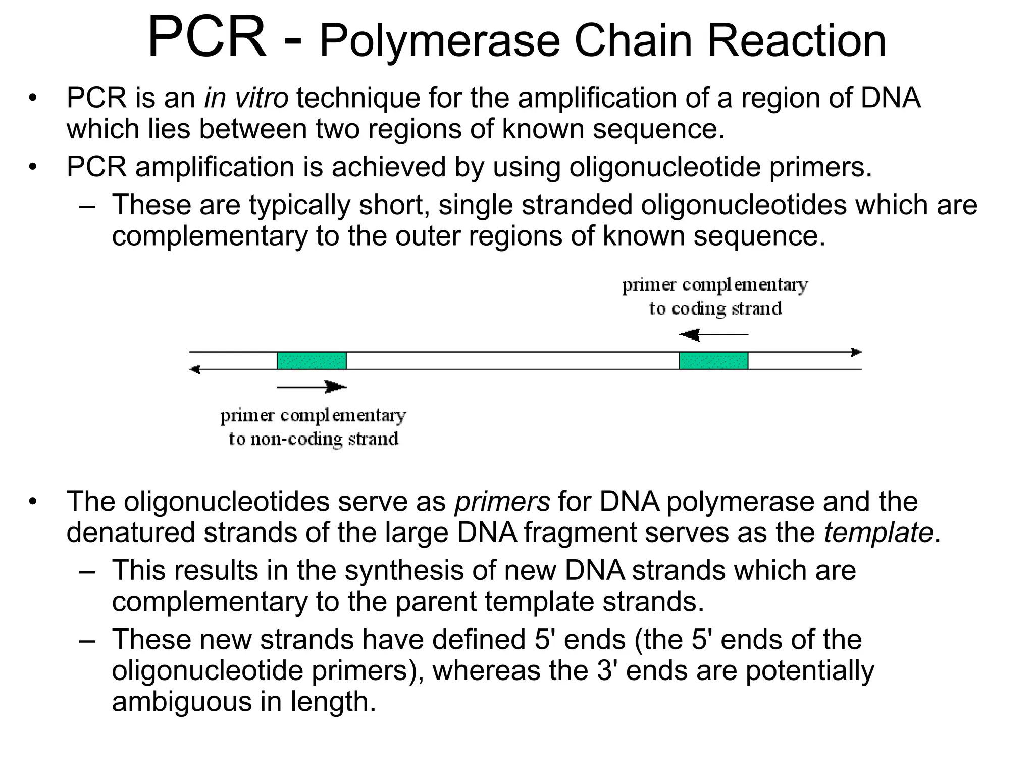
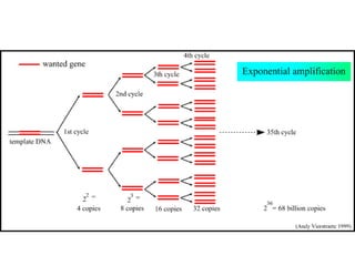
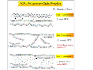
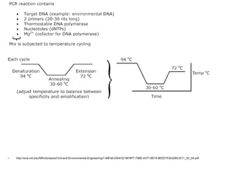
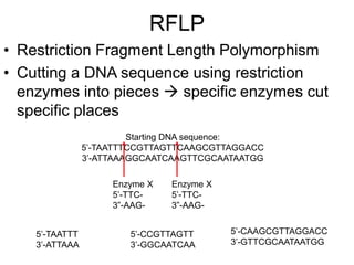
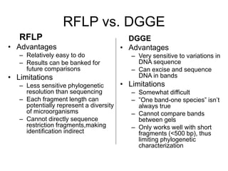
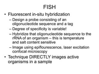
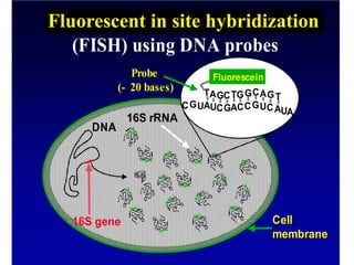
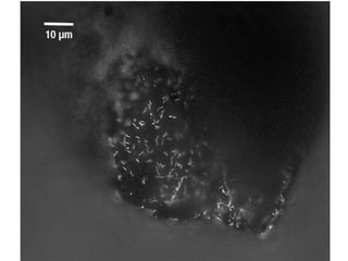
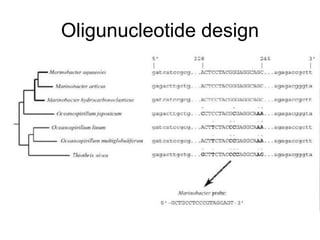
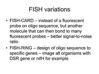
PCR - Polymerase Chain Reaction
• PCR is an in vitro technique for the amplification of a region of DNA
which lies between two regions of known sequence.
• PCR amplification is achieved by using oligonucleotide primers.
– These are typically short, single stranded oligonucleotides which are
complementary to the outer regions of known sequence.
• The oligonucleotides serve as primers for DNA polymerase and the
denatured strands of the large DNA fragment serves as the template.
– This results in the synthesis of new DNA strands which are
complementary to the parent template strands.
– These new strands have defined 5' ends (the 5' ends of the
oligonucleotide primers), whereas the 3' ends are potentially
ambiguous in length.
• http://ocw.mit.edu/NR/rdonlyres/Civil-and-Environmental-Engineering/1-89Fall-2004/321BF8FF-75BE-4377-8D74-8EEE753A328C/0/11_02_04.pdf
Primer selection
• Primer is an oligonucleotide sequence – will target
a specific sequence of opposite base pairing (A-T,
G-C only) of single-stranded nucleic acids
• For example, there is a
– ¼ chance (4-1) of finding an A, G, C or T in any given DNA
sequence; there is a
– 1/16 chance (4-2) of finding any dinucleotide sequence (eg.
AG); a
– 1/256 chance of finding a given 4-base sequence.
• Thus, a sixteen base sequence will statistically be
present only once in every 416 bases (=4 294 967
296, or 4 billion): this is about the size of the human or
maize genome, and 1000x greater than the genome
size of E. coli.
Primer Specificity
• Universal – amplifies ALL bacterial DNA
for instance
• Group Specific – amplify all denitrifiers for
instance
• Specific – amplify just a given sequence
Forward and reverse primers
• If you know the sequence targeted for
amplification, you know the size which the
primers should be anealing across
• If you don’t know the sequence… What
do you get?
DNA Polymerase
• DNA Polymerase is the enzyme responsible for
copying the sequence starting at the primer from
the single DNA strand
• Commonly use Taq, an enzyme from the
hyperthermophilic organisms Thermus aquaticus,
isolated first at a thermal spring in Yellowstone
National Park
• This enzyme is heat-tolerant  useful both because
it is thermally tolerant (survives the melting T of
DNA denaturation) which also means the process is
more specific, higher temps result in less mismatch
– more specific replication
RFLP
• Restriction Fragment Length Polymorphism
• Cutting a DNA sequence using restriction
enzymes into pieces  specific enzymes cut
specific places
Starting DNA sequence:
5’-TAATTTCCGTTAGTTCAAGCGTTAGGACC
3’-ATTAAAGGCAATCAAGTTCGCAATAATGG
Enzyme X
5’-TTC-
3”-AAG-
Enzyme X
5’-TTC-
3”-AAG-
5’-TAATTT
3’-ATTAAA
5’-CCGTTAGTT
3’-GGCAATCAA
5’-CAAGCGTTAGGACC
3’-GTTCGCAATAATGG
RFLP
• DNA can be processed by RFLP either directly (if
you can get enough DNA from an environment) or
from PCR product
• T-RFLP (terminal-RFLP) is in most respects
identical except for a marker on the end of the
enzyme
• Works as fingerprinting technique because
different organisms with different DNA sequences
will have different lengths of DNA between
identical units targeted by the restriction enzymes
– specificity can again be manipulated with PCR primers
Liu et al. (1997) Appl Environ Microbiol 63:4516-4522
Electrophoresis
• Fragmentation products of differing length
are separated – often on an agarose gel
bed by electrophoresis, or using a
capilarry electrophoretic separation
DGGE
• Denaturing gradient gel electrophoresis
– The hydrogen bonds formed between complimentary base
pairs, GC rich regions ‘melt’ (melting=strand separation or
denaturation) at higher temperatures than regions that are AT
rich.
• When DNA separated by electrophoresis through a gradient of
increasing chemical denaturant (usually formamide and urea), the
mobility of the molecule is retarded at the concentration at which
the DNA strands of low melt domain dissociate.
– The branched structure of the single stranded moiety of the
molecule becomes entangled in the gel matrix and no further
movement occurs.
– Complete strand separation is prevented by the presence of a
high melting domain, which is usually artificially created at one
end of the molecule by incorporation of a GC clamp. This is
accomplished during PCR amplification using a PCR primer
with a 5' tail consisting of a sequence of 40 GC.
Run DGGE animation here – from http://www.charite.de/bioinf/tgge/
RFLP vs. DGGE
DGGE
• Advantages
– Very sensitive to variations in
DNA sequence
– Can excise and sequence
DNA in bands
• Limitations
– Somewhat difficult
– ”One band-one species” isn’t
always true
– Cannot compare bands
between gels
– Only works well with short
fragments (<500 bp), thus
limiting phylogenetic
characterization
RFLP
• Advantages
– Relatively easy to do
– Results can be banked for
future comparisons
• Limitations
– Less sensitive phylogenetic
resolution than sequencing
– Each fragment length can
potentially represent a diversity
of microorganisms
– Cannot directly sequence
restriction fragments,making
identification indirect
FISH
• Fluorescent in-situ hybridization
– Design a probe consisting of an
oligonucleotide sequence and a tag
– Degree of specificity is variable!
– Hybridize that oligonucleotide sequence to the
rRNA of an organism – this is temperature
and salt content sensitive
– Image using epiflourescence, laser excitation
confocal microscopy
• Technique DIRECTLY images active
organisms in a sample
16S gene
16S rRNA
Cell
Cell
membrane
membrane
DNA
16S gene
*
*
*
*
*
*
*
*
*
* *
* *
*
* * *
Fluorescentin situhybridisation
(FISH) using DNA probes
TAGCTGGCAGT
AUCGACCGUCA
CGU
Fluorescein
A
U
Probe
Probe
(
(-
- 20 bases)
20 bases)
Fluorescent in site hybridization
10 µm
DAPI FER656
B Drift Slime Streamer
Oligunucleotide design
FISH variations
• FISH-CARD – instead of a fluorescent
probe on oligo sequence, but another
molecule that can then bond to many
fluorescent probes – better signal-to-noise
ratio
• FISH-RING – design of oligo sequence to
specific genes – image all organisms with
DSR gene or nifH for example
Clone Library
• http://ocw.mit.edu/NR/rdonlyres/Civil-and-Environmental-Engineering/1-89Fall-2004/321BF8FF-75BE-4377-8D74-8EEE753A328C/0/11_02_04.pdf
http://www.ifa.hawaii.edu/UHNAI/NAIweb/presentations/astrobiol6.pdf
Polymerase Chain Reaction (PCR) Lecture3.ppt

Polymerase Chain Reaction (PCR) Lecture3.ppt

  • 1.
    PCR - PolymeraseChain Reaction • PCR is an in vitro technique for the amplification of a region of DNA which lies between two regions of known sequence. • PCR amplification is achieved by using oligonucleotide primers. – These are typically short, single stranded oligonucleotides which are complementary to the outer regions of known sequence. • The oligonucleotides serve as primers for DNA polymerase and the denatured strands of the large DNA fragment serves as the template. – This results in the synthesis of new DNA strands which are complementary to the parent template strands. – These new strands have defined 5' ends (the 5' ends of the oligonucleotide primers), whereas the 3' ends are potentially ambiguous in length.
  • 4.
  • 5.
    Primer selection • Primeris an oligonucleotide sequence – will target a specific sequence of opposite base pairing (A-T, G-C only) of single-stranded nucleic acids • For example, there is a – ¼ chance (4-1) of finding an A, G, C or T in any given DNA sequence; there is a – 1/16 chance (4-2) of finding any dinucleotide sequence (eg. AG); a – 1/256 chance of finding a given 4-base sequence. • Thus, a sixteen base sequence will statistically be present only once in every 416 bases (=4 294 967 296, or 4 billion): this is about the size of the human or maize genome, and 1000x greater than the genome size of E. coli.
  • 6.
    Primer Specificity • Universal– amplifies ALL bacterial DNA for instance • Group Specific – amplify all denitrifiers for instance • Specific – amplify just a given sequence
  • 7.
    Forward and reverseprimers • If you know the sequence targeted for amplification, you know the size which the primers should be anealing across • If you don’t know the sequence… What do you get?
  • 8.
    DNA Polymerase • DNAPolymerase is the enzyme responsible for copying the sequence starting at the primer from the single DNA strand • Commonly use Taq, an enzyme from the hyperthermophilic organisms Thermus aquaticus, isolated first at a thermal spring in Yellowstone National Park • This enzyme is heat-tolerant  useful both because it is thermally tolerant (survives the melting T of DNA denaturation) which also means the process is more specific, higher temps result in less mismatch – more specific replication
  • 9.
    RFLP • Restriction FragmentLength Polymorphism • Cutting a DNA sequence using restriction enzymes into pieces  specific enzymes cut specific places Starting DNA sequence: 5’-TAATTTCCGTTAGTTCAAGCGTTAGGACC 3’-ATTAAAGGCAATCAAGTTCGCAATAATGG Enzyme X 5’-TTC- 3”-AAG- Enzyme X 5’-TTC- 3”-AAG- 5’-TAATTT 3’-ATTAAA 5’-CCGTTAGTT 3’-GGCAATCAA 5’-CAAGCGTTAGGACC 3’-GTTCGCAATAATGG
  • 10.
    RFLP • DNA canbe processed by RFLP either directly (if you can get enough DNA from an environment) or from PCR product • T-RFLP (terminal-RFLP) is in most respects identical except for a marker on the end of the enzyme • Works as fingerprinting technique because different organisms with different DNA sequences will have different lengths of DNA between identical units targeted by the restriction enzymes – specificity can again be manipulated with PCR primers Liu et al. (1997) Appl Environ Microbiol 63:4516-4522
  • 11.
    Electrophoresis • Fragmentation productsof differing length are separated – often on an agarose gel bed by electrophoresis, or using a capilarry electrophoretic separation
  • 13.
    DGGE • Denaturing gradientgel electrophoresis – The hydrogen bonds formed between complimentary base pairs, GC rich regions ‘melt’ (melting=strand separation or denaturation) at higher temperatures than regions that are AT rich. • When DNA separated by electrophoresis through a gradient of increasing chemical denaturant (usually formamide and urea), the mobility of the molecule is retarded at the concentration at which the DNA strands of low melt domain dissociate. – The branched structure of the single stranded moiety of the molecule becomes entangled in the gel matrix and no further movement occurs. – Complete strand separation is prevented by the presence of a high melting domain, which is usually artificially created at one end of the molecule by incorporation of a GC clamp. This is accomplished during PCR amplification using a PCR primer with a 5' tail consisting of a sequence of 40 GC. Run DGGE animation here – from http://www.charite.de/bioinf/tgge/
  • 14.
    RFLP vs. DGGE DGGE •Advantages – Very sensitive to variations in DNA sequence – Can excise and sequence DNA in bands • Limitations – Somewhat difficult – ”One band-one species” isn’t always true – Cannot compare bands between gels – Only works well with short fragments (<500 bp), thus limiting phylogenetic characterization RFLP • Advantages – Relatively easy to do – Results can be banked for future comparisons • Limitations – Less sensitive phylogenetic resolution than sequencing – Each fragment length can potentially represent a diversity of microorganisms – Cannot directly sequence restriction fragments,making identification indirect
  • 15.
    FISH • Fluorescent in-situhybridization – Design a probe consisting of an oligonucleotide sequence and a tag – Degree of specificity is variable! – Hybridize that oligonucleotide sequence to the rRNA of an organism – this is temperature and salt content sensitive – Image using epiflourescence, laser excitation confocal microscopy • Technique DIRECTLY images active organisms in a sample
  • 16.
    16S gene 16S rRNA Cell Cell membrane membrane DNA 16Sgene * * * * * * * * * * * * * * * * * Fluorescentin situhybridisation (FISH) using DNA probes TAGCTGGCAGT AUCGACCGUCA CGU Fluorescein A U Probe Probe ( (- - 20 bases) 20 bases) Fluorescent in site hybridization
  • 17.
    10 µm DAPI FER656 BDrift Slime Streamer
  • 19.
  • 20.
    FISH variations • FISH-CARD– instead of a fluorescent probe on oligo sequence, but another molecule that can then bond to many fluorescent probes – better signal-to-noise ratio • FISH-RING – design of oligo sequence to specific genes – image all organisms with DSR gene or nifH for example
  • 21.
  • 22.