Clinical Correlate: Dyspnea
•RL, 59/M,
• Known Hypertensive and Diabetic on maintenance meds x 5 years
• Cc: dyspnea.
• 3 days PTA (+) productive cough with whitish sputum, worse in the morning,
no medications taken at the time.
• 1 day PTA, fever relieved by paracetamol intake, increased sputum production
• ODA (+) tachypnea and SOB
• PMHX/SHX: s/p debridement DM foot L, previous smoker 40 pack years
6.
Clinical Correlate: Dyspnea
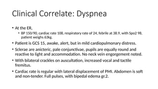
•At the ER,
• BP 150/90, cardiac rate 108, respiratory rate of 24, febrile at 38.9, with Spo2 98,
patient weighs 63kg.
• Patient is GCS 15, awake, alert, but in mild cardiopulmonary distress.
• Sclerae are anicteric, pale conjunctivae, pupils are equally round and
reactive to light and accommodation. No neck vein engorgement noted.
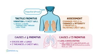
• With bilateral crackles on auscultation, increased vocal and tactile
fremitus.
• Cardiac rate is regular with lateral displacement of PMI. Abdomen is soft
and non-tender. Full pulses, with bipedal edema gr.2.
7.
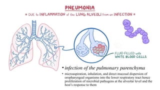
• infection ofthe pulmonary parenchyma
• microaspiration, inhalation, and direct mucosal dispersion of
oropharyngeal organisms into the lower respiratory tract hence
proliferation of microbial pathogens at the alveolar level and the
host’s response to them
8.
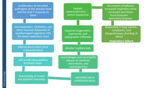
proliferation of microbial
pathogensat the alveolar level
and the host’s response to
them
microaspiration, inhalation, and
direct mucosal dispersion of
oropharyngeal organisms into
the lower respiratory tract
inflammatory event (viral,
macroaspiration)
self-accelerating positive
feedback loops
Overcoming of innate
and adaptive immunity
epithelial and or
endothelial injury
macrophages and neutrophils
release of cytokines,
chemokines, and
catecholamines
alveolar capillary leak
impaired oxygenation,
hypoxemia, and
radiographic infiltrates
hypoxic
vasoconstriction
severe hypoxemia
Decreased compliance,
increased respiratory drive,
increased secretions,
bronchospasm
worsening dyspnea.
Reductions in lung volume,
compliance, and
intrapulmonary shunting of
blood
respiratory failure
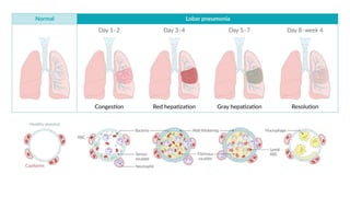
Pathophysiology
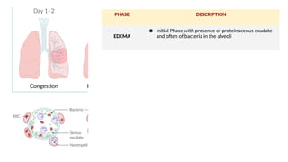
PHASE DESCRIPTION
EDEMA
● InitialPhase with presence of proteinaceous exudate
and often of bacteria in the alveoli
RED
HEPATIZATION
● Erythrocytes in the cellular intra alveolar exudates
● Neutrophil influx is more important from the
standpoint of host defense
12.
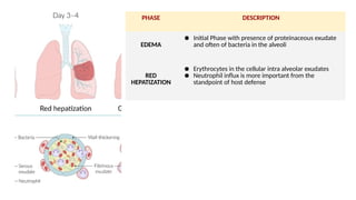
PHASE DESCRIPTION
EDEMA
● InitialPhase with presence of proteinaceous exudate
and often of bacteria in the alveoli
RED
HEPATIZATION
● Erythrocytes in the cellular intra alveolar exudates
● Neutrophil influx is more important from the
standpoint of host defense
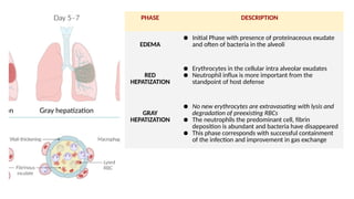
GRAY
HEPATIZATION
● No new erythrocytes are extravasating with lysis and
degradation of preexisting RBCs
● The neutrophils the predominant cell, fibrin
deposition is abundant and bacteria have disappeared
● This phase corresponds with successful containment
of the infection and improvement in gas exchange
13.
PHASE DESCRIPTION
EDEMA
● InitialPhase with presence of proteinaceous exudate
and often of bacteria in the alveoli
RED
HEPATIZATION
● Erythrocytes in the cellular intra alveolar exudates
● Neutrophil influx is more important from the
standpoint of host defense
GRAY
HEPATIZATION
● No new erythrocytes are extravasating with lysis and
degradation of preexisting RBCs
● The neutrophils is the predominant cell, fibrin
deposition is abundant and bacteria have disappeared
● This phase corresponds with successful containment
of the infection and improvement in gas exchange
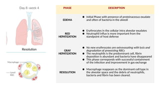
RESOLUTION
● Macrophage reappears as the dominant cell type in
the alveolar space and the debris of neutrophils,
bacteria and fibrin has been cleared.
14.
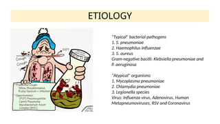
“Typical” bacterial pathogens
1.S. pneumoniae
2. Haemophilus influenzae
3. S. aureus
Gram-negative bacilli: Klebsiella pneumoniae and
P. aeruginosa
“Atypical” organisms
1. Mycoplasma pneumoniae
2. Chlamydia pneumoniae
3. Legionella species
Virus: Influenza virus, Adenovirus, Human
Metapneumoviruses, RSV and Coronavirus
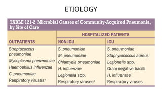
ETIOLOGY
Physical Exam
• increasedrespiratory rate
• use of accessory muscles
• increased or decreased tactile fremitus
• percussion note can vary from dull (consolidated
lung) to flat (pleural fluid)
• Crackles, bronchial breath sounds, and possibly a
pleural friction rub
22.
Diagnostics
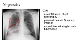
CXR
• new infiltrateon chest
radiography
• pneumatoceles in S. aureus
infection
• upper-lobe cavitating lesion in
tuberculosis
23.
Diagnostics
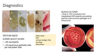
SPUTUM GSCS
suitable sputumsample:
• >25 neutrophils
• <10 squamous epithelial cells
per low-power field
BLOOD CULTURES
• Only 5–14% of cultures from
hospitalized CAP patients are positive,
and the most common pathogen is S.
Pneumonia
Other tests:
PCR
Urinary Antigen Test
Serology
Biomarkers
24.
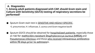
A. Diagnostics
1. Amongadult patients diagnosed with CAP, should Gram stain and
Culture with Sensitivity (GS/CS) testing of respiratory secretions be
performed?
● Sputum Gram stain test is SENSITIVE AND HIGHLY SPECIFIC.
-S. pneumoniae, H. influenzae, S. aureus and Gram-negative bacilli
● Sputum GS/CS should be obtained for hospitalized patients, especially those
at risk for methicillin-resistant Staphylococcus aureus (MRSA) and
Pseudomonas infectious and those who received intravenous antibiotics
within 90 days prior to admission.
26.
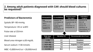
2. Among adultpatients diagnosed with CAP, should blood cultures
be requested?
Predictors of Bacteremia
Systolic BP <90 mmHg
Temperature <35 or 40’C
≥
Pulse rate 125/min
≥
Liver disease
Blood urea nitrogen 30 mg/dL
≥
Serum sodium <130 mmol/L
WBC <5,000/mm3 or > 20,000/mm3
27.
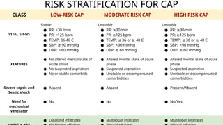
RISK STRATIFICATION FORCAP
CLASS LOW-RISK CAP MODERATE RISK CAP HIGH RISK CAP
VITAL SIGNS
Stable:
● RR: <30 /min
● PR: <125 bpm
● TEMP: 36-40 C
● SBP: 90 mmHg
≥
● DBP: > 60 mmHg
Unstable
● RR: 30/min
≥
● PR: 125 bpm
≥
● TEMP: 36 or 40 C
≤ ≥
● SBP: <90 mmHg
● DBP: 60 mmHg
≤
Unstable
● RR: 30/min
≥
● PR: 125 bpm
≥
● TEMP: 36 or 40 C
≤ ≥
● SBP: <90 mmHg
● DBP: 60 mmHg
≤
FEATURES
● No altered mental state of
acute onset
● No suspected aspiration
● No or stable comorbids
● Altered mental state of acute
phase
● Suspected aspiration
● Unstable or decompensated
comorbidities
● Altered mental state of acute
phase
● Suspected aspiration
● Unstable or decompensated
comorbidities
Severe sepsis and
Septic shock
● Absent ● Absent ● Present/Absent
Need for
mechanical
ventilator
● No ● No ● No/Yes
● Localized infiltrates ● Multilobar infiltrates ● Multilobar infiltrates
28.
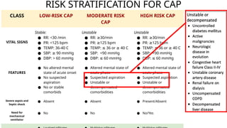
RISK STRATIFICATION FORCAP
CLASS LOW-RISK CAP MODERATE RISK
CAP
HIGH RISK CAP
VITAL SIGNS
Stable:
● RR: <30 /min
● PR: <125 bpm
● TEMP: 36-40 C
● SBP: 90 mmHg
≥
● DBP: > 60 mmHg
Unstable
● RR: 30/min
≥
● PR: 125 bpm
≥
● TEMP: 36 or 40 C
≤ ≥
● SBP: <90 mmHg
● DBP: 60 mmHg
≤
Unstable
● RR: 30/min
≥
● PR: 125 bpm
≥
● TEMP: 36 or 40 C
≤ ≥
● SBP: <90 mmHg
● DBP: 60 mmHg
≤
FEATURES
● No altered mental
state of acute onset
● No suspected
aspiration
● No or stable
comorbids
● Altered mental state of
acute phase
● Suspected aspiration
● Unstable or
decompensated
comorbidities
● Altered mental state of
acute phase
● Suspected aspiration
● Unstable or
decompensated
comorbidities
Severe sepsis and
Septic shock
● Absent ● Absent ● Present/Absent
Need for
mechanical
ventilator
● No ● No ● No/Yes
30.
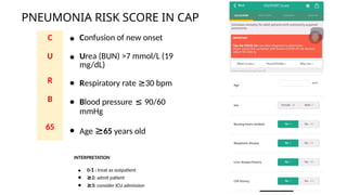
PNEUMONIA RISK SCOREIN CAP
C ● Confusion of new onset
U ● Urea (BUN) >7 mmol/L (19
mg/dL)
R ● Respiratory rate ≥30 bpm
B ● Blood pressure ≤ 90/60
mmHg
65 ● Age ≥65 years old
INTERPRETATION
● 0-1 : treat as outpatient
● ≥2: admit patient
● ≥3: consider ICU admission
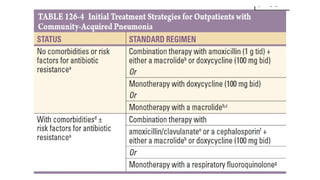
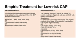
Empiric Treatment forLow-risk CAP
Recommendation 7:
The following antibiotics should be started for
empiric treatment of patients with low risk CAP
without co-morbidities:
Amoxicillin 1 gram, three times daily
OR
Clarithromycin 500mg, twice daily
OR
Azithromycin 500mg once daily
Recommendation 8:
The following antibiotics should be started for
empiric treatment of patients with low risk CAP with
stable co-morbidities:
Beta-lactam
Co-amoxiclav (amoxicillin/clavulanate 500 mg/125
mg three times daily, OR amoxicillin/ clavulanate
875 mg/125 mg twice daily)
OR
Cefuroxime 500mg, twice daily
PLUS OR MINUS (+/-)
Macrolide
Clarithromycin 500mg, twice daily
OR
Azithromycin 500mg once daily
OR
Doxycycline 100mg, twice daily
35.
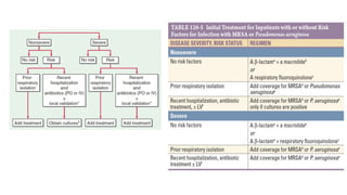
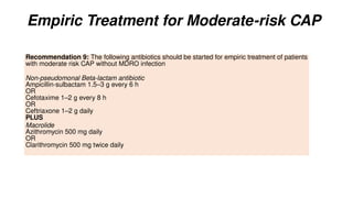
Empiric Treatment forModerate-risk CAP
Recommendation 9: The following antibiotics should be started for empiric treatment of patients
with moderate risk CAP without MDRO infection
Non-pseudomonal Beta-lactam antibiotic
Ampicillin-sulbactam 1.5–3 g every 6 h
OR
Cefotaxime 1–2 g every 8 h
OR
Ceftriaxone 1–2 g daily
PLUS
Macrolide
Azithromycin 500 mg daily
OR
Clarithromycin 500 mg twice daily
36.
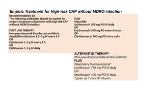
Empiric Treatment forHigh-risk CAP without MDRO infection
Recommendation 10:
The following antibiotics should be started for
empiric treatment of patients with high risk CAP
without MDRO infection:
FIRST LINE THERAPY
Non-pseudomonal Beta-lactam antibiotic
Ampicillin-sulbactam 1.5–3 g IV every 6 h
OR
Cefotaxime 1–2 g IV every 8 h
OR
Ceftriaxone 1–2 g IV daily
PLUS
Macrolide
Azithromycin 500 mg PO/IV daily
OR
Erythromycin 500 mg PO every 6 hours
OR
Clarithromycin 500 mg PO twice daily
ALTERNATIVE THERAPY
Non-pseudomonal Beta-lactam antibiotic
PLUS
Respiratory fluoroquinolone*
Levofloxacin 750 mg PO/IV daily
OR
Moxifloxacin 400 mg PO/IV daily
* given as 1 hour IV infusion
37.
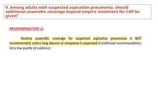
9. Among adultswith suspected aspiration pneumonia, should
additional anaerobic coverage beyond empiric treatment for CAP be
given?
38.
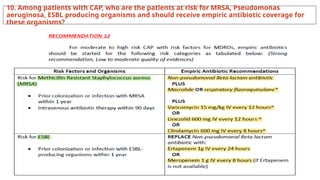
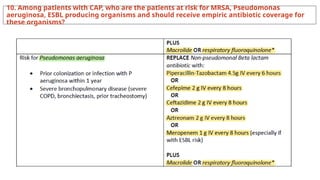
10. Among patientswith CAP, who are the patients at risk for MRSA, Pseudomonas
aeruginosa, ESBL producing organisms and should receive empiric antibiotic coverage for
these organisms?
39.
10. Among patientswith CAP, who are the patients at risk for MRSA, Pseudomonas
aeruginosa, ESBL producing organisms and should receive empiric antibiotic coverage for
these organisms?
40.
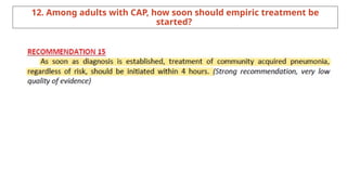
12. Among adultswith CAP, how soon should empiric treatment be
started?
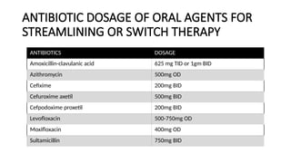
ANTIBIOTIC DOSAGE OFORAL AGENTS FOR
STREAMLINING OR SWITCH THERAPY
ANTIBIOTICS DOSAGE
Amoxicillin-clavulanic acid 625 mg TID or 1gm BID
Azithromycin 500mg OD
Cefixime 200mg BID
Cefuroxime axetil 500mg BID
Cefpodoxime proxetil 200mg BID
Levofloxacin 500-750mg OD
Moxifloxacin 400mg OD
Sultamicillin 750mg BID
43.
How long isthe duration of treatment for CAP?
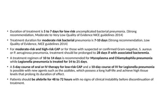
• Duration of treatment is 5 to 7 days for low risk uncomplicated bacterial pneumonia. (Strong
recommendation, Moderate to Very Low Quality of Evidence NICE guidelines 2014)
• Treatment duration for moderate risk bacterial pneumonia is 7-10 days (Strong recommendation, Low
Quality of Evidence, NICE guidelines 2014)
• For moderate-risk and high-risk CAP or for those with suspected or confirmed Gram-negative, S. aureus
or P. aeruginosa pneumonia, treatment should be prolonged to 28 days if with associated bacteremia.
• A treatment regimen of 10 to 14 days is recommended for Mycoplasma and Chlamydophila pneumonia
while Legionella pneumonia is treated for 14 to 21 days.
• A 5-day course of oral or IV therapy for low-risk CAP and a 10-day course of IV for Legionella pneumonia
is possible with new agents such as the azalides, which possess a long half-life and achieve high tissue
levels that prolong its duration of effect.
• Patients should be afebrile for 48 to 72 hours with no signs of clinical instability before discontinuation of
treatment.
44.
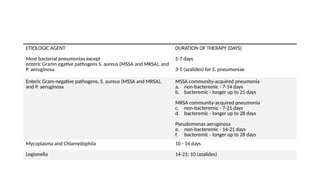
Duration of AntibioticUse BASED ON ETIOLOGY
ETIOLOGIC AGENT DURATION OF THERAPY (DAYS)
Most bacterial pneumonias except
enteric Gramn egative pathogens S. aureus (MSSA and MRSA), and
P. aeruginosa
5-7 days
3-5 (azalides) for S. pneumoniae
Enteric Gram-negative pathogens, S. aureus (MSSA and MRSA),
and P. aeruginosa
MSSA community-acquired pneumonia
a. non-bacteremic - 7-14 days
b. bacteremic - longer up to 21 days
MRSA community-acquired pneumonia
c. non-bacteremic - 7-21 days
d. bacteremic - longer up to 28 days
Pseudomonas aeruginosa
e. non-bacteremic - 14-21 days
f. bacteremic - longer up to 28 days
Mycoplasma and Chlamydophila 10 - 14 days
Legionella 14-21; 10 (azalides)
45.
What should
be donefor
patients who
are not
improving
after 72 hours
of empiric
antibiotic
therapy?
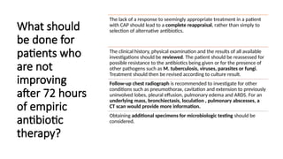
The lack of a response to seemingly appropriate treatment in a patient
with CAP should lead to a complete reappraisal, rather than simply to
selection of alternative antibiotics.
The clinical history, physical examination and the results of all available
investigations should be reviewed. The patient should be reassessed for
possible resistance to the antibiotics being given or for the presence of
other pathogens such as M. tuberculosis, viruses, parasites or fungi.
Treatment should then be revised according to culture result.
Follow-up chest radiograph is recommended to investigate for other
conditions such as pneumothorax, cavitation and extension to previously
uninvolved lobes, pleural effusion, pulmonary edema and ARDS. For an
underlying mass, bronchiectasis, loculation , pulmonary abscesses, a
CT scan would provide more information.
Obtaining additional specimens for microbiologic testing should be
considered.
46.
REASONS
FOR
A
LACK
OF
RESPONSE
TO
TREATMENT
OF
CAP
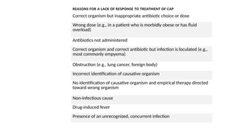
REASONS FOR ALACK OF RESPONSE TO TREATMENT OF CAP
Correct organism but inappropriate antibiotic choice or dose
Wrong dose (e.g., in a patient who is morbidly obese or has fluid
overload)
Antibiotics not administered
Correct organism and correct antibiotic but infection is loculated (e.g.,
most commonly empyema)
Obstruction (e.g., lung cancer, foreign body)
Incorrect identification of causative organism
No identification of causative organism and empirical therapy directed
toward wrong organism
Non-infectious cause
Drug-induced fever
Presence of an unrecognized, concurrent infection
47.
When can ahospitalized
patient with CAP be
discharged?
• In the absence of any unstable coexisting illness or other
life-threatening complication, the patient may be
discharged once clinically stable and oral therapy is
initiated.
• A repeat chest radiograph prior to hospital discharge is
not needed in a patient who is clinically improving.
• A repeat chest radiograph is recommended during a
follow-up visit, approximately 4 to 6 weeks after
hospital discharge to establish a new radiographic
baseline and to exclude the possibility of malignancy
associated with CAP, particularly in older smokers.
48.
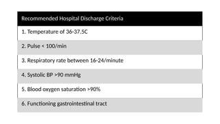
Recommended Hospital DischargeCriteria
1. Temperature of 36-37.5C
2. Pulse < 100/min
3. Respiratory rate between 16-24/minute
4. Systolic BP >90 mmHg
5. Blood oxygen saturation >90%
6. Functioning gastrointestinal tract
49.
What other informationshould be explained
and discussed with the patient?
• Explain to patients with CAP that after starting treatment
their symptoms are expected to steadily improve,
although the rate of improvement will vary with the
severity of the pneumonia. Most people can expect that
by:
• 1 week: fever should have resolved
• 4 weeks: chest pain and sputum production should
have substantially reduced
• 6 weeks: cough and breathlessness should have
substantially reduced
• 3 months: most symptoms should have resolved but
fatigue may still be present 6 months: most people
will feel back to normal.
50.
Clinical Correlate: Dyspnea
•RL, 59/M,
• Known Hypertensive and Diabetic on maintenance meds x 5 years
• Cc: dyspnea.
• 3 days PTA (+) productive cough with whitish sputum, worse in the morning,
no medications taken at the time.
• 1 day PTA, fever relieved by paracetamol intake, increased sputum production
• ODA (+) tachypnea and SOB
• PMHX/SHX: s/p debridement DM foot L, previous smoker 40 pack years
51.
Clinical Correlate: Dyspnea
•At the ER,
• BP 150/90, cardiac rate 108, respiratory rate of 24, febrile at 38.9, with Spo2 98,
patient weighs 63kg.
• Patient is GCS 15, awake, alert, but in mild cardiopulmonary distress.
• Sclerae are anicteric, pale conjunctivae, pupils are equally round and
reactive to light and accommodation. No neck vein engorgement noted.
• With bilateral crackles on auscultation, increased vocal and tactile
fremitus.
• Cardiac rate is regular with lateral displacement of PMI. Abdomen is soft
and non-tender. Full pulses, with bipedal edema gr.2.
Reference
• Philippine ClinicalPractice Guidelines: Diagnosis, Empiric Management and Prevention
of Community-Acquired Pneumonia in Immunocompetent Adults 2020 Update
Treatment. Joint Statement of PSMID • PCCP • PAFP • PCR
• Harrison’s Internal Medicine 21st
edition
Editor's Notes
#6 CAP MR
CHF FC II 2 HCVD
DM T2, DM foot L s/p Debridement
#8 proliferation of microbial pathogens at the alveolar level and the host’s response to them
microaspiration, inhalation, and direct mucosal dispersion of oropharyngeal organisms into the lower respiratory tract
Loss of barriers and echanical protection such as the hairs and turbinates of the nares, the branching tracheobronchial tree, mucociliary clearance, and gag and cough reflexes
Regional growth conditions for bacteria, such as pH, oxygen tension, and temperature.
inflammatory event (viral, macroaspiration)
self-accelerating positive feedback loops
(cycle of inflammation, enhanced nutrient availability, and release of potential bacterial growth factors)
Overcoming of innate and adaptive immunity
epithelial and or endothelial injury
macrophages and neutrophils release of cytokines, chemokines, and catecholamines
interleukin 6 and tumor necrosis factor fever,
chemokines such (IL-8, GCSF) increase local neutrophil numbers
alveolar capillary leak
impaired oxygenation, hypoxemia, and radiographic infiltrates
hypoxic vasoconstriction severe hypoxemia
Decreased compliance, increased respiratory drive, increased secretions, bronchospasm worsening dyspnea.
Reductions in lung volume, compliance, and intrapulmonary shunting of blood respiratory failure
#9 proliferation of microbial pathogens at the alveolar level and the host’s response to them
microaspiration, inhalation, and direct mucosal dispersion of oropharyngeal organisms into the lower respiratory tract
Loss of barriers and echanical protection such as the hairs and turbinates of the nares, the branching tracheobronchial tree, mucociliary clearance, and gag and cough reflexes
Regional growth conditions for bacteria, such as pH, oxygen tension, and temperature.
inflammatory event (viral, macroaspiration)
self-accelerating positive feedback loops
(cycle of inflammation, enhanced nutrient availability, and release of potential bacterial growth factors)
Overcoming of innate and adaptive immunity
epithelial and or endothelial injury
macrophages and neutrophils release of cytokines, chemokines, and catecholamines
interleukin 6 and tumor necrosis factor fever,
chemokines such (IL-8, GCSF) increase local neutrophil numbers
alveolar capillary leak
impaired oxygenation, hypoxemia, and radiographic infiltrates
hypoxic vasoconstriction severe hypoxemia
Decreased compliance, increased respiratory drive, increased secretions, bronchospasm worsening dyspnea.
Reductions in lung volume, compliance, and intrapulmonary shunting of blood respiratory failure
#17 “Typical” bacterial pathogens
1. S. pneumoniae
2. Haemophilus influenzae
3. S. aureus
Gram-negative bacilli: Klebsiella pneumoniae and P. aeruginosa
“Atypical” organisms
1. Mycoplasma pneumoniae
2. Chlamydia pneumoniae
3. Legionella species
Virus: Influenza virus, Adenovirus, Human Metapneumoviruses, RSV and Coronavirus
#18 The extensive list of potential etiologic agents in CAP includes bacteria, fungi, viruses, and protozoa.
Newly identified pathogens include metapneumovirus, coronaviruses.
*respiratory viruses: influenza A and B viruses, human metapneumovirus, adenoviruses, respiratory syncytial viruses, parainfluenza viruses
Bacterial pathogen
Typical - S.pneumoniae, Haemophilus influenzae, S.aureus; gram neg bacilli - Klebsiella pneumoniae, Pseudomonas aeruginosa
Atypical - Mycoplasma pneumoniae, Chlamydia pneumoniae, and Legionella species
Anaerobes - play a significant role only when an episode of aspiration has occurred days to weeks before presentation for pneumonia
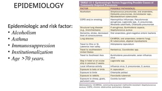
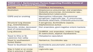
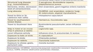
#20 Epidemiologic and risk factors may suggest the involvement of certain pathogens
alcoholism, asthma, immunosuppression, institutionalization, and age
>70 years.
In the elderly, decreased cough and gag reflexes and reduced
antibody and Toll-like receptor responses increase the likelihood
of pneumonia.
CA-MRSA and after viral infection.
Enterobacteriaceae tend to infect patients who have recently been hospitalized or given antibiotics or who have comorbidities
such as alcoholism, heart failure, or renal failure.
P. Aeruginosa is a particular problem in patients with severe structural lung disease
(e.g., bronchiectasis, cystic fibrosis, or severe COPD).
Legionella infection include diabetes, hematologic malignancy, cancer,
severe renal disease, HIV infection, smoking, male gender, and a recent
hotel stay or trip on a cruise ship
#21 Epidemiologic and risk factors may suggest the involvement of certain pathogens
Epidemiologic and risk factors may suggest the involvement of certain pathogens
alcoholism, asthma, immunosuppression, institutionalization, and age
>70 years.
In the elderly, decreased cough and gag reflexes and reduced
antibody and Toll-like receptor responses increase the likelihood
of pneumonia.
CA-MRSA and after viral infection.
Enterobacteriaceae tend to infect patients who have recently been hospitalized or given antibiotics or who have comorbidities
such as alcoholism, heart failure, or renal failure.
P. Aeruginosa is a particular problem in patients with severe structural lung disease
(e.g., bronchiectasis, cystic fibrosis, or severe COPD).
Legionella infection include diabetes, hematologic malignancy, cancer,
severe renal disease, HIV infection, smoking, male gender, and a recent
hotel stay or trip on a cruise ship
#22 Epidemiologic and risk factors may suggest the involvement of certain pathogens
alcoholism, asthma, immunosuppression, institutionalization, and age
>70 years.
In the elderly, decreased cough and gag reflexes and reduced
antibody and Toll-like receptor responses increase the likelihood
of pneumonia.
CA-MRSA and after viral infection.
Enterobacteriaceae tend to infect patients who have recently been hospitalized or given antibiotics or who have comorbidities
such as alcoholism, heart failure, or renal failure.
P. Aeruginosa is a particular problem in patients with severe structural lung disease
(e.g., bronchiectasis, cystic fibrosis, or severe COPD).
Legionella infection include diabetes, hematologic malignancy, cancer,
severe renal disease, HIV infection, smoking, male gender, and a recent
hotel stay or trip on a cruise ship
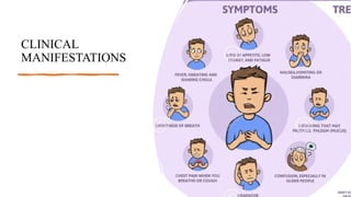
#24 ■CLINICAL MANIFESTATIONS
vary from indolent to fulminant and from mild to fatal in severity
■■DIAGNOSIS
the sensitivity and specificity of findings on physical examination are only 58% and 67%, respectively.
Febrile and/or tachycardic
Chills and/or sweats
Cough may be nonproductive or productive of mucoid, purulent, or blood-tinged sputum
Gross hemoptysis is suggestive of necrotizing pneumonia (e.g., that due to CA-MRSA)
Depending on severity, the patient may be able to speak in full sentences or may be short of breath
Pleuritic chest pain if with pleural involvement
20% of patients may have gastrointestinal symptoms such as nausea, vomiting, or diarrhea
Other symptoms may include fatigue, headache, myalgias, and arthralgias
febrile
tachycardic
chills and/or sweats
Cough - nonproductive or productive of mucoid, purulent, or blood-tinged sputum
Gross hemoptysis necrotizing pneumonia (e.g., that due to CA-MRSA)
short of breath.
pleuritic chest pain
gastrointestinal symptoms such as nausea, vomiting, or diarrhea
fatigue, headache,
myalgias, and arthralgias
#26 CXR
new infiltrate on chest radiography
pneumatoceles in S. aureus infection
upper-lobe cavitating lesion in tuberculosis
Pneumonia-right-middle-lobe-4.jpg
SPUTUM GSCS
Main purpose of the sputum Gram’s stain is to ensure suitability of a specimen for culture
suitable sputum sample: >25 neutrophils and <10 squamous epithelial cells per low-power field
identify certain pathogens (e.g., S. pneumoniae, S. aureus, and gram-negative bacteria)
yield of positive cultures from sputum is ≤50%
ICU and intubated: deep-suction aspirate or bronchoalveolar lavage sample has a high yield on culture when sent ASAP
#27 SPUTUM GSCS
Main purpose of the sputum Gram’s stain is to ensure suitability of a specimen for culture
suitable sputum sample: >25 neutrophils and <10 squamous epithelial cells per low-power field
identify certain pathogens (e.g., S. pneumoniae, S. aureus, and gram-negative bacteria)
yield of positive cultures from sputum is ≤50%
ICU and intubated: deep-suction aspirate or bronchoalveolar lavage sample has a high yield on culture when sent ASAP
BLOOD CULTURES
• Only 5–14% of cultures from hospitalized CAP patients are positive, and the most common pathogen is S.
Pneumonia
Certain high-risk patients should have blood cultured:
neutropenia secondary to pneumonia
asplenia
complement deficiencies
chronic liver disease
severe CAP
those at risk of MRSA or P. aeruginosa infection
#28 The study showed that sputum GS is HIGHLY SPECIFIC for identifying S. pneumoniae, H. influenzae, S. aureus and Gram-negative bacilli infection. A positive sputum GS result can confirm the causative pathogen of CAP.
Based from these 2 studies, sputum Gram stain test is SENSITIVE AND HIGHLY SPECIFIC for identifying causative pathogens in adult patients with CAP.
Negative GS results cannot be used to conclude absence of respiratory pathogen; hence, discontinuation of antimicrobials in GS-negative sputum may be inappropriate.
The Infectious Diseases Society of America (IDSA) guidelines for the treatment of CAP recommend that sputum GS/CS be obtained for hospitalized patients, especially those at risk for methicillin-resistant Staphylococcus aureus (MRSA) and Pseudomonas infectious and those who received intravenous antibiotics within 90 days prior to admission (Metlay JP et al. 2019). Similarly, the National Institute for Health and Care Excellence (NICE) guidelines recommend that sputum cultures be done only for individuals with moderate or high severity CAP (National Clinical Guideline Centre 2014).
Similarly, the National Institute for Health and Care Excellence (NICE) guidelines recommend that sputum cultures be done only for individuals with moderate or high severity CAP (National Clinical Guideline Centre 2014).
#30 A 2004 retrospective cohort study involving 13,043 patients with pneumonia found that predictors of bacteremia include systolic BP <90 mmHg, temperature <35 or ≥40oC, pulse rate ≥125/min, liver disease, blood urea nitrogen ≥30 mg/dL, serum sodium <130 mmol/L, and WBC <5,000/mm3 or > 20,000/mm3. These predictors of bacteremia are more often found in individuals with severe illness.
A 2001 prospective cohort study of 209 patients with pneumonia found a statistically significant trend towards bacteremia among patients with higher Pneumonia Severity Index (PSI) grade. The PSI is an early prediction rule that uses a combination of demographic factors, co-morbid illnesses, laboratory and chest x-ray findings to determine prognosis.
This study demonstrated that the risk of bacteremia is higher in patients with severe illness (Falguera et al. 2009).
The IDSA guidelines for the treatment of CAP pneumonia recommend that blood cultures be obtained for hospitalized patients. Similarly, the NICE guidelines for the same condition recommend that blood cultures be done only for individuals with moderate- or high-severity CAP.
#31 Unstable or decompensated comorbidities
• Uncontrolled diabetes mellitus
• Active malignancies
• Neurologic disease in evolution
• Congestive heart failure Class II-IV
• Unstable coronary artery disease
• Renal failure on dialysis
• Uncompensated COPD
• Decompensated liver disease
High risk CAP: Any of the clinical feature of moderate risk CAP plus any of the following: Severe sepsis and Septic shock OR need for mechanical ventilator
#32 Unstable or decompensated comorbidities
• Uncontrolled diabetes mellitus
• Active malignancies
• Neurologic disease in evolution
• Congestive heart failure Class II-IV
• Unstable coronary artery disease
• Renal failure on dialysis
• Uncompensated COPD
• Decompensated liver disease
High risk CAP: Any of the clinical feature of moderate risk CAP plus any of the following: Severe sepsis and Septic shock OR need for mechanical ventilator
#34 When a clinical diagnosis of community-acquired pneumonia is made in primary care, determine whether patients are at low, intermediate or high risk of death using the CRB65 score.
CURB-65 Criteria include 5 variables: Confusion of new onset
Urea (BUN) >7 mmol/L (19 mg/dL)
Respiratory rate ≥30 bpm
Blood pressure ≤ 90/60 mmHg
Age ≥65 years old
Patients with a score of 0 or 1, can be treated outside the hospital
Patient score 2 or more, the patient should be hospitalized unless the score is entirely or in part attributable to an age of more than or equal to 65 years old.
Among patients with scores of ≥ 3, these patients may require ICU admission
age of ≥65 year
score of 0 (a 30-day mortality rate of 1.5%) can be treated as outpatients
score of 1 or 2, the patient should be hospitalized unless the score is entirely or in part
attributable to an age of ≥65 years; in such cases, hospitalization may not be necessary
scores of ≥3, mortality rates are 22% overall; these patients may require ICU admission.
*Neither PSI nor CURB-65 is accurate in determining the need for ICU admission
*Patients with septic shock requiring vasopressors or with acute respiratory failure requiring
intubation and mechanical ventilation should be admitted directly to an ICU
#42 Influenza infection is a self-limited disease which causes uncomplicated, acute febrile respiratory symptoms but may also cause significant morbidity and mortality (Uyeki et al. 2019). Influenza virus can result in pneumonia which may be severe or fatal. Individuals infected with influenza are also at risk for co-infection or secondary infection by bacterial pathogens. The defined influenza season in the Philippines is from June to November.
Shown in Figure 1 is a guide from the U.S. Centers for Disease Control website for influenza testing when influenza virus is circulating in the community.
The benefits of antiviral therapy support testing of patients during periods of high influenza activity.
IDSA recommends the use of rapid influenza molecular assays over rapid influenza diagnostic tests (RIDTs) for detection of influenza viruses in respiratory specimens of outpatients, and the use of Reverse Transcription-Polymerase Chain Reaction (RT-PCR) or other molecular assays for hospitalized patients.
We recommend testing of respiratory secretions for influenza through rapid molecular testing using rapid nucleic acid amplification tests during periods of high influenza activity (July to January) for patients with high risk CAP preceded by influenza-like illness symptoms (sore throat, rhinorrhea, body malaise, joint pains) and any of the following risk factors:
• Aged 60 years and above
• Pregnant
• Asthmatic
• Other co-morbidities: uncontrolled diabetes mellitus, active malignancies, neurologic disease in evolution, congestive heart failure class II-IV, unstable coronary artery disease, renal failure on dialysis, uncompensated COPD, decompensated liver disease.
#43 The 2018 NICE clinical practice guidelines for the diagnosis and management of pneumonia in adults recommends considering the use of Legionella urine antigen tests (UATs) in moderate to severe CAP.
The 2019 American Thoracic Society (ATS) and IDSA guidelines suggest not routinely testing urine for Legionella antigen in adults with CAP unless indicated by epidemiological factors such as in Legionella outbreaks, patients with history of recent travel, or patients with severe CAP.
A multicenter, prospective, surveillance study of hospitalized patients with CAP in 2018 evaluated the sensitivity and specificity of the IDSA/ATS indications for performing UATs in identifying Legionella. These indications include ICU admission, failure of outpatient antibiotic therapy, active alcohol abuse, recent travel, and pleural effusion.
A major issue with the use of UAT is whether positive results will significantly alter therapy, since most guidelines recommend that patients with severe CAP be given empiric treatment with antibiotics active against this pathogen.
A randomized control trial was conducted in 2009 on 177 hospitalized patients with CAP who were given empiric guideline-directed treatment or pathogen-directed treatment based on UAT results. Out of the 88 patients given pathogen-directed treatment, 25 (28%) had positive UAT results, with 22 patients positive for Streptococcal pneumoniae and 3 patients positive for Legionella. There were no statistical differences in death, clinical relapse, ICU admission, length of hospitalization, and length of antibiotic treatment in the 2 treatment groups.
In summary, RCTs do not demonstrate benefit for Legionella UAT. This finding is accompanied by concerns that narrowing the spectrum of antibiotic therapy in response to positive UATs could lead to increased risk of clinical relapse. Current empiric treatment recommendations for patients with severe CAP already include the use of antibiotics with activity against Legionella.
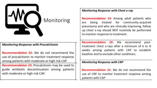
#44 One open-label pragmatic RCT conducted in 2017 evaluated the impact of routine point-of-care testing for respiratory viruses using multiplex polymerase chain reaction (PCR) compared to routine clinical care among adults with acute respiratory illness. Results of the study showed no significant reductions in antibiotic use and duration of antibiotic use.
These 2 studies demonstrated similar trends toward benefit of multiplex PCR in the reduction in antibiotic use, duration of antibiotic use, length of hospital stay, and use of hospital isolation facilities, and multiplex PCR guided-antiviral use. However, the results were not statistically significant.
#45 To this date there are no clinical trials available comparing treatment regimens with and without anaerobic coverage for patients hospitalized with suspected aspiration. However, in the background of increasing prevalence of antibiotic resistant pathogens and antibiotic complications, judicious use of antibiotics is encouraged, such that IDSA 2019 CAP guideline does not recommend routinely adding anaerobic coverage for suspected aspiration pneumonia unless lung abscess or empyema is suspected.
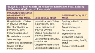
#47 The IDSA 2019 CAP guidelines abandoned the use of the categorization of Healthcare-associated pneumonia (HCAP). Many studies showed that the risk factors that defined HCAP did not predict higher prevalence of pathogens resistant to standard first-line antibiotic therapy. More importantly, the use of HCAP only resulted in a significant increased use of broad-spectrum antibiotics (especially vancomycin and antipseudomonal beta-lactams) without improvement in patient outcomes. As a replacement, the IDSA 2019 CAP guidelines proposed obtaining local data on the prevalence of multi-drug resistant organisms (MDRO) in patients with CAP, along with identification of risk factors for these infections at a local level.
MRSA
The most strongly and consistently associated risk factors for CAP due to MRSA were previous MRSA colonization or infection, especially of the respiratory tract, within 1 year [(OR 6.21, 95% CI 3.25-11.85), Aliberti 2016; (OR 6.05, 95% CI 2.99-12.22), Jung 2013], and intravenous antibiotic therapy within 90 days.
P. aeruginosa
Previous P. aeruginosa colonization or infection of the respiratory tract (OR 16.10, 95%CI 9.48-27.35) and severe bronchopulmonary disease [very severe chronic obstructive pulmonary disease {COPD} (OR 2.76, 95% CI 1.25-6.06)], bronchiectasis (OR 2.88, 95% CI 1.65-5.05), prior tracheostomy (OR 6.5, 95% CI 2.61-16.19) were independent risk factors for CAP due to P. aeruginosa (Restrepo 2018).
#48 The CAP guideline of the ATS/IDSA favors the use of antiviral therapy for adults with CAP who test positive for influenza virus. For inpatients, use of antiviral therapy is a strong recommendation based on moderate quality of evidence. For outpatients, use of antiviral therapy is a conditional recommendation based on low quality of evidence (Metlay et al. 2019).
The IDSA influenza guideline recommends giving antibiotic and antiviral treatment for patients with suspected or laboratory-confirmed influenza with bacterial coinfection who present with severe disease such as extensive pneumonia, respiratory failure, hypotension, and fever (Uyeki et al. 2019).
A randomized, open label, trial evaluated the effect of providing oseltamivir compared to standard of care on clinical failure. Clinical failure was defined as failure to reach clinical improvement within 7 days, transfer to the intensive care unit after 24 hours in a ward, or need for re-hospitalization within 30 days.
#50 Antibiotics, the mainstay for the treatment of pneumonia, should be initiated as soon as a diagnosis of CAP is made. Time of the first antimicrobial dose (TFAD) is defined as the time in hours from arrival at the emergency department (ED) to the intravenous infusion of the antimicrobial. (Bordon 2013) NICE CPG 2019 recommends that antibiotic therapy be started as soon as possible after diagnosis, and within 4 hours of admission (Strong Recommendation, Low Quality of Evidence).
The NICE CAP Guideline Development Group (GDG) acknowledged that making an early confident diagnosis of CAP is not always straightforward. They concluded that when a diagnosis of CAP is made with reasonable confidence, it is desirable to administer antibiotic therapy as soon as possible. However, this has to be balanced with avoiding inappropriate antibiotic prescribing for patients who do not have CAP, but in whom this is considered a potential differential diagnosis. Earlier antibiotic prescribing could be associated with higher rates of misdiagnosis and inappropriate prescribing, which could result in harm to patients (such as adverse events due to antibiotic therapy) and to the wider population (such as increased antibiotic resistance) as well as being wasteful from an economic standpoint. However, it was considered that the cost of adverse events and inappropriate prescribing were likely to be outweighed by the additional risk of mortality associated with inappropriately delayed antibiotic therapy.
Swift diagnostic procedures should be encouraged as part of the timing recommendation wherever possible, without discouraging clinical judgment. In patients with suspected CAP who are severely ill, antibiotic therapy should not be withheld until investigations such as chest X-ray are performed.
Antibiotic therapy ≥ 4 hours vs ≤4 hours
For the key outcome of mortality, the majority of the studies (mainly retrospective chart reviews) suggested that administering antibiotic therapy within the first 4 hours of admission was beneficial in reducing mortality.
Suggested that the benefit of antibiotic administration within the first 4 hours of admission was slightly greater for patients with low-to moderate-severity CAP compared with the high-severity group for the outcomes of (1) 30-day mortality- AOR: 0.62 (95% CI 0.42-0.92) for low-to-mod-severity vs 0.87 (95% CI 0.78-0.97) for high-severity), (2) length of hospital stay AOR 0.86 (95% CI 0.75 - 0.99) for low-to-mod-severity vs 0.92 (95% CI 0.84 - 1.01) for high-severity and (3) re-admission after discharge AOR 0.87 (95% CI 0.70-1.08) for low-to-mod-severity vs 0.99 (95% CI 0.88-1.11) for high-severity.
The clinical events in CAP go from establishment of infection, to onset of symptoms and arrival in the ED to TFAD. The priority of the management of patients with presumptive pneumonia should be to increase the accuracy of the diagnosis of CAP for appropriate and timely antimicrobial therapy. (Bordon 2013) Rather than designating a specific window in which to initiate treatment, the 2007 IDSA guidelines committee felt that hospitalized patients with CAP should receive the first antibiotic dose in the ED. The committee does feel that therapy should be administered as soon as possible after the diagnosis is considered likely.
#61 CAP MR
CHF FC II 2 HCVD
DM T2, DM foot L s/p Debridement


![ANIMAL_CELL_,_TISSUE_AND_ORGAN_CULTURE[1].pptx](https://cdn.slidesharecdn.com/ss_thumbnails/animalcelltissueandorganculture1-260204172026-4462b440-thumbnail.jpg?width=640&height=640&fit=bounds)


