PARTURITION is definedas the process of bringing forth of
young which comprises of multiple transformations in
both uterine and cervical functions
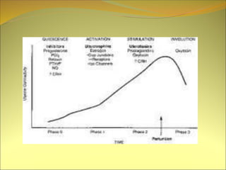
There are four phases :
Quiescence
Activation phase
Stimulation phase
Involution phase.
3.
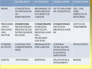
PHASES OF PARTURITION
QUIESCENCEACTIVATION STIMULATION INVOLUTION
FROM CONCEPTION
TO INITIATION
OF
PARTURITION
BEGINNING OF
PARTURITION
TO ONSET OF
LABOUR
UP TO DELIVERY
OF CONCEPTUS
TILL THE
TIME
FERTILITY IS
RESTORED
PREDOMIN
-ANTLY
INFLUENC
-ING
FACTOR
INHIBITORS
PROGESTRONE ,
PROSTACYCLIN,
NITROUSOXIDE,
RELAXIN
UTEROTROPIC
ESTROGEN,
OXYTOCIN ,
PROSTAGLAND
INS->
INCREASED
GAP JUNC.
UTEROTONICS
OXYTOCIN
PROSTAGLANDI
NS
OXYTOCIN
THROMBINS
UTERINE
ACTIVITY
CONTRACTILE
UNRESPONSIVE
NESS.
PREPARATION
FOR LABOUR
CONTRAC
TIONS ALONG
WITH FETAL &
PLACENTAL
EXPULSION
INVOLUTION
CERVIX SOFTENING RIPENING DILATATION &
EFFACEMENT
REPAIR
5.
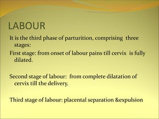
LABOUR
It is thethird phase of parturition, comprising three
stages:
First stage: from onset of labour pains till cervix is fully
dilated.
Second stage of labour: from complete dilatation of
cervix till the delivery.
Third stage of labour: placental separation &expulsion
6.
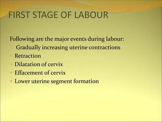
FIRST STAGE OFLABOUR
Following are the major events during labour:
Gradually increasing uterine contractions
Retraction
Dilatation of cervix
Effacement of cervix
Lower uterine segment formation
7.
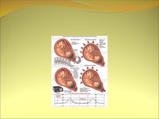
UTERINE CONTRACTIONS IN
LABOUR
Characteristicsof normal uterine contractions:
Pace maker: situated in the region of tubal ostia from where
wave of contraction spread downwards.
Sometimes there is emergence of multiple pace maker foci
leading to less efficient contractions and hence causing
primary dysfunction labour
Fundal dominance with gradual diminishing contractions
towards the lower segment.
Polarity of uterus : when upper segment contracts, retracts
and pushes the fetus down the lower uterine segment and
cervix dilates in response.
Lack of fundal dominance and the reverse polarity leads to
spastic lower uterine segment. Here pacemaker does not
work in rhythm.
8.
Good synchronizationof contraction waves from
both sides of uterus.
Regular pattern of contractions
Good relaxation in between the contractions
Intra amniotic pressure during relaxation is 8mm
rising beyond 20mm during contraction
10.
INTENSITY: describes degreeof uterine systole.
increases with progress of labour.Maximum during 2nd
stage of labour
DURATION: initially last for 10-15 seconds gradually
increases up to 40-45 sec.
FREQUENCY: in the early stage of labour, contractions
come at the interval of 10-15min and increases to maximum
in 2nd stage of labour.
Clinically contractions are said to be good when they come
after interval of 3-5minutes and at the height of
contractions uterine wall can not be indented by fingers.
11.
TONUS : intrauterine pressure in between the
contractions.
During Quiscent stage- 2-3mm Hg
During first stage of labour 8-10mmHg.
Factors governing tonus are:
Contractility of uterine muscles
Intra abdominal pressure
Over distension of uterus as in twins and
hydramnios.
12.
If the intensitydiminishes, duration is shortened and
period between the increases it leads to hypotonic
uterine dysfunction. Here intrauterine pressure
during the contractions remains below 25mm of Hg.
if there is increased frequency and duration without
adequate relaxation in between it leads to inco-
ordinate uterine action.
It comprises a rise in the base line tone which and hence
diminishing the circulation in the intervillous space of
placenta
13.
LABOUR PAINS
Pain duringcontractions is along the cutaneous nerve
distribution of T10 to L1
Pain of cervical dilatation is radiated to back through sacral
plexus
Causes of pain:
Myometrial hypoxia
Streching of peritonium over the fundus
Streching of cervix during dilatation
Compression of nerve ganglia
14.
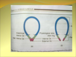
Retraction
Permanent shortening ofuterine muscle.
net effects are :
Formation of lower uterine segment.
Maintain advancement of presenting part made during
contractions
Reduce the surface area of uterus and hence favouring
placental separation.
Effective haemostasis after separation of placenta.
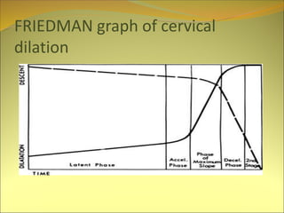
Latent phase :during which there is little dilatation
occurs with considerable changes taking place in the
connective tissue component of cervix which include:
Breaking down of collagen by collagease and elastases.
Accumulation of fluid between collagen fibres.
Fibro- muscular glandular hypertrophy.
Increased vascularity
Acceleration phase with cervical dilatation 2.5-4 cm.
Phase of maximum slope: between 4-9cm
Phase of decelaration: 9-10cm
17.
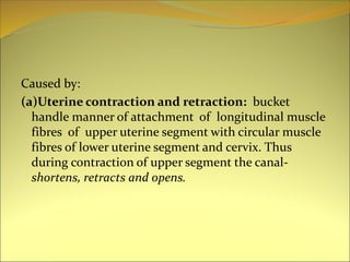
Caused by:
(a)Uterine contractionand retraction: bucket
handle manner of attachment of longitudinal muscle
fibres of upper uterine segment with circular muscle
fibres of lower uterine segment and cervix. Thus
during contraction of upper segment the canal-
shortens, retracts and opens.
19.
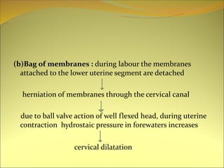
(b)Bag of membranes: during labour the membranes
attached to the lower uterine segment are detached
herniation of membranes through the cervical canal
due to ball valve action of well flexed head, during uterine
contraction hydrostaic pressure in forewaters increases
cervical dilatation
21.
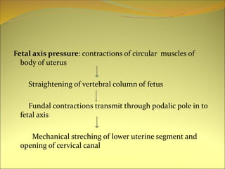
Fetal axis pressure:contractions of circular muscles of
body of uterus
Straightening of vertebral column of fetus
Fundal contractions transmit through podalic pole in to
fetal axis
Mechanical streching of lower uterine segment and
opening of cervical canal
22.
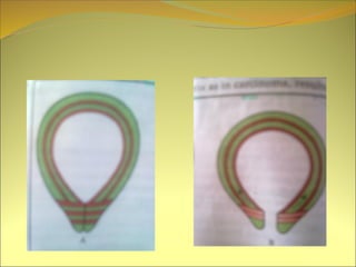
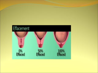
Effacement of cervix
MuscularFibres of cervix are pulled upwards and merge with
lower uterine segment.
Effacement precedes the dilatation in primegravidae
While it occurs simultaneously with dilatation in multiparae
24.
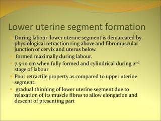
Lower uterine segmentformation
During labour lower uterine segment is demarcated by
physiological retraction ring above and fibromuscular
junction of cervix and uterus below.
formed maximally during labour.
7.5-10 cm when fully formed and cylindrical during 2nd
stage of labour
Poor retractile property as compared to upper uterine
segment.
gradual thinning of lower uterine segment due to
relaxation of its muscle fibres to allow elongation and
descent of presenting part
25.
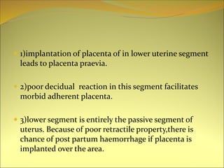
1)implantation ofplacenta of in lower uterine segment
leads to placenta praevia.
2)poor decidual reaction in this segment facilitates
morbid adherent placenta.
3)lower segment is entirely the passive segment of
uterus. Because of poor retractile property,there is
chance of post partum haemorrhage if placenta is
implanted over the area.
27.
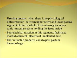
Uterine tetany:when there is no physiological
differentiation between upper active and lower passive
segment of uterus whole of the uterus goes in to a
tonic muscular spasm holding the fetus inside.
Poor decidual reaction in this segments facilitates
morbid adherent placenta if implanted here
Poor retractile property leads to post partum
haemorrhage.
28.
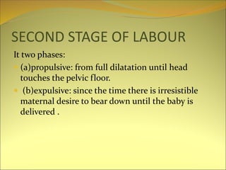
SECOND STAGE OFLABOUR
It two phases:
(a)propulsive: from full dilatation until head
touches the pelvic floor.
(b)expulsive: since the time there is irresistible
maternal desire to bear down until the baby is
delivered .
29.
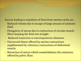
Factors leading toexpulsion of fetus from uterine cavity are :
Reduced volume due to escape of large amount of amniotic
fluid.
Elongation of uterus due to contraction of circular muscle
fibers keeping the fetal axis straight.
Reduced transverse or anterioposterior diameter.
Downward thurst offered by uterine contractions
supplimented by voluntary contractions of abdominal
muscle.
Retraction of uterus which counterbalance the resistance
offered by pelvic floor.
30.
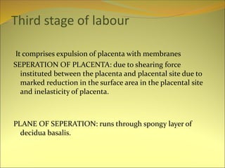
Third stage oflabour
It comprises expulsion of placenta with membranes
SEPERATION OF PLACENTA: due to shearing force
instituted between the placenta and placental site due to
marked reduction in the surface area in the placental site
and inelasticity of placenta.
PLANE OF SEPERATION: runs through spongy layer of
decidua basalis.
31.
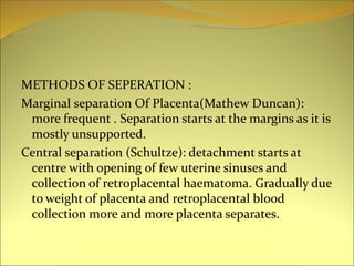
METHODS OF SEPERATION:
Marginal separation Of Placenta(Mathew Duncan):
more frequent . Separation starts at the margins as it is
mostly unsupported.
Central separation (Schultze): detachment starts at
centre with opening of few uterine sinuses and
collection of retroplacental haematoma. Gradually due
to weight of placenta and retroplacental blood
collection more and more placenta separates.
32.
SEPARATION OFMEMBRANES: The membranes in
the upper part are thrown in to folds while those in the
lower part are already detached due to stretching.
Expulsion of placenta : After complete separation the
placenta is forced in to the lower uterine segment and
then in the vagina.
Complete expulsion occures due bearing down efforts
of by manual procedure.
33.
HAEMOSTASIS
Living ligature: as the arterioles pass tortuously through
interlacing intermediate layers of myometrium they are
actually clamped during uterine contractions.
Thrombosis: occlude torn sinuses as pregnancy is
hypercoagulation state.
Myotamponade: apposition of walls of uterus after
expulsion of placenta.