1. Introduction
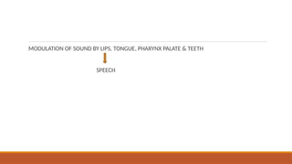
Phonation is the physiological process by which the human larynx generates sound, forming the basis for speech and vocal communication. It involves a complex interplay between respiratory airflow, laryngeal muscle activity, vocal fold vibration, and resonance in the vocal tract. The phenomenon of phonation transforms aerodynamic energy from exhaled air into acoustic energy, producing the fundamental tone of the voice. Subsequent modification by supralaryngeal structures—the pharynx, oral cavity, and nasal cavity—results in articulated speech.
Phonation is both a neuromuscular and aerodynamic process. It depends on precise coordination among the respiratory system (power source), the larynx (vibrator), and the supraglottic vocal tract (resonator and articulator). Understanding its physiology is fundamental to medicine, speech pathology, linguistics, and voice sciences.
2. Functional Anatomy of the Larynx
2.1. Position and Structure
The larynx, located in the anterior neck opposite the C3–C6 vertebrae, serves as the organ of phonation. It connects the pharynx above to the trachea below. Structurally, it consists of cartilages, intrinsic and extrinsic muscles, ligaments, and a mucosal lining.
2.2. Cartilages of the Larynx
The laryngeal framework is composed of nine cartilages—three unpaired and three paired:
Unpaired: Thyroid, Cricoid, Epiglottis
Paired: Arytenoid, Corniculate, Cuneiform
1. Thyroid Cartilage
Largest laryngeal cartilage.
Comprises two laminae meeting at the laryngeal prominence (“Adam’s apple”).
Provides anterior attachment for vocal folds via the thyroid angle.
2. Cricoid Cartilage
Signet-ring shaped, forming the base of the laryngeal framework.
Articulates with the thyroid cartilage (cricothyroid joint) and arytenoids (cricoarytenoid joints).
3. Arytenoid Cartilages
Pyramid-shaped, perched atop the cricoid’s posterior lamina.
Each arytenoid has a vocal process (anteriorly) and muscular process (laterally), serving as attachment points for vocal ligaments and intrinsic muscles.
4. Epiglottis
Leaf-shaped elastic cartilage that guards the laryngeal inlet during swallowing.
5. Corniculate and Cuneiform Cartilages
Small nodules within the aryepiglottic folds, supporting the laryngeal inlet.
2.3. Joints and Ligaments
Cricothyroid joint allows rotation and gliding movements between the cricoid and thyroid cartilages, crucial for pitch modulation.
Cricoarytenoid joints permit the arytenoids to rotate and glide, enabling vocal fold abduction and adduction.
Ligaments and membranes—such as the conus elasticus, quadrangular membrane, and vocal ligament—connect these cartilages and contribute to the vibratory system.
Vocal Folds
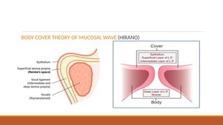
The vocal folds (true vocal cords) are paired structures extending from the thyroid cartilage anteriorly to the arytenoid cartilages posteriorly. Each fold consists of:
Epithelium – stratified squamous, protecting against friction.
Lamina propria – divided into:
Superficial layer (Reinke’s space) – loose